
Mycoplasma pneumoniae: Walking Pneumonia, No Cell Wall, Diagnosis, Treatment
Why Mycoplasma pneumoniae has no cell wall and beta-lactams cannot work, how it causes walking pneumonia and cold-agglutinin hemolysis, and how it is diagnosed and treated.

On this page
A school-age child has a mild cough, low-grade fever, and headache for over a week, but is still up and about, the classic picture of "walking pneumonia." The doctor prescribes amoxicillin. The child stays unwell, a chest x-ray shows patchy infiltrates, and paired serology shows rising Mycoplasma pneumoniae antibody.
The doctor switches to azithromycin, and within a couple of days the child improves. The amoxicillin did not make things worse; it did nothing at all. M. pneumoniae has no cell wall, and beta-lactam antibiotics work by attacking the cell wall, so there was no target for the drug to act on. That single fact, no cell wall, explains almost everything about this organism.
About Mycoplasma
Mycoplasma is the smallest free-living bacterium (about 0.2 to 0.8 micrometers), so small that it is below the reliable limit of the light microscope and can pass through filters that hold back most bacteria. It also has one of the smallest genomes of any free-living organism (roughly 500 to 1000 genes).
The single most important feature is that Mycoplasma has no cell wall. Almost every distinctive property follows from this one fact.
- No cell wall means no fixed shape, so M. pneumoniae is pleomorphic: round, pear-shaped, or filamentous.
- No cell wall means beta-lactam antibiotics (which target the cell wall) have nothing to act on, so M. pneumoniae is intrinsically resistant to all penicillins and cephalosporins. This is the point in the hook.
- No cell wall also means the organism does not Gram stain usefully and cannot be seen on a routine Gram film.
- Without the protection of a cell wall, M. pneumoniae is fragile and dries out easily, so it spreads person to person only through close contact by respiratory droplets. This is why outbreaks cluster in families, schools, and other close groups.
Most mycoplasmas are facultative anaerobes, but M. pneumoniae is a strict aerobe.
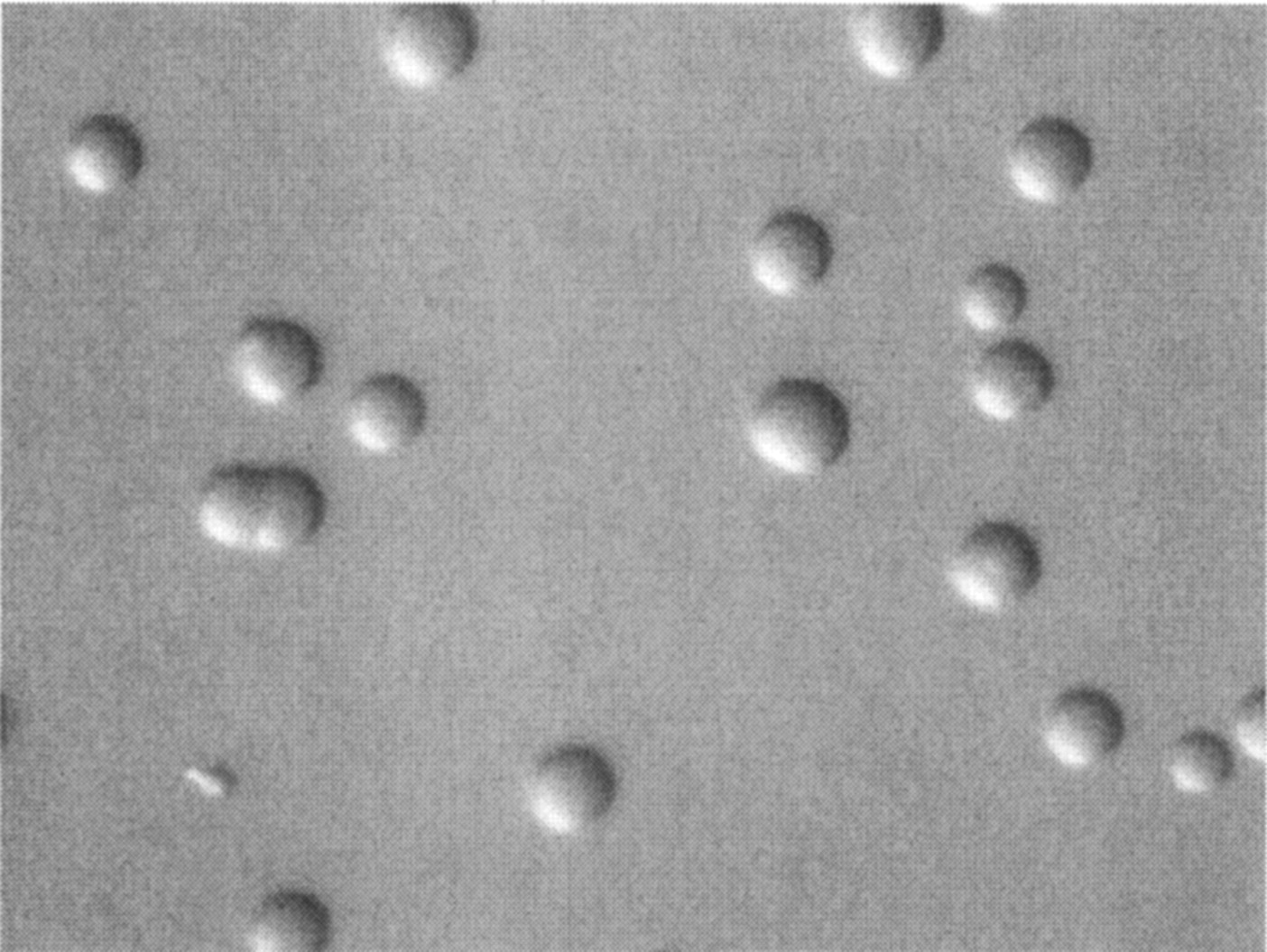 Figure: Spherical colonies of M. pneumoniae growing on SP4 agar(Source)
M. pneumoniae grows very slowly by binary fission, and its tiny colonies can take up to about three weeks to appear. On special media the colonies can show a "fried egg" shape (a denser center with a lighter edge), although M. pneumoniae colonies are often more granular and less classically "fried egg" than those of some other mycoplasmas.
Mycoplasmas are nutritionally demanding. Because they have no cell wall, they need sterols (such as cholesterol) to stabilize their membrane, so growth media must supply sterols. This sterol requirement is a defining feature of the group.
Extrapulmonary and autoimmune features
M. pneumoniae infection is well known for causing problems outside the lungs, through an autoimmune mechanism. The immune response against the organism can cross-react with the body's own cells.
- Cold agglutinins and hemolysis: many patients develop cold agglutinins, which are antibodies that clump red cells at low temperatures. In a minority of patients (around 10 percent may develop clinically significant hemolysis), these can cause cold-antibody hemolytic anemia. Cold agglutinins are a classic clue pointing to M. pneumoniae.
- Neurological disease: M. pneumoniae can trigger Guillain-Barré syndrome and other neurological complications, when antibodies against the organism cross-react with nerve tissue.
- Skin and mucous membranes: it can cause skin rashes and, in some cases, severe mucocutaneous disease (erythema multiforme and Stevens-Johnson-like reactions, sometimes called Mycoplasma-induced rash and mucositis).
These extrapulmonary features are exam favorites and are useful clinical clues, because they point to M. pneumoniae rather than an ordinary bacterial pneumonia.
As a separate practical point, mycoplasmas are a notorious contaminant of cell cultures in research and biotechnology laboratories. They are hard to detect and eliminate, which is why they are nicknamed the "crab grass" of cell culture.
Pathogenesis and Virulence
M. pneumoniae is an exclusively human pathogen, spread by respiratory droplets among close contacts. It causes a usually mild pneumonia, often called "walking pneumonia" because most patients are not sick enough to need hospital care. The pathogenesis has three parts.
Adhesion. M. pneumoniae has a specialized tip attachment organelle, with an adhesin protein called P1, that binds tightly to the respiratory epithelium. This close attachment lets the organism resist being swept away by the mucociliary clearance system, so it stays in the airway.
Direct injury and CARDS toxin. Once attached, it damages the ciliated epithelium and interferes with ciliary action. It also produces the Community-Acquired Respiratory Distress Syndrome (CARDS) toxin, which contributes to inflammation and airway dysfunction and is linked to more severe disease.
Immune-mediated damage. Much of the illness comes not from the organism destroying tissue directly, but from the host inflammatory and autoimmune response it provokes. This is the same cross-reacting immune response that produces the cold agglutinins and the extrapulmonary features above. It also helps explain why the x-ray can look worse than the patient (patchy infiltrates in a patient who is still walking around), a classic feature of atypical pneumonia.
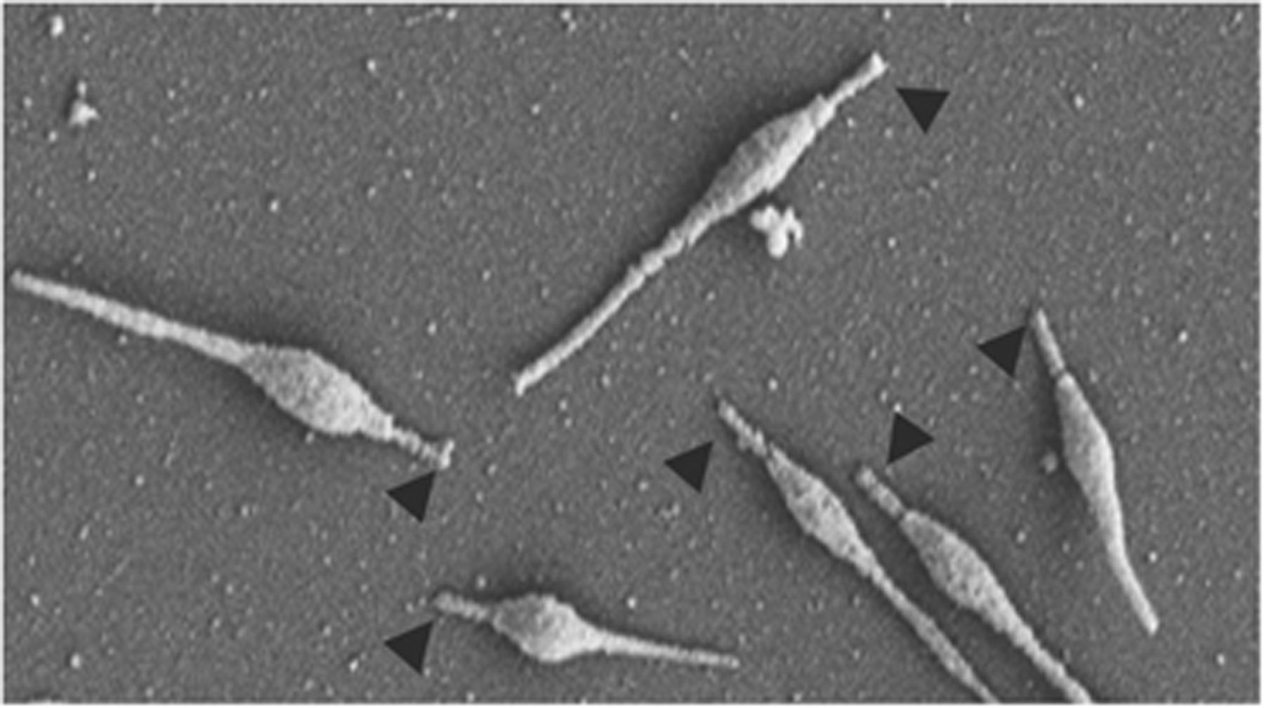 Figure: Scanning electron micrograph of Mycoplasma pneumoniae cells
Laboratory Diagnosis
Diagnosing M. pneumoniae is not done by routine Gram stain or culture, and understanding why makes the whole approach clear.
Why not Gram stain? It has no cell wall, so it does not take the Gram stain and cannot be seen on a routine film.
Culture: possible but impractical. It is slow (up to about three weeks), needs special sterol-containing media, and is used only by some reference laboratories, not for everyday diagnosis.
Molecular testing (PCR): the primary method for laboratory identification, including in CDC guidance. Nucleic acid amplification (real-time PCR) is fast, sensitive, and specific, and works directly on respiratory samples. This is the modern first-line test.
Serology: detecting a rising antibody titer between paired acute and convalescent samples supports the diagnosis. Its main drawback is that a single early sample is often not enough, because the antibody rise takes time, so serology is more useful in retrospect than for immediate management.
Cold agglutinins: a positive cold-agglutinin test is a non-specific but classic bedside clue that supports M. pneumoniae when the clinical picture fits, though it is not diagnostic on its own.
Because it is a cause of atypical pneumonia, M. pneumoniae is compared with the other atypical-pneumonia organisms in the table on the Legionella pneumophila article.
Treatment of Mycoplasma pneumoniae infection
Treatment follows from the biology. Because M. pneumoniae has no cell wall, cell-wall-active antibiotics (all penicillins and cephalosporins) are useless. Effective drugs are those that act on other targets, mainly protein synthesis.
The first-line drugs are macrolides (such as azithromycin), which are usually preferred, especially in children. The alternatives, used when a macrolide cannot be used or is not working, are tetracyclines (such as doxycycline) and respiratory fluoroquinolones. Many M. pneumoniae infections are mild and self-limiting, so not every case needs antibiotics.
An important and current point: macrolide resistance in M. pneumoniae has become common, and it varies widely by region. Reported resistance is relatively low in some parts of the world (for example, low double digits in parts of the United States and Europe) but very high in parts of East Asia, where a large majority of strains can be resistant. Where macrolide resistance is common or when a patient is not responding to a macrolide, doxycycline or a fluoroquinolone is used instead. This regional difference is worth knowing, because it directly affects the right first choice of drug.
How to Remember
| Device | The memory hook |
|---|---|
| No wall, no beta-lactam | The whole organism is "the one with no cell wall." No wall means no target for penicillins. If a walking pneumonia does not respond to amoxicillin, think Mycoplasma. |
| No wall, no Gram stain | No cell wall means it does not Gram stain and is not seen on a routine film. So "invisible on Gram stain" fits Mycoplasma. |
| Needs sterols | No wall means the membrane needs sterols (cholesterol) to stay stable, so it needs sterol-rich media. Unique among common bacteria. |
| Walking pneumonia | Patient is mildly ill and still "walking around," often with an x-ray that looks worse than the patient. Classic atypical pneumonia. |
| Cold agglutinins | Mycoplasma triggers cold agglutinins that can clump red cells in the cold and cause hemolytic anemia. "Cold" clue for Mycoplasma. |
| Fried egg colony | Tiny colonies with a dense center and lighter edge, like a fried egg, on special media. |
| Treatment = protein-synthesis drugs | Since the wall is out, hit protein synthesis: macrolides first, then doxycycline or a fluoroquinolone, especially where macrolide resistance is high. |
Key exam facts
| Feature | Mycoplasma pneumoniae |
|---|---|
| Cell wall | Absent (defining feature) |
| Size | Smallest free-living bacterium (~0.2–0.8 μm) |
| Genome | One of the smallest of free-living organisms |
| Shape | Pleomorphic (no fixed shape) |
| Gram stain | Not useful (no cell wall) |
| Membrane requirement | Needs sterols (cholesterol) |
| Oxygen | Strict aerobe |
| Colony | "Fried egg" on special media; grows up to ~3 weeks |
| Adhesion | Tip organelle with P1 adhesin |
| Toxin | CARDS toxin |
| Transmission | Respiratory droplets, close contact; human-only |
| Main disease | Walking pneumonia (mild atypical pneumonia) |
| Extrapulmonary | Cold-agglutinin hemolytic anemia, Guillain-Barré, mucocutaneous disease |
| Beta-lactams | Intrinsically resistant (no cell wall target) |
| Diagnosis | PCR (primary); serology (paired titers); cold agglutinins (clue) |
| Treatment | Macrolide first-line; doxycycline or fluoroquinolone (esp. if macrolide-resistant) |
Where Students Get Confused
| Confusion | The clarification |
|---|---|
| Why don't beta-lactams work? | M. pneumoniae has no cell wall, and beta-lactams act on the cell wall. There is no target, so penicillins and cephalosporins do nothing. |
| Why can't you Gram stain it? | Gram staining depends on the cell wall. With no cell wall, the organism does not stain and is not seen on a routine film. |
| Is it a virus? | No. It is a true bacterium (it is free-living and grows on cell-free media), it is just very small and wall-less. Its size and filterability once caused confusion with viruses. |
| Why does the x-ray look worse than the patient? | Much of the illness is from the immune response rather than direct destruction, so patients often feel relatively well ("walking") despite patchy x-ray changes. |
| What do cold agglutinins tell you? | They are a classic clue to M. pneumoniae and can cause cold hemolytic anemia, but they are not specific, so they support the diagnosis rather than prove it. |
| Is a single serology enough? | Often not. Antibody takes time to rise, so paired acute and convalescent samples are usually needed. PCR is better for immediate diagnosis. |
| Is a macrolide always the right treatment? | Not always. Macrolide resistance is high in some regions, so where resistance is common or the patient is not improving, doxycycline or a fluoroquinolone is used. |
References
- Tille, P. M. (2022). Bailey & Scott's Diagnostic Microbiology (15th ed.). Elsevier.
- Procop, G. W., & Koneman, E. W. (2017). Koneman's Color Atlas and Textbook of Diagnostic Microbiology (7th ed.). Wolters Kluwer.
- Carroll, K. C., Pfaller, M. A., et al. (2020). Murray's Medical Microbiology (9th ed.). Elsevier.
- Centers for Disease Control and Prevention. Mycoplasma pneumoniae infections: clinical and laboratory guidance. CDC (current version).
- Pereyre, S., Goret, J., & Bébéar, C. (2016). Mycoplasma pneumoniae: current knowledge on macrolide resistance and treatment. Frontiers in Microbiology, 7, 974.
Frequently Asked Questions
Why don't penicillins work against Mycoplasma pneumoniae?
Why don't penicillins work against Mycoplasma pneumoniae?
M. pneumoniae has no cell wall, and penicillins and other beta-lactam antibiotics work by attacking the bacterial cell wall. With no wall, there is no target, so these drugs have no effect. This is why treatment uses macrolides, doxycycline, or fluoroquinolones instead.
What is "walking pneumonia"?
What is "walking pneumonia"?
It is a mild pneumonia, often caused by M. pneumoniae, in which the patient feels only moderately unwell and can stay up and about rather than needing hospital care. The chest x-ray can look worse than the patient feels, which is a classic feature.
Why can't Mycoplasma pneumoniae be seen on a Gram stain?
Why can't Mycoplasma pneumoniae be seen on a Gram stain?
Gram staining depends on the bacterial cell wall, and M. pneumoniae has no cell wall. So it does not take the stain and is not visible on a routine Gram film. Diagnosis relies on PCR and serology instead.
What are cold agglutinins and how do they relate to Mycoplasma?
What are cold agglutinins and how do they relate to Mycoplasma?
Cold agglutinins are antibodies that make red blood cells clump together at low temperatures. M. pneumoniae infection often triggers them, and in some patients they cause a cold-type hemolytic anemia. A positive cold-agglutinin test is a classic clue to M. pneumoniae, though it is not specific.
How is Mycoplasma pneumoniae infection diagnosed?
How is Mycoplasma pneumoniae infection diagnosed?
The main laboratory method is PCR on a respiratory sample, which is fast, sensitive, and specific. Serology can support the diagnosis when paired acute and convalescent samples show a rising antibody titer. Routine Gram stain and culture are not useful for everyday diagnosis.
Can Mycoplasma pneumoniae cause problems outside the lungs?
Can Mycoplasma pneumoniae cause problems outside the lungs?
Yes. Through cross-reacting immune responses it can cause cold-agglutinin hemolytic anemia, neurological complications such as Guillain-Barré syndrome, and skin and mucous membrane disease. These extrapulmonary features are useful clues to the diagnosis.
Does Mycoplasma pneumonia always need antibiotics?
Does Mycoplasma pneumonia always need antibiotics?
No. Many infections are mild and self-limiting. When treatment is needed, a macrolide such as azithromycin is usually first-line, but because macrolide resistance is high in some regions, doxycycline or a fluoroquinolone may be used instead.

Tankeshwar Acharya, MSc (Medical Microbiology)
Tankeshwar Acharya is an Assistant Professor in the Department of Microbiology at Patan Academy of Health Sciences (PAHS), Nepal, where he has been teaching and practicing clinical microbiology for over 14 years. He is the founder of Microbe Online, one of the leading free microbiology education resources on the web, covering bacteriology, mycology, parasitology, immunology, and clinical laboratory diagnostics written from direct experience in both the classroom and the diagnostic laboratory.
Comments
No comments yet. Be the first to share your thoughts.
Leave a comment
All comments are reviewed before they appear.