
Bacteriology MCQs (1-10) With Answers and Explanations
Ten bacteriology MCQs with answer key and explanations: Gram stain, coagulase test, streptococcal classification, UTI and diarrhea pathogens, ID50, and serologic diagnosis.

On this page
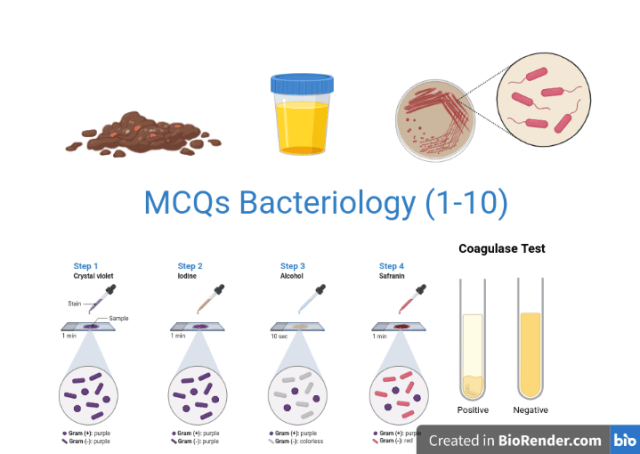 1. The identification of bacteria by serologic tests is based on the presence of specific antigens. Which of the following bacterial components is least likely to contain useful antigens?
a. Capsule
b. Cell wall
c. Flagella
d. Ribosomes
2. Each of the following statements concerning the Gram stain is correct except:
a. Escherichia coli stains pink because it has a thin peptidoglycan layer
b. Streptococcus pyogenes stains blue because it has a thick peptidoglycan layer
c Mycoplasma pneumoniae is not visible in the Gram’s stain because it does not have a cell wall
d. Mycobacterium tuberculosis stain blue because it has a thick lipid layer
3. An outbreak of sepsis caused by Staphylococcus aureus has occurred in the newborn nursery. You are called upon to investigate. According to your knowledge of the normal flora, what is the most likely source of the organism?
a. Nose
b. Colon
c. Hand
d. Throat
4. Each of the following organisms is an important cause of urinary tract infections except:
a. Klebsiella pneumoniae
b. Escherichia coli
c. Bacteroides fragilis
d. Proteus mirabilis
5. A 30-year woman has non-bloody diarrhea for the past 14 hours. Which one of the following organisms is least likely to cause this illness?
a. Streptococcus pyogenes
b. Clostridioides difficile
c. Shigella dysenteriae
d. Salmonella enteritidis
6. Each of the following agents is a recognized cause of diarrhea except
a. Clostridium perfringens
b. Vibrio cholerae
c. Enterococcus faecalis
d. Escherichia coli
7. Each of the following statements about the classification of streptococci is correct except
a. Pneumococci (Streptococcus pneumoniae) are alpha-hemolytic and can be serotyped on the basis of their polysaccharide capsule
b. Enterococci are group D streptococci and can be classified by their ability to grow 6.5% sodium chloride
c. Viridans streptococci are identified by Lancefield grouping, which is based on the C carbohydrate in the cell wall
d. Although pneumococci and the viridans streptococci are alpha-hemolytic, they can be differentiated by the bile solubility test and their susceptibility to optochin
8. Which of the following bacteria has the lowest 50% infective dose (ID50)?
a. Campylobacter jejuni
b. Salmonella typhi
c. Vibrio cholerae
d. Shigella sonnei
9. Which of the following disease is best diagnosed by serologic means?
a. Pulmonary tuberculosis
b. Gonorrhea
c. Actinomycosis
d. Q Fever
10. The coagulase test is used to differentiate
a. Staphylococcus epidermidis from Neisseria meningitidis
b. Staphylococcus aureus from Staphylococcus epidermidis
c. Streptococcus pyogenes from Staphylococcus aureus
d. Streptococcus pyogenes from Enterococcus faecalis
Answer Key
- d (Ribosomes)
- d (Mycobacterium tuberculosis stains blue because it has a thick lipid layer)
- a (Nose)
- c (Bacteroides fragilis)
- a (Streptococcus pyogenes)
- c (Enterococcus faecalis)
- c (Viridans streptococci are identified by Lancefield grouping)
- d (Shigella sonnei)
- d (Q fever)
- b (Staphylococcus aureus from Staphylococcus epidermidis)
Why these are the answers
- Ribosomes. Serologic identification relies on surface structures the immune system and antisera can reach: the capsule (K antigen), the cell wall O antigen, and the flagella (H antigen) are all classic serotyping targets. Ribosomes sit inside the cell and are highly conserved across species, so they carry no useful species-level antigenic signal. The pattern to remember: serology reads the outside of the cell, not the inside.
- The false statement is that Mycobacterium tuberculosis stains blue because of a thick lipid layer. In fact the lipid (mycolic acid) layer is exactly why M. tuberculosis resists the Gram stain and does not stain reliably at all; it needs the acid-fast (Ziehl-Neelsen) stain instead. Options a, b, and c are all true: E. coli is gram-negative (thin peptidoglycan, stains pink), S. pyogenes is gram-positive (thick peptidoglycan, stains purple), and Mycoplasma has no cell wall so it cannot be seen on Gram stain at all. The trap is that "thick lipid layer" sounds like a real explanation; it is the reason for non-staining, not for blue staining.
- Nose. This question asks for the reservoir in the normal flora, and the anterior nares are the primary carriage site for S. aureus: roughly 30% of healthy people carry it there. The clinically important subtlety, and why "Hand" is such a tempting distractor: in a nursery outbreak the organism usually reaches the newborns on the hands of staff, but those hands are contaminated from the carrier's own nose. The nose is the source (reservoir); the hand is the vehicle (transmission route). The question wording, "according to your knowledge of the normal flora," points at the reservoir.
- Bacteroides fragilis. The question asks which is NOT an important cause of UTI. E. coli, Klebsiella pneumoniae, and Proteus mirabilis are all leading uropathogens; Proteus especially, because its urease raises urine pH and forms struvite stones. Bacteroides fragilis is a strict anaerobe of the gut and rarely causes UTI, because urine and the urinary tract are not an anaerobic environment. Remember: UTIs are overwhelmingly aerobic and facultative gram-negative rods; a strict anaerobe in the list is almost always the odd one out.
- Streptococcus pyogenes. The question asks which is LEAST likely to cause non-bloody diarrhea. Clostridioides difficile, Shigella dysenteriae, and Salmonella Enteritidis are all enteric pathogens that cause diarrhea. S. pyogenes is a throat and skin pathogen (pharyngitis, cellulitis, impetigo); it is not an enteric pathogen and does not cause gastroenteritis. The clue is anatomical: three of the four live in or attack the gut, one does not.
- Enterococcus faecalis. The question asks which is NOT a recognized cause of diarrhea. Clostridium perfringens (toxin-mediated food poisoning), Vibrio cholerae (watery cholera), and diarrheagenic E. coli are all established causes. Enterococcus faecalis is a normal gut commensal and a cause of UTI, endocarditis, and wound infection, but it is not a recognized diarrheal pathogen.
- The false statement is that viridans streptococci are identified by Lancefield grouping. Lancefield grouping is based on the C carbohydrate in the cell wall and applies to beta-hemolytic streptococci (groups A, B, and so on). Viridans streptococci are alpha-hemolytic and lack a groupable C carbohydrate, so they are not Lancefield-typed; they are identified physiologically and biochemically instead. The other three statements are correct, including the key practical pair in option d: pneumococci and viridans strep are both alpha-hemolytic but are separated by the bile solubility test and optochin susceptibility (pneumococci are bile-soluble and optochin-sensitive; viridans are neither).
- Shigella sonnei. This asks for the lowest 50% infective dose. Shigella is famous for needing only a tiny inoculum, on the order of 10 to 100 organisms, because it survives stomach acid well and is highly efficient at invading the colonic mucosa. Salmonella Typhi, Campylobacter jejuni, and Vibrio cholerae all require far larger doses (typically thousands to millions) to establish infection. This low infective dose is exactly why Shigella spreads readily by person-to-person contact, unlike the food-and-water-dose-dependent spread of the others.
- Q fever. Coxiella burnetii, the cause of Q fever, is diagnosed serologically because it is hazardous to culture (a biosafety risk) and does not grow on routine media. Pulmonary tuberculosis is diagnosed by smear, culture, and molecular tests; gonorrhea by culture or NAAT; actinomycosis by identifying sulfur granules and anaerobic culture. The rule of thumb: serology is the go-to when the organism is dangerous or impractical to culture, which fits Coxiella precisely.
- The coagulase test separates Staphylococcus aureus from Staphylococcus epidermidis. Both are gram-positive cocci in clusters and both are catalase-positive, so they need a further test to tell apart: S. aureus is coagulase-positive (it clots plasma), while S. epidermidis and the other skin staphylococci are coagulase-negative. The other pairings are wrong because those organisms are already separated by simpler tests: staphylococci versus streptococci by catalase, and Neisseria is a gram-negative diplococcus, distinguishable on Gram stain alone.

Tankeshwar Acharya, MSc (Medical Microbiology)
Tankeshwar Acharya is an Assistant Professor in the Department of Microbiology at Patan Academy of Health Sciences (PAHS), Nepal, where he has been teaching and practicing clinical microbiology for over 14 years. He is the founder of Microbe Online, one of the leading free microbiology education resources on the web, covering bacteriology, mycology, parasitology, immunology, and clinical laboratory diagnostics written from direct experience in both the classroom and the diagnostic laboratory.
Comments
No comments yet. Be the first to share your thoughts.
Leave a comment
All comments are reviewed before they appear.