
Modified Oxidase (Microdase) Test: The DMSO Trick That Separates Micrococcus From Staphylococcus

On this page
Why It Matters
Two Gram-positive cocci in clusters, both catalase-positive, sit on a plate: a Staphylococcus and a Micrococcus. Under the microscope and on routine media they look alike, but the distinction matters. Staphylococcus includes some of the most important human pathogens (S. aureus, and the coagulase-negative species that infect prosthetic devices). Micrococcus is an environmental organism and skin commensal that rarely causes disease. Calling a Micrococcus a Staphylococcus wastes a workup; missing a true Staphylococcus is worse.
The ordinary oxidase test, the one used every day on Gram-negative rods, would seem the obvious tool, since Micrococcus has cytochrome c oxidase and most staphylococci do not. But there is a problem: the standard oxidase reagent does not work reliably on Gram-positive cocci. Their thick cell walls keep the reagent from reaching the cytochrome inside, giving weak or false results.
The modified oxidase (microdase) test solves this with one change: it dissolves the oxidase reagent in dimethyl sulfoxide (DMSO), a solvent that makes the cells permeable so the reagent can reach the cytochrome c. With that single modification, the test becomes reliable on Gram-positive cocci: Micrococcus turns the disk blue within two minutes, Staphylococcus stays colorless.
This article covers why the DMSO modification is necessary, the cytochrome-c mechanism behind the color, and the handful of staphylococci that break the rule. For the full set of tests that separate Staphylococcus from Micrococcus, see the Staphylococcus vs Micrococcus comparison.
Modified oxidase test (microdase) is recommended for Gram-positive, catalase-positive cocci, only. The microdase test is a rapid method to differentiate Staphylococcus (-ve) from Micrococcus +ve) by detection of the enzyme oxidase. Filter paper disks impregnated with tetramethyl-p-phenylenediamine dihydrochloride (oxidase reagent) in dimethyl sulfoxide (DMSO) are used.
Principle: detecting cytochrome c, and why DMSO is essential
The modified oxidase test detects the same enzyme as the standard oxidase test: cytochrome c oxidase, part of the electron transport chain. The reagent, tetramethyl-p-phenylenediamine (TMPD), is an artificial electron donor. If the organism has cytochrome c oxidase, it oxidizes the TMPD to a blue-purple compound, indophenol. Blue disk = cytochrome c oxidase present; no color = absent.
Among the catalase-positive Gram-positive cocci, this maps cleanly onto genus: Micrococcus has cytochrome c and is positive; most Staphylococcus lack it and are negative.
So why not just use the ordinary oxidase reagent? Because it does not work reliably on Gram-positive cocci. Their cell walls are thick enough to block the water-based reagent from reaching the cytochrome inside the cell, giving weak, slow, or false-negative results. This is the problem Faller and Schleifer solved in 1981: they dissolved the TMPD in dimethyl sulfoxide (DMSO) instead of water. DMSO is a penetrating solvent, it renders the cells permeable, carrying the reagent through the cell wall to the cytochrome. It also keeps the reagent stable against auto-oxidation.
That single change, TMPD in DMSO rather than in water, is the entire "modification" in "modified oxidase." It is what makes a reliable oxidase reading possible on Gram-positive cocci, where the standard test fails.
Modified oxidase test is performed in the same manner as the oxidase test, except that the reagent is 6% tetramethyl phenylenediamine hydrochloride in dimethyl sulfoxide. The original Faller-Schleifer formulation uses 6% TMPD in DMSO; commercial microdase disks are typically impregnated at a lower concentration (around 1%). Both work; the 6% figure is the original reference method.
Procedure
- Using sterile forceps, transfer a microdase disk from the stock bottle to a petri dish. Return the bottle of disks to the refrigerator, promptly.
- Using a wooden applicator stick, rub a small amount of several colonies of an 18-24 hour pure culture grown on blood agar onto the top of microdase disk.
- Incubate at room temperate for 2 minutes
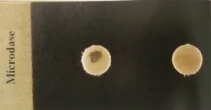 Figure: Microdase Test (Modified Oxidase Test). Right: Negative Left: Positive
Result
- Positive test: A blue or purple-blue color change within 2 minutes
- Negative test: No change in color
Quality Control
- Positive control: Micrococcus luteus ATCC 10240 (or ATCC 381) — blue within 2 minutes
- Negative control: Staphylococcus aureus ATCC 25923 — no color change
Run controls with each new lot of disks. For oxidase-variable isolates that give equivocal results, the Tarrand-Gröschel modification (a longer reaction read or a modified reagent) can clarify the reading.
Where the microdase test fits
The microdase test is one of several tests used to separate Staphylococcus from Micrococcus. The others include the bacitracin (Taxo A) disk (Staphylococcus resistant, Micrococcus susceptible), furazolidone susceptibility (Staphylococcus susceptible, Micrococcus resistant), and lysostaphin (Staphylococcus susceptible, Micrococcus resistant). No single test is infallible, and they are often read together. The microdase test is the most direct of them, because it detects the underlying biochemical difference (cytochrome c) rather than an antibiotic response. For the full comparison and when to reach for each test, see the Staphylococcus vs Micrococcus article.
How to remember
Micrococcus makes it blue; Staph stays clear. The microdase disk goes blue for Micrococcus (which has cytochrome c) and stays colorless for Staphylococcus (which usually lacks it). Blue = Micrococcus, the harmless one. Clear = Staphylococcus, the one you care about clinically.
The whole trick is DMSO. The standard oxidase reagent cannot get through a Gram-positive cell wall. Dissolving it in DMSO lets it penetrate. "Modified" oxidase just means "oxidase reagent in DMSO so it works on Gram-positive cocci." If you remember one thing, remember the solvent.
Three staphs break the rule: sciuri, lentus, vitulinus. A few staphylococci carry c-type cytochromes and give a positive (blue) microdase, the exceptions to "Staph is negative." S. sciuri, S. lentus, S. vitulinus. If a "Staph" is microdase-positive, think of these three (or reconsider whether it is really a Micrococcus).
Detects the cause, not a symptom. Unlike bacitracin or furazolidone (which measure an antibiotic response), microdase detects the actual biochemical difference, cytochrome c. That is why it is the most direct of the Staph/Micrococcus tests.
Key exam facts in one table
| Question | Answer | The reason behind it |
|---|---|---|
| What does the test detect? | Cytochrome c oxidase | Via TMPD → indophenol (blue) |
| What organisms is it for? | Catalase-positive Gram-positive cocci | Separates Micrococcus from Staphylococcus |
| Reagent | Tetramethyl-p-phenylenediamine (TMPD) in DMSO | DMSO penetrates the Gram-positive cell wall |
| Original reagent concentration | 6% TMPD in DMSO (Faller-Schleifer) | Commercial disks ~1% |
| Why DMSO (not water)? | Standard reagent can't reach cytochrome in GPC | DMSO makes cells permeable |
| Positive result | Blue/purple-blue within 2 minutes | Indophenol formed |
| Negative result | No color change | No cytochrome c oxidase |
| Micrococcus result | Positive (blue) | Has cytochrome c |
| Staphylococcus result | Negative (no color) | Usually lacks cytochrome c |
| Exceptions (positive staphs) | S. sciuri, S. lentus, S. vitulinus | Carry c-type cytochromes |
| Read time | Within 2 minutes | Later reads risk auto-oxidation false positives |
| Culture age | 18-24h (or 24-36h) on blood agar | Too young/old gives inaccurate results |
| Positive QC | Micrococcus luteus ATCC 10240 | Reliable positive |
| Negative QC | S. aureus ATCC 25923 | Reliable negative |
| Prerequisite | Gram-positive cocci, catalase-positive | The test is meaningless outside this group |
| vs standard oxidase test | Same enzyme; DMSO added for GPC | Standard test is for Gram-negative rods |
| Sibling separation tests | Bacitracin, furazolidone, lysostaphin | See the Staph vs Micrococcus comparison |
Where students get confused
Using the standard oxidase reagent on Gram-positive cocci. The standard (water-based) oxidase reagent gives unreliable results on Gram-positive cocci because it cannot penetrate the cell wall. That is the whole reason the modified (DMSO) version exists. Do not substitute the ordinary oxidase reagent here.
Reading too late. TMPD auto-oxidizes in air, turning blue on its own over time. A color that appears after 2 minutes may be reagent auto-oxidation, not a true positive. Read within 2 minutes; disregard later color.
Forgetting the S. sciuri group exception. A microdase-positive result usually means Micrococcus, but S. sciuri, S. lentus, and S. vitulinus are staphylococci that give a positive. So a positive result on an organism you suspect is a staphylococcus should prompt considering these three, not an automatic Micrococcus call.
Running it on the wrong organisms. The test is only for catalase-positive Gram-positive cocci. It is not an oxidase test for Gram-negative rods (that is the standard oxidase test), and it is meaningless on catalase-negative cocci (streptococci, enterococci). Confirm Gram stain and catalase first.
Treating microdase as a standalone genus ID. Even done correctly, microdase distinguishes Micrococcus from Staphylococcus at the genus level, not to species. And because of the exceptions, it is often read alongside bacitracin, furazolidone, or lysostaphin. Use it as one line of evidence, not the whole answer.
Culture age. Too young or too old a culture gives inaccurate results. Use an 18-24 hour (up to 36 hour) pure culture from blood agar.
References
- Faller A, Schleifer KH. Modified oxidase and benzidine tests for separation of staphylococci from micrococci. J Clin Microbiol. 1981;13(6):1031-1035. doi:10.1128/jcm.13.6.1031-1035.1981
- Tarrand JJ, Gröschel DH. Rapid, modified oxidase test for oxidase-variable bacterial isolates. J Clin Microbiol. 1982;16(4):772-774. doi:10.1128/jcm.16.4.772-774.1982
- von Rheinbaben KE, et al. Comparison of various methods for differentiation of staphylococci and micrococci. J Clin Microbiol. 1984;19(6):875-879. doi:10.1128/jcm.19.6.875-879.1984
- Tille PM. Bailey and Scott's Diagnostic Microbiology. 15th ed. St. Louis: Elsevier; 2022.
Frequently Asked Questions
What is the difference between the oxidase test and the modified oxidase (microdase) test?
Why is DMSO used in the microdase test?
What does a positive microdase test indicate?
Which staphylococci give a positive microdase test?
Why must the microdase test be read within 2 minutes?

Tankeshwar Acharya, MSc (Medical Microbiology)
Tankeshwar Acharya is an Assistant Professor in the Department of Microbiology at Patan Academy of Health Sciences (PAHS), Nepal, where he has been teaching and practicing clinical microbiology for over 14 years. He is the founder of Microbe Online, one of the leading free microbiology education resources on the web, covering bacteriology, mycology, parasitology, immunology, and clinical laboratory diagnostics written from direct experience in both the classroom and the diagnostic laboratory.
Comments
No comments yet. Be the first to share your thoughts.
Leave a comment
All comments are reviewed before they appear.