

Why It Matters
Late in pregnancy, usually around 36 to 37 weeks, women are screened for one organism carried harmlessly in the vagina and rectum: Group B Streptococcus, Streptococcus agalactiae. The mother is unaffected. The concern is the newborn. If GBS is present during delivery it can pass to the baby and cause early-onset neonatal sepsis, pneumonia, and meningitis, and it remains a leading cause of life-threatening infection in the first week of life. A positive screen means intrapartum antibiotics, which sharply reduce that risk.
So a lab that grows a beta-hemolytic Streptococcus from a vaginal-rectal swab needs to answer one question fast: is this Group B Strep? Hippurate hydrolysis is one of the classic ways to answer it. S. agalactiae hydrolyzes hippurate; the other beta-hemolytic streptococci (Groups A, C, F, G) do not.
The test does a second job among the curved Gram-negative rods that cause gastroenteritis: Campylobacter jejuni hydrolyzes hippurate and C. coli does not, making hippurate the standard way to separate the two commonest Campylobacter species. Both calls are presumptive, confirmed by further testing, but both are the kind of fast answer that changes what happens next.
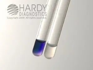 Figure: Hippurate hydrolysis test-Left (positive tube), Right (Negative tube)
Hippurate hydrolysis occurs in an organism that can form glycine and benzoic acid.
In the classical test (48 hours), the production of benzoic acid was detected using ferric chloride as an indicator. The gas-liquid chromatography (GLC) method is also available to detect benzoic acid.
In contrast, the rapid test (2 hours) detects glycine, the second byproduct of Hippurate hydrolysis. It uses ninhydrin as the indicator. Rapid Hippurate hydrolysis is more specific and as sensitive as the classical method.
Principle of Hippurate Hydrolysis
Hippurate is a single molecule made of two parts joined by an amide bond: benzoic acid and the amino acid glycine. Think of it as benzoic acid with a glycine attached. Hippuricase cuts that bond and releases both halves. This is why there are two detection methods that look completely different but test the same thing: the classical ferric chloride method watches for the benzoic acid half, and the rapid ninhydrin method watches for the glycine half. Two windows onto one cut.
Hippuric acid is hydrolyzed to benzoic acid and glycine by the enzymatic action of hippuricase. The addition of a ninhydrin reagent detects glycine. Ninhydrin reacts with glycine to form a deep blue or purple color (Ruhemann’s purple). Hwang and Ederer described this rapid test for detecting glycine.
Similarly, the traditional method detects benzoic acid using ferric chloride. The ferric chloride dissolves the benzoic acid present as residue after centrifugation of hippurate solution with the test organism.
Uses of Hippurate Hydrolysis
The hippurate test identifies Campylobacter jejuni, Listeria monocytogenes, Gardnerella vaginalis, and Streptococcus agalactiae.
- The hippurate hydrolysis test is critical for separating Campylobacter jejuni (hydrolyzes hippurate) and Campylobacter coli (negative) strains.
- It also aids in differentiating β-hemolytic Streptococcus agalactiae from other β-hemolytic streptococci.
Test Procedure
Classical Method
- Firstly, take or prepare sterile sodium hippurate broth and inoculate it with the test organism.
- After that incubate it overnight at 35°C.
- Then, centrifuge the broth and remove the sediment.
- Finally, add ferric chloride reagent to the supernatant.
- If the residue remains after 10 minutes, benzoic acid is present. The test is positive for hippurate hydrolysis.
Rapid Method (Ninhydrin)
- Add 0.2-0.4 mL of sodium hippurate reagent to a sterile tube.
- Make a heavy suspension of the test organism from an 18-24 hour culture, taking care not to pick up agar, which contains protein that can interfere with the reaction.
- Cap and incubate at 35-37°C for 2 hours, a water bath is preferred for even heating.
- Add 2-4 drops (about 0.2 mL) of ninhydrin reagent.
- Re-incubate at 35-37°C for an additional 10-30 minutes.
- Observe at 10-minute intervals for a deep blue or purple color (Ruhemann's purple).
Positive: deep blue/purple color within 30 minutes.
Negative: no color change, or a faint blue-gray tint.
Quality Control used in Hippurate Hydrolysis Test
Test each new lot or shipment of the reagent with known positive and negative controls, and retest at least monthly. Discard all reagents and prepare new ones if the reagents do not pass QC. Grow the ATCC strains in sodium hippurate broth for quality control and look for the results.
- Streptococcus agalactiae (ATCC 4768): Moderate to heavy growth and hydrolysis of hippurate.
- Streptococcus pyogenes (ATCC 19615): Moderate to heavy growth; hippurate not hydrolyzed.
Hippurate Hydrolysis Test Positive Organisms :
The hippurate hydrolysis test is used in the presumptive identification of various bacteria. Organisms giving positive (+ve) tests are:
- Gardnerella vaginalis,
- Campylobacter jejuni,
- Listeria monocytogenes and
- Group B streptococci (Streptococcus agalactiae)
Limitations
- False-positive results can occur if incubation with ninhydrin exceeds 30 min.
- A negative test does not rule out the identification of G. vaginalis since the biotypes that cause bacterial vaginosis can be hippurate negative.
- Viridans group streptococci can be hippurate positive; performing other tests to confirm the identification of non-hemolytic colonies is essential.
- A small number of enterococci are beta-hemolytic and may hydrolyze hippurate, but they are pyrrolidinyl-β-naphthylamide (PYR) positive (S. agalactiae is PYR negative)
- Furthermore a small percentage of C. jejuni organisms are hippurate negative and use of other methods for complete identification is necessary.
Where students actually get confused
- The rapid method is more sensitive with enterococci than the classical method, not less. A published evaluation found 95.4% of enterococcal Group D strains positive by the rapid method versus only 9.3% by the classical method. If you're using the rapid method, expect more Group D positives, that's the method being accurate, not a contamination problem.
- A weak hippurate-positive Group D streptococcus isn't automatically GBS. Bile esculin is the test that separates them, Group D hydrolyzes esculin, GBS doesn't.
- Viridans streptococci and PYR are the other tiebreaker. A small number of enterococci hydrolyze hippurate too, but they're PYR-positive while S. agalactiae is PYR-negative, exactly the same logic already covered on the PYR test page.
- Ninhydrin timing is not flexible. Reading past 30 minutes risks a false positive. Set a timer rather than checking back "whenever convenient."
Key exam facts in one table
| Feature | Detail | Memory hook |
|---|---|---|
| Detects | Hippuricase activity | Splits hippurate into glycine + benzoic acid |
| Classical method | Ferric chloride detects benzoic acid | 48 hours |
| Rapid method | Ninhydrin detects glycine | 2 hours, deep blue/purple |
| Identifies | S. agalactiae, C. jejuni, L. monocytogenes, G. vaginalis | |
| GAS/GBS panel role | The one result that doesn't flip | GBS positive, almost everything else negative |
| Group D false positive | Resolved by bile esculin | Group D hydrolyzes esculin, GBS doesn't |
| Enterococcus false positive | Resolved by PYR | Enterococcus PYR-positive, GBS PYR-negative |
| Ninhydrin read window | Within 30 minutes | Longer risks false positive |
References and further readings
- Hwang MN, Ederer GM. Rapid hippurate hydrolysis method for presumptive identification of group B streptococci. J Clin Microbiol. 1975;1(1):114-115. doi:10.1128/jcm.1.1.114-115.1975
- Wilkinson HW, Thacker LG, Facklam RR. Evaluation of the rapid hippurate hydrolysis test with enterococcal group D streptococci. J Clin Microbiol. 1977;5(3):290-292. doi:10.1128/jcm.5.3.290-292.1977
- Leber AL, editor. Clinical Microbiology Procedures Handbook. 4th ed. Washington, DC: ASM Press; 2016. doi:10.1128/9781555818814
- Procop GW, Church DL, Hall GS, Janda WM, Koneman EW, Schreckenberger PC, Woods GL. Koneman's Color Atlas and Textbook of Diagnostic Microbiology. 7th ed. Philadelphia: Wolters Kluwer; 2017.
- Tille PM. Bailey and Scott's Diagnostic Microbiology. 15th ed. St. Louis: Elsevier; 2022.

Comments
No comments yet. Be the first to share your thoughts.
Leave a comment
All comments are reviewed before they appear.