
Amoebic vs Bacillary Dysentery: Differences in Stool, Symptoms, and Microscopy
Amoebic and bacillary dysentery differ in stool appearance and microscopy. Compare them side by side, and learn which stool findings, ghost cells, pyknotic bodies, and Charcot-Leyden crystals, point to which cause.

On this page
Dysentery means bloody diarrhea. Two causes account for most cases, and they are treated very differently, so telling them apart matters.
Bacillary dysentery is caused by bacteria, most often Shigella. Amoebic dysentery is caused by a parasite, Entamoeba histolytica. A patient with bloody, mucoid stool could have either, and the clinical picture alone does not always separate them. The stool sample does.
This is one of the few places in clinical microbiology where simple stool microscopy, done well, can point straight to the cause and change treatment the same day: an antibiotic for Shigella, an antiparasitic for Entamoeba.
This article compares the two side by side, first the clinical picture, then the stool findings under the microscope. Bacteria other than Shigella can also cause bloody diarrhea, including enteroinvasive E. coli (EIEC), enterohemorrhagic E. coli (EHEC), Campylobacter, Salmonella, and Vibrio parahaemolyticus, but the classic comparison, and the one that turns up in exams and on the bench, is Shigella versus Entamoeba.
For the full biology of each organism, see the dedicated articles on Shigella and Entamoeba histolytica. This article is about telling the two apart.
The clinical picture: how they differ at the bedside
Before the stool reaches the microscope, the bedside picture gives a first steer, though it is not decisive on its own.
Fever. Bacillary dysentery usually comes with fever, sometimes high, because it is an acute invasive bacterial infection. Amoebic dysentery typically causes little or no fever. A dysentery with high fever and toxic appearance leans bacterial.
Onset and course. Bacillary dysentery tends to start abruptly and run an acute course over days. Amoebic dysentery often comes on more gradually and can become chronic or relapsing.
Stool volume and frequency. Bacillary dysentery produces frequent, small-volume stools (often more than ten a day) with intense straining (tenesmus). Amoebic dysentery produces fewer, more copious motions (typically six to eight a day).
The catch. These are tendencies, not rules. Either infection can present atypically, and the two can even occur together. This is exactly why the stool examination matters: it moves the diagnosis from a guess based on the clinical picture to a finding you can act on.
Amoebic vs bacillary dysentery: differences at a glance
Character | Amoebic dysentery | Bacillary dysentery |
Macroscopic | ||
Number | 6-8 motions a day | Over 10 motions a day |
Amount (Volume) | Relatively copious | Small amount |
Appearance and Amount | Blood mucus, semi formed | Blood mucus, mainly watery |
Odor | Offensive (foul-smelling) | Odorless |
Color | Dark red (altered blood) | Bright red (fresh blood) |
Reaction (stool pH) | Acidic | Alkaline |
Consistency | Not adherent to the container | Adherent to the container |
Microscopic | ||
RBCs | In clumps (typical of amoebic stool) | Discrete, sometimes in clumps due to rouleaux formation |
Pus Cells | Few | Numerous |
Macrophages | Few | Numerous; many contain ingested RBCs and can be mistaken for E. histolytica trophozoites (tell them apart by the nucleus and motility, see below) |
Eosinophils | Present | Scarce |
Charcot-Leyden (C-L) crystals | Present | Absent |
Pyknotic bodies | Present | Absent |
Ghost Cells | Absent | Present |
Parasites Seen | Trophozoites of E. histolytica | Absent |
Bacteria Seen | Mixed commensal flora, some motile (not a diagnostic feature) | Scanty; the invading organism (Shigella) is non-motile |
Growth on MacConkey Agar | Various intestinal flora may grow | Pure growth of Shigella spp. may be seen |
Further notes: Charcot-Leyden crystals, pyknotic bodies, and ghost cells are each explained in full, what they are, why they form, and which dysentery they point to, in the stool smear section below.
Reading the stool smear: which findings point to which dysentery on microscopy
The comparison table above lists the microscopy findings. This section explains what each one means at the bench, because a few specific findings can settle the diagnosis on their own.
The findings that point to amoebic dysentery
Trophozoites of Entamoeba histolytica with ingested red blood cells. This is the single most decisive finding. A motile trophozoite that has eaten red cells confirms invasive amoebic dysentery. Nothing in bacillary dysentery looks like this. The stool must be examined fresh (within about an hour) and kept warm, or the trophozoite stops moving and is easy to miss.
Charcot-Leyden crystals.
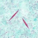 Figure: Charcot Leyden Crystals
These are hexagonal, bipyramidal (needle-like, pointed at both ends) crystals formed from the breakdown products of eosinophils. They point to a parasitic or allergic process rather than a bacterial one, so in a dysentery smear they favor amoebic over bacillary. They are not specific to amoebiasis (they appear in other parasitic and allergic conditions), but in the amoebic-versus-bacillary question they sit on the amoebic side.
Pyknotic bodies.
These are the shrunken, condensed nuclear remains of dead tissue cells and leukocytes. Their presence, alongside a relatively scanty cellular exudate, fits the amoebic pattern, where tissue is destroyed by the parasite's cytolytic action but the brisk neutrophil flood of bacterial infection is absent.
Few pus cells, eosinophils present.
Amoebic dysentery provokes less neutrophil infiltration than bacterial dysentery. A smear with only a few pus cells and some eosinophils, against a bloody background, fits amoebic disease.
The findings that point to bacillary dysentery
Numerous pus cells (neutrophils).
Bacterial invasion triggers an intense neutrophil response. Sheets of pus cells in the stool are the hallmark of bacillary dysentery and are the clearest cellular difference from the amoebic smear.
Ghost cells.
A ghost cell is a swollen epithelial cell that has lost its nucleus, leaving only the faint outline of its cytoplasm. These appear in bacillary dysentery and are absent in amoebic dysentery. This is a genuinely useful and often-forgotten discriminator: if you see ghost cells, think bacterial.
Many macrophages, some containing red cells.
Bacillary dysentery shows numerous macrophages. This is the classic trap: a macrophage that has engulfed red blood cells can look very much like an E. histolytica trophozoite with ingested red cells. The way to tell them apart is the nucleus. The E. histolytica trophozoite has a small central nucleolus (karyosome) and fine, evenly distributed peripheral chromatin, and it is often still motile in a fresh warm sample. A macrophage has a larger, irregular nucleus and does not move like a trophozoite. Mistaking one for the other is the most common serious error on a dysentery smear.
Scanty, non-motile bacteria.
Shigella is non-motile, so the bacterial background in bacillary dysentery lacks the swarming motile organisms seen in some other diarrheal stools. This absence of motile bacteria, combined with sheets of pus cells, fits Shigella.
The one-look summary
If the smear shows a motile trophozoite with ingested red cells, or Charcot-Leyden crystals with few pus cells, read it as amoebic. If it shows sheets of pus cells, ghost cells, and red-cell-containing macrophages, read it as bacillary. When a red-cell-containing cell is present, decide whether it is a trophozoite (amoebic) or a macrophage (bacterial) by its nucleus and its motility, because that single call often decides the diagnosis.
How to remember amoebic vs bacillary dysentery
Bacteria bring the pus and the fever. Bacillary dysentery is the loud, acute one: high fever, sheets of pus cells (neutrophils), many small stools, bright red fresh blood. Bacteria trigger a strong neutrophil response, so the smear is full of pus cells. Tie the two "B" words together: Bacillary means Bacterial, and Bacterial means Busy with pus cells.
Amoeba is quieter and leaves crystals and trophozoites. Amoebic dysentery is the milder, more chronic one: little fever, fewer but more copious stools, darker altered blood, and a smear with few pus cells but Charcot-Leyden crystals, eosinophils, and, if you are lucky, a motile trophozoite that has eaten red cells. Parasite equals crystals and trophozoites, not pus.
Ghost cells haunt the bacterial smear. Ghost cells (nucleus gone, only the cytoplasm outline left) appear in bacillary dysentery, not amoebic. A small memory hook: ghosts are pale and empty, and they show up on the bacterial side.
The trophozoite-vs-macrophage trap: look at the nucleus. Both can hold red cells. The E. histolytica trophozoite has a small central karyosome and fine peripheral chromatin and it moves; the macrophage has a big irregular nucleus and sits still. When in doubt, warm fresh stool and watch: only the trophozoite crawls.
Key exam facts
| Feature | Amoebic dysentery | Bacillary dysentery |
|---|---|---|
| Cause | Entamoeba histolytica (parasite) | Shigella and other bacteria |
| Fever | Little or none | Often present, sometimes high |
| Onset / course | Gradual, can be chronic/relapsing | Abrupt, acute |
| Stool frequency | Fewer, ~6 to 8 a day | Many, often >10 a day |
| Stool volume | Relatively copious | Small amount |
| Blood color | Dark red (altered) | Bright red (fresh) |
| Stool reaction (pH) | Acidic | Alkaline |
| Pus cells | Few | Numerous (sheets of neutrophils) |
| Charcot-Leyden crystals | Present | Absent |
| Pyknotic bodies | Present | Absent |
| Ghost cells | Absent | Present |
| Macrophages | Few | Numerous, may contain RBCs (trophozoite mimic) |
| Parasite on smear | Trophozoite with ingested RBCs | Absent |
| Decisive finding | Motile trophozoite with ingested RBCs | Sheets of pus cells + ghost cells |
| Treatment direction | Antiparasitic (metronidazole then a luminal agent) | Antibiotic per susceptibility; avoid anti-motility drugs |
Where Students Get Confused
A macrophage with red cells looks like an E. histolytica trophozoite. This is the classic trap and the most consequential one. In bacillary dysentery, macrophages that have engulfed red cells mimic the amoebic trophozoite. Separate them by the nucleus (trophozoite: small central karyosome, fine peripheral chromatin; macrophage: large irregular nucleus) and by motility (only the trophozoite moves in fresh warm stool). Calling a macrophage a trophozoite can send a bacterial dysentery down the wrong treatment path.
Bloody stool alone does not mean amoebiasis, and it does not mean Shigella either. Both cause bloody, mucoid stool. The blood is not the discriminator; the cellular findings are. Reaching for "bloody equals amoebic" is a common shortcut that fails often.
Charcot-Leyden crystals are not proof of amoebiasis. They point to a parasitic or allergic process and favor amoebic over bacillary in a dysentery smear, but they appear in other parasitic and allergic conditions too. They are a strong hint, not a stamp.
High fever leans bacterial, but its absence does not rule bacterial out. Bacillary dysentery usually brings fever and amoebic usually does not, so fever is a useful first steer. But atypical presentations happen, and the two can coexist. The stool, not the temperature, decides.
Amoebic dysentery is not just "a worse version" of bacillary. They are a parasite and a bacterium with different mechanisms, different smears, and opposite treatments. An antibiotic will not clear amoebiasis, and an antiparasitic will not clear Shigella. Getting the category right is the whole point of the comparison.
Further reading
This article is about telling the two dysenteries apart. For the full biology of each cause:
- Shigella: identification, pathogenesis, and bacillary dysentery covers how Shigella invades the colon, its lab identification, and how it is separated from Salmonella.
- Entamoeba histolytica: life cycle, diseases, and laboratory diagnosis covers the parasite's life cycle, the flask-shaped ulcer, amoebic liver abscess, and how E. histolytica is distinguished from E. dispar.
Frequently Asked Questions
What is the main difference between amoebic and bacillary dysentery?
What is the main difference between amoebic and bacillary dysentery?
Bacillary dysentery is caused by bacteria, most often Shigella, and typically brings fever with many small, bright-red bloody stools and sheets of pus cells on microscopy.
Amoebic dysentery is caused by the parasite Entamoeba histolytica, usually causes little fever, produces fewer but more copious dark-red stools, and shows few pus cells but Charcot-Leyden crystals and, when present, a motile trophozoite that has eaten red blood cells. They are treated differently: an antibiotic for Shigella, an antiparasitic for Entamoeba.
How can you tell amoebic from bacillary dysentery on stool microscopy?
How can you tell amoebic from bacillary dysentery on stool microscopy?
Look at the cells. Bacillary dysentery shows numerous pus cells (neutrophils), ghost cells, and macrophages. Amoebic dysentery shows few pus cells, eosinophils, Charcot-Leyden crystals, pyknotic bodies, and trophozoites of E. histolytica with ingested red cells. The single most decisive finding is a motile trophozoite containing red blood cells, which confirms amoebic dysentery.
What are ghost cells in stool, and which dysentery do they indicate?
What are ghost cells in stool, and which dysentery do they indicate?
A ghost cell is a swollen epithelial cell that has lost its nucleus, leaving only the faint outline of its cytoplasm. Ghost cells appear in bacillary dysentery and are absent in amoebic dysentery, so seeing them points to a bacterial cause.
What are pyknotic bodies in stool?
What are pyknotic bodies in stool?
Pyknotic bodies are the shrunken, condensed nuclear remains of dead tissue cells and leukocytes. They appear in the stool in amoebiasis, alongside a relatively scanty cellular exudate, and fit the amoebic pattern rather than the pus-cell-rich bacterial pattern.
What do Charcot-Leyden crystals in stool mean?
What do Charcot-Leyden crystals in stool mean?
Charcot-Leyden crystals are hexagonal, bipyramidal crystals formed from the breakdown of eosinophils. In a dysentery smear they favor an amoebic (parasitic) cause over a bacterial one. They are not specific to amoebiasis, since they also appear in other parasitic and allergic conditions, but in the amoebic-versus-bacillary question they sit on the amoebic side.
Why can a macrophage be mistaken for an Entamoeba histolytica trophozoite?
Why can a macrophage be mistaken for an Entamoeba histolytica trophozoite?
In bacillary dysentery, macrophages often engulf red blood cells, which makes them resemble an E. histolytica trophozoite that has also eaten red cells. They are separated by the nucleus (the trophozoite has a small central karyosome and fine peripheral chromatin; the macrophage has a larger, irregular nucleus) and by motility (only the trophozoite moves in fresh, warm stool). This is the most common serious error on a dysentery smear.
Does bloody stool always mean amoebic dysentery?
Does bloody stool always mean amoebic dysentery?
No. Both amoebic and bacillary dysentery produce bloody, mucoid stool, so blood alone does not identify the cause. The distinction comes from the cellular findings on microscopy and the clinical picture, not from the presence of blood.
References
- Garcia, L. S. (2016). Diagnostic Medical Parasitology (6th ed.). ASM Press.
- Sastry, A. S., & Bhat, S. (2014). Essentials of Medical Parasitology. Jaypee Brothers Medical Publishers.
- Tille, P. M. (2022). Bailey & Scott's Diagnostic Microbiology (15th ed.). Elsevier.
- Haque, R., Huston, C. D., Hughes, M., Houpt, E., & Petri, W. A. Jr. (2003). Amebiasis. New England Journal of Medicine, 348(16), 1565–1573. https://doi.org/10.1056/NEJMra022710
- Dans, L. F., & Martinez, E. G. (2007). Amoebic dysentery. BMJ Clinical Evidence, 2007, 0918.

Tankeshwar Acharya, MSc (Medical Microbiology)
Tankeshwar Acharya is an Assistant Professor in the Department of Microbiology at Patan Academy of Health Sciences (PAHS), Nepal, where he has been teaching and practicing clinical microbiology for over 14 years. He is the founder of Microbe Online, one of the leading free microbiology education resources on the web, covering bacteriology, mycology, parasitology, immunology, and clinical laboratory diagnostics written from direct experience in both the classroom and the diagnostic laboratory.
Comments
No comments yet. Be the first to share your thoughts.
Leave a comment
All comments are reviewed before they appear.