
Corynebacterium diphtheriae: Diphtheria Toxin, Pseudomembrane, Diagnosis, Treatment
How diphtheria toxin shuts down protein synthesis to cause the pseudomembrane and myocarditis, why antitoxin timing decides survival, and how C. diphtheriae is diagnosed and confirmed toxigenic.

On this page
A 6-year-old who never completed her childhood vaccinations is brought in with a sore throat, low fever, and a swollen neck. Looking into her throat, the doctor sees something that a simple sore throat never produces: a thick, gray, sheet-like membrane stuck firmly over the tonsils. When a swab is used to lift its edge, the surface underneath bleeds.
The child is not just fighting a throat infection. A bacterium growing on that membrane is releasing a toxin that is already traveling to her heart and nerves. The membrane itself can grow into the airway and block it. This is diphtheria, caused by Corynebacterium diphtheriae, and the priority is not the culture result but neutralizing the toxin without delay.
Corynebacterium diphtheriae, also known as the Klebs-Löffler bacillus, is a gram-positive rod that causes diphtheria. Once a major killer of children, diphtheria became rare after widespread vaccination, but it re-emerges wherever immunization coverage falls. C. diphtheriae barely invades tissue at all, and nearly all the damage, the throat membrane, the heart failure, the nerve palsies, comes from a diphtheria toxin. Once you understand what that toxin does, the clinical picture, the diagnosis, and the treatment all follow.
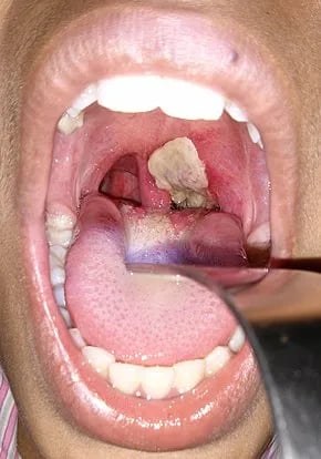 Figure: Characteristics Pseudomembrane of C. diphtheriae
The names capture the two most testable features. Korynee is Greek for "club," describing the club-shaped rods seen under the microscope, and diphthera means "leather hide," describing the tough pseudomembrane that is the hallmark of respiratory diphtheria.
General properties
- Gram-positive rods that are club-shaped, because of metachromatic (volutin) granules stored at one or both ends. After dividing, the cells snap into sharp angles and stay partly attached, so they line up as parallel "palisades" or as V and L shapes, the "Chinese letter" arrangement. This distinctive arrangement and the granules are what let you suspect C. diphtheriae on a stained smear, though they do not by themselves prove the organism is toxigenic.
- Nonmotile, non-capsulated, and non-sporing.
- Aerobes or facultative anaerobes,
- Fastidious organisms; grows best at 37°C on blood or serum-containing media such as Loeffler’s medium, tellurite medium, etc.
- Ferment glucose and glycogen.
- Catalase positive, non-pigmented, oxidase negative, indole negative, and do not form phosphatase.
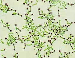 Figure: Albert Staining of Corynebacterium diphtheriae
Pathogenesis
Disease Transmission
The source of infection is carriers who harbor the organisms in the oropharynx or skin. Human to human transmission is spread by respiratory droplets, secretions, or direct contact with infected cutaneous lesions.
C. diphtheriae is not invasive. It stays on the surface of the throat or skin. Almost the entire disease is caused by one potent exotoxin, diphtheria toxin. A crucial point for exams and for understanding the organism: only strains infected by a specific bacteriophage (a virus that carries the tox gene) can make the toxin. The toxin gene belongs to the phage, not the bacterium itself. This is why a C. diphtheriae isolate is not automatically dangerous, and why the laboratory must prove toxin production, not just identify the organism.
Corynebacterium diphtheriae is not the part of the normal flora of humans but coryneforms bacteria are normal flora and may mistaken for C. diphtheriae in gram staining)
Mechanism of Diphtheria toxin
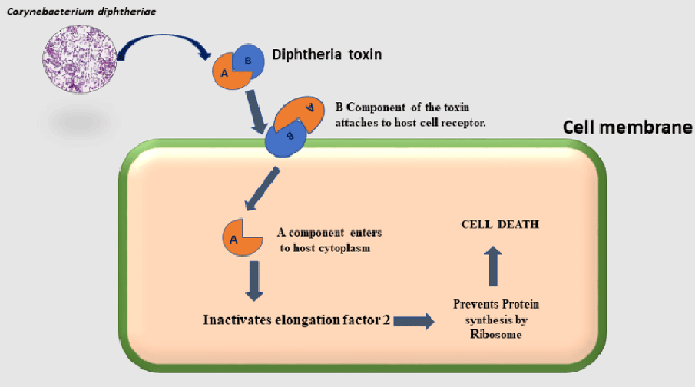 Figure: Mechanism of Diphtheria toxin (Image source: Ref-3)
Diphtheria toxin is a classic A-B toxin. The B (binding) subunit attaches the toxin to receptors on the host cell, and the A (active) subunit is the enzyme that does the damage once it is inside.
The mechanism explains the whole disease:
- The B subunit binds the host cell and the toxin is taken in.
- The A subunit enters the cytoplasm and acts as an enzyme. It catalyzes ADP-ribosylation of elongation factor 2 (EF-2), a protein the cell needs to build proteins.
- ADP-ribosylation switches EF-2 off. With EF-2 disabled, the cell can no longer make proteins.
- Without protein synthesis, the cell dies.
Two features make this toxin especially dangerous. It is extremely potent: a single molecule of the A subunit can inactivate enough EF-2 to kill the cell, because the A subunit works catalytically (it is not used up). And it acts on all cells, so once it spreads through the blood it damages distant organs, especially the heart and nerves.
Putting it together: how the toxin builds the disease
At the throat, toxin kills the surface epithelial cells. The dead cells, together with fibrin, inflammatory cells, and bacteria, pile up into the thick, adherent gray pseudomembrane. Because the membrane is stuck to living tissue underneath, trying to peel it off makes the raw surface bleed, and if the membrane extends into the larynx it can obstruct the airway. Toxin that reaches the bloodstream then damages the heart muscle (myocarditis) and the nerves (palsies), which are the dangerous late complications. Every major feature of diphtheria traces back to this one toxin.
In addition to C. diphtheriae, two other corynebacteria species can produce diphtheria toxin and thus also cause diphtheria: C. ulcerans and very rarely C. pseudotuberculosis.
Clinical manifestations
There are two types of clinical diphtheria: nasopharyngeal (respiratory) and cutaneous.
Respiratory diphtheria
Respiratory diphtheria begins with non-specific fever, sore throat, and swollen neck glands. The specific sign is the thick, gray, adherent pseudomembrane over the tonsils and throat. Two dangers follow, and both come from the toxin. Locally, the membrane can extend into the larynx and block the airway, causing suffocation. A markedly swollen neck ("bull neck") signals severe disease.
Systemically, toxin carried in the blood causes myocarditis (which can be fatal) and neurological damage, classically palsy of the palate and later of other cranial and peripheral nerves. These complications can appear even after the throat looks better, which is why neutralizing the toxin early matters so much.
Cutaneous diphtheria
Cutaneous diphtheria occurs by skin contact with other infected persons and appears as ulcerating skin lesions covered by a gray membrane. These lesions do not invade surrounding tissues and the systemic symptoms rarely occur.
Lab Diagnosis
Laboratory diagnosis has two distinct jobs: first, identify the organism as C. diphtheriae, and second, prove that the isolate actually produces the toxin. Identifying the organism is not enough, because non-toxigenic strains exist and do not cause classic diphtheria. This is why every step below leads toward the toxigenicity test as the definitive answer.
One point that must sit above all the laboratory detail: do not wait for the laboratory to start treatment. Diphtheria is treated on clinical suspicion, because antitoxin only works before the toxin binds tissue (explained under treatment). The laboratory confirms the diagnosis; it does not gate the decision to treat.
Sample: Swabs (preferably two) from the lesion of throat, larynx or nasal cavity; one for direct examination and another for culture or a portion of the pseudomembrane.
Direct examination
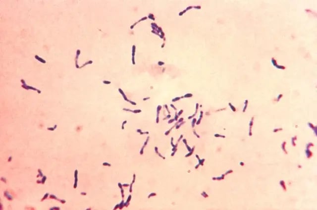 Figure: Club shaped Corynebacterium diphtheriae in Methylene Blue Staining Source: Source: PHIL Photo ID# 7323
Throat-swab smears are stained with a Gram stain and with methylene blue or Albert stain. Irregularly stained, tapered, pleomorphic gram-positive rods in Chinese-letter arrangement suggest C. diphtheriae, and the metachromatic (volutin) granules stand out on methylene blue or Albert stain. In Albert stain, the bacilli appear green with bluish-black granules. Remember, the smear supports a clinical suspicion quickly, but it cannot distinguish toxigenic from non-toxigenic strains, and harmless coryneform "diphtheroids" can look similar. For the full method, see the article on the Albert stain.
Culture
Corynebacterium diphtheriae is a fastidious organism, so it does not grow on the ordinary medium. To avoid the growth of commensals and to differentiate among various biotypes, the sample should be cultured on a selective and differential medium such as cystine-tellurite blood agar and modified Tinsdale’s medium.
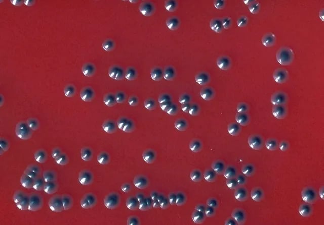 Figure: Colonies of Corynebacterium diphtheriae biotype gravis in tellurite blood agar (Image source: CDC/ Dr. W.A. Clark )
In addition, Loeffler’s medium, containing serum and eggs, stimulates the growth of C. diphtheriae and stimulates the production of metachromatic granules within the cells. Primary plating in Loeffler’s medium is not recommended due to the growth of commensals.
Blood tellurite agar: It is a selective and differential medium for C. diphtheriae. After 48-72 hours, colonies of C. diphtheriae appear as small, grey, or black with a raised center. Biotypes (gravis, intermedius, mitis, and belfanti) can be differentiated on the basis of colony morphology on tellurite blood agar and biochemical tests such as;
- Urease
- Nitrate reduction
- Esculin hydrolysis
- Fermentation of Glycogen
- Lipophilic characteristics
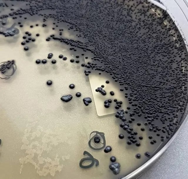 Figure: Colonies of C. diphtheriae in Tinsdale Agar
Tinsdale’s Agar: After incubation for at least 48 hours, colonies of Corynebacterium diphtheriae appear black with dark brown halos.
Rapid identification methods
API Coryne strip and RapID CB Plus are commercial products available for the rapid identification of Corynebacterium diphtheriae.
Virulence test
This is the decisive step. An isolate is only a true diphtheria pathogen if it produces toxin, so toxigenicity must be demonstrated. The available tests:
- Elek immunodiffusion test: It is the most common in vitro assay for determining toxigenicity of C. diphtheriae. This test is based on the double diffusion of diphtheria toxin and antitoxin in an agar medium. A sterile, antitoxin-saturated filter paper strip is embedded in the culture medium, and C diphtheriae isolates are streak-inoculated at a 90° angle to the filter paper.
The production of diphtheria toxin is shown within 18 to 48 hours by lines of precipitation where toxin from a toxigenic strain meets the antitoxin. A toxigenic isolate produces these precipitin lines; a non-toxigenic one does not. For the full method and how to read it, see the article on the Elek test. - Detection of toxin gene by polymerase chain reaction (PCR). PCR detects the toxin gene quickly, but carrying the gene is not identical to actively expressing toxin, so PCR is often paired with the Elek test, which shows that functional toxin is actually made.
- Guinea pig lethality test
Treatment of diphtheria
Treatment has two parts, and their order matters.
1. Diphtheria antitoxin (the priority). The single most important treatment is diphtheria antitoxin, and it must be given as early as possible on clinical suspicion, without waiting for laboratory confirmation. The reason follows directly from the mechanism: antitoxin can only neutralize toxin that is still free in the blood. Once the toxin has entered and bound host cells, antitoxin cannot pull it back out. So every hour of delay lets more toxin bind irreversibly to the heart and nerves. This is why diphtheria is a "treat on suspicion" disease. The antitoxin is an animal-derived (equine) product, so it is given with care for hypersensitivity.
2. Antibiotics (supportive). Penicillin or a macrolide (such as erythromycin or azithromycin) is given to kill the organism and stop further toxin production, to help clear the pseudomembrane, and to reduce spread to others. But antibiotics do nothing to the toxin already released, so they support the antitoxin rather than replace it.
Supportive care includes protecting the airway (the membrane can obstruct breathing) and monitoring for myocarditis. Close contacts and carriers are given antibiotics and their immunization is checked.
Prevention and immunization
Prevention is by active immunization with diphtheria toxoid, which is diphtheria toxin inactivated with formaldehyde. The toxoid triggers antibodies that neutralize the toxin, so it protects against the effects of the toxin rather than against carriage of the organism. It is given in childhood in combination with tetanus and pertussis vaccines (as DTP/DTaP), with boosters, because immunity fades over time. This is the same "toxin is the disease, so vaccinate against the toxin" logic seen in tetanus.
How to Remember
| Device | The memory hook |
|---|---|
| Toxin is the disease | C. diphtheriae barely invades. One toxin causes the membrane, the heart failure, and the nerve palsies. Learn the toxin and you know the disease. |
| A-B, and A stops protein synthesis | B = Binds. A = Active enzyme. The A subunit ADP-ribosylates EF-2, switching off protein synthesis, so the cell dies. |
| EF-2 target (and its cousin) | Diphtheria toxin targets EF-2. (Pseudomonas exotoxin A does the same, ADP-ribosylates EF-2, an easy pair to remember together.) |
| Phage makes it toxigenic | The tox gene comes from a bacteriophage. No phage, no toxin, no classic diphtheria. This is why the lab must prove toxigenicity. |
| Chinese letters + granules | V and L "Chinese letter" arrangement with metachromatic (volutin) granules at the ends. Club-shaped rods stacked at angles. |
| Membrane that bleeds | The gray pseudomembrane is stuck to living tissue, so scraping it makes it bleed. That adherence separates it from ordinary throat exudate. |
| Antitoxin first, don't wait | Antitoxin only neutralizes unbound toxin. Give it early on suspicion, before the lab result. Antibiotics are the backup, not the main event. |
| Vaccinate against the toxin | The vaccine is the toxoid (inactivated toxin). You are immunizing against the toxin, not the bacterium. |
Key exam facts
| Feature | Corynebacterium diphtheriae |
|---|---|
| Other name | Klebs-Löffler bacillus |
| Morphology | Gram-positive club-shaped rod; metachromatic (volutin) granules |
| Arrangement | Palisades and V/L "Chinese letter" |
| Motility/spores/capsule | Non-motile, non-sporing, non-capsulated |
| Growth | Fastidious; Loeffler's, tellurite, Tinsdale media |
| Invasiveness | Non-invasive; disease is toxin-mediated |
| Toxin gene source | Bacteriophage (tox gene); only lysogenized strains are toxigenic |
| Toxin type | A-B exotoxin |
| Toxin mechanism | A subunit ADP-ribosylates EF-2 → protein synthesis stops → cell death |
| Same-mechanism cousin | Pseudomonas exotoxin A (also targets EF-2) |
| Local disease | Pseudomembrane; risk of airway obstruction; "bull neck" |
| Systemic complications | Myocarditis, cranial nerve and peripheral palsies |
| Staining | Albert (green bacilli, bluish-black granules), methylene blue |
| Selective/differential media | Cystine-tellurite blood agar; Tinsdale (black colonies, brown halo) |
| Toxigenicity test | Elek test (in vitro); PCR for tox gene; guinea-pig test |
| Priority treatment | Diphtheria antitoxin, early, on clinical suspicion |
| Antibiotics | Penicillin or macrolide (supportive) |
| Prevention | Diphtheria toxoid (with DTP/DTaP) |
Where Students Get Confused
| Confusion | The clarification |
|---|---|
| Identifying the organism vs. confirming disease | Finding C. diphtheriae is not enough. Only toxigenic strains cause diphtheria, so the toxin must be demonstrated (Elek test or PCR plus expression). |
| Why is toxigenicity phage-dependent? | The toxin gene is carried by a bacteriophage. Strains without the phage cannot make toxin, so they do not cause classic diphtheria. |
| What exactly does the toxin do? | Its A subunit ADP-ribosylates EF-2, which halts protein synthesis and kills the cell. This one action explains the membrane, myocarditis, and nerve palsies. |
| Antitoxin or antibiotics first? | Antitoxin first, and early. It only neutralizes toxin not yet bound to cells. Antibiotics kill the organism and stop further toxin but cannot undo bound toxin. |
| Why not wait for the culture? | Because delay lets toxin bind irreversibly. Diphtheria is treated on clinical suspicion; the lab confirms afterward. |
| Why does the pseudomembrane bleed when removed? | It is firmly attached to the living, damaged tissue underneath, so peeling it exposes a raw, bleeding surface. This adherence is a diagnostic clue. |
| What is the Schick test? | An old skin test of whether a person is immune or susceptible to diphtheria toxin. It is not a treatment and not a pre-antitoxin allergy test. |
| Diphtheroids vs C. diphtheriae | Harmless coryneform "diphtheroids" are normal flora and can resemble C. diphtheriae on a smear. Culture on selective media and toxigenicity testing separate them. |
References and further readings
- Tille, P. M. (2022). Bailey & Scott's Diagnostic Microbiology (15th ed.). Elsevier.
- Procop, G. W., & Koneman, E. W. (2017). Koneman's Color Atlas and Textbook of Diagnostic Microbiology (7th ed.). Wolters Kluwer.
- Carroll, K. C., Pfaller, M. A., et al. (2020). Murray's Medical Microbiology (9th ed.). Elsevier.
- Jamal, S. B., Tiwari, S., Silva, A., & Azevedo, V. (2017). Pathogenesis of Corynebacterium diphtheriae and available vaccines: an overview. Global Journal of Infectious Diseases and Clinical Research, 3(1), 20–24.
- Centers for Disease Control and Prevention. Diphtheria: clinical information. CDC (current version).
Frequently Asked Questions
Why is Corynebacterium diphtheriae dangerous if it does not invade the body?
Why is Corynebacterium diphtheriae dangerous if it does not invade the body?
Because nearly all the damage comes from a toxin, not from invasion. The organism stays on the throat or skin surface, but its toxin kills local cells to form the pseudomembrane and then spreads through the blood to harm the heart and nerves. The bacterium barely moves; the toxin does the damage.
How does diphtheria toxin work?
How does diphtheria toxin work?
It is an A-B toxin. The B subunit binds the host cell, and the A subunit enters and acts as an enzyme that adds ADP-ribose to elongation factor 2 (EF-2). This switches off EF-2, so the cell can no longer make proteins and it dies. Because one A subunit can inactivate many EF-2 molecules, the toxin is extremely potent.
Why must the laboratory test whether a strain is toxigenic?
Why must the laboratory test whether a strain is toxigenic?
Because only strains that carry a specific bacteriophage can make the toxin, and non-toxigenic strains do not cause classic diphtheria. Identifying the organism as C. diphtheriae is not enough; the toxin itself must be demonstrated, usually with the Elek test or by detecting and confirming expression of the toxin gene.
Why is diphtheria antitoxin given before the laboratory result?
Why is diphtheria antitoxin given before the laboratory result?
Because antitoxin can only neutralize toxin that is still free in the blood. Once the toxin binds to heart and nerve cells, antitoxin cannot remove it. Waiting for confirmation allows more toxin to bind irreversibly, so treatment is started on clinical suspicion.
What is the difference between the diphtheria antitoxin and the diphtheria vaccine?
What is the difference between the diphtheria antitoxin and the diphtheria vaccine?
The antitoxin is ready-made antibody given during illness to neutralize circulating toxin (passive, immediate, short-term). The vaccine is the toxoid, an inactivated form of the toxin that makes the body produce its own antibodies for long-term protection (active, preventive). One treats, the other prevents.
Why does the throat membrane in diphtheria bleed when you try to remove it?
Why does the throat membrane in diphtheria bleed when you try to remove it?
The pseudomembrane is firmly attached to the living tissue underneath, which has been damaged by the toxin. Peeling it off exposes a raw surface that bleeds. This tight adherence helps distinguish diphtheria from an ordinary throat infection with removable exudate.
Do antibiotics cure diphtheria on their own?
Do antibiotics cure diphtheria on their own?
No. Antibiotics kill the organism and stop it making more toxin, which helps clear the infection and reduce spread, but they cannot neutralize toxin that has already been released. Antitoxin is the key treatment, and antibiotics support it.

Tankeshwar Acharya, MSc (Medical Microbiology)
Tankeshwar Acharya is an Assistant Professor in the Department of Microbiology at Patan Academy of Health Sciences (PAHS), Nepal, where he has been teaching and practicing clinical microbiology for over 14 years. He is the founder of Microbe Online, one of the leading free microbiology education resources on the web, covering bacteriology, mycology, parasitology, immunology, and clinical laboratory diagnostics written from direct experience in both the classroom and the diagnostic laboratory.
Comments
No comments yet. Be the first to share your thoughts.
Leave a comment
All comments are reviewed before they appear.