
Carbohydrate Fermentation Test: Reading Acid, Gas, and the Sugar Panel That Fingerprints a Species
Feed an organism a single sugar and watch two things: does the broth turn yellow (acid), and does a bubble collect in the Durham tube (gas)? Run a panel of sugars and the pattern of acid and gas becomes a species fingerprint. Here is how to read acid vs acid-plus-gas, why some organisms make acid but no gas, and how the sugar panel narrows an identification.
Why It Matters
A single carbohydrate fermentation tube answers a small question: can this organism turn this one sugar into acid, and does it make gas doing so? On its own, that is a modest result. The power comes from running several sugars at once.
Feed an unknown organism a panel of sugars, glucose, lactose, sucrose, maltose, mannitol, and record which ones it turns to acid and which produce gas, and the pattern becomes a fingerprint. Two organisms that look identical on a Gram stain can have completely different sugar patterns, and that pattern is often what names the species. This is the principle underneath the older manual identification schemes and the modern strip kits like API 20E: a row of sugar reactions read as a code.
The classic teaching example lives one click away. Among the oxidase-positive Gram-negative diplococci, Neisseria gonorrhoeae ferments glucose only, Neisseria meningitidis ferments glucose and maltose, and Neisseria lactamica adds lactose. Three species, three sugar patterns, one panel. That separation is important enough to have its own dedicated method, the rapid carbohydrate utilization test, covered in full in its own article.
(See the RCUT and the N. gonorrhoeae vs N. meningitidis articles for that workflow.)
This article is about the test underneath all of that: how a single sugar tube works, how to read acid and gas, and why the gas result is a real identification feature in its own right.
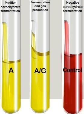 The carbohydrate fermentation test is used to determine whether or not bacteria can ferment a specific carbohydrate. Carbohydrate fermentation patterns are useful in differentiating among bacterial groups or species.
It tests for the presence of acid and/or gas produced from carbohydrate fermentation. Basal medium containing a single carbohydrate source such as glucose, lactose, sucrose, or any other carbohydrate is used for this purpose. A pH indicator (such as Andrade’s solution, bromocresol purple (BCP), bromothymol blue (BTB), or phenol red) is also present in the medium; which will detect the lowering of the pH of the medium due to acid production. Small inverted tubes called Durham tube is also immersed in the medium to test for the production of the gas (hydrogen or carbon dioxide).
The term fermentation is often used to describe the breaking down or catabolism of a carbohydrate under anaerobic conditions. Therefore, bacteria capable of fermenting a carbohydrate are usually facultative anaerobes.
A note on what this test does and does not distinguish.
The carbohydrate fermentation test detects acid (and gas) production from a sugar, but it does not, by itself, separate true anaerobic fermentation from aerobic oxidation of the sugar. An organism that only oxidizes glucose aerobically can still produce enough acid to turn the indicator in an open tube. To distinguish genuine fermentation from oxidation, the oxidative-fermentative (OF) test is used, which compares a sealed and an open tube. See the Oxidative-Fermentative (OF) Test for that distinction. This article focuses on the sugar-panel pattern and gas production, which are read the same way regardless of that mechanistic question.
Uses of Carbohydrate Fermentation Test
Carbohydrate fermentation patterns can be used to differentiate among bacterial groups or species.
- All members of Enterobacteriaceae family are glucose fermenters (they can metabolize glucose anaerobically).
- Maltose fermentation differentiates Proteus vulgaris (positive) from Proteus mirabilis (negative).
- Both Neisseria gonorrhoeae (gonococci) and Neisseria meningitides (meningococci) ferments glucose, but only meningococci ferments maltose.
- Rapid carbohydrate utilization tests (RCUTs) can be performed to identify Corynebacterium diphtheriae and other Corynebacterium species.
Principle
When microorganisms ferment carbohydrate an acid or acid with gas are produced. Depending upon the organisms involved and the substrate being fermented, the end products may vary. Common end-products of bacterial fermentation include lactic acid, formic acid, acetic acid, butyric acid, butyl alcohol, acetone, ethyl alcohol, carbon dioxide, and hydrogen. The production of the acid lowers the pH of the test medium, which is detected by the color change of the pH indicator. Color change only occurs when a sufficient amount of acid is produced, as bacteria may utilize the peptone producing alkaline by-products.
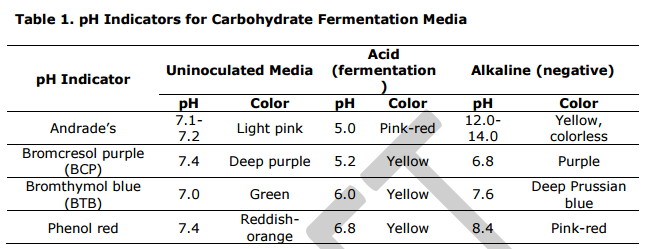 Figure: Common pH Indicators for Carbohydrate Fermentation Media (Source: ASMCUE)
Phenol red is commonly used as a pH indicator in carbohydrate fermentation tests. Other pH indicators such as bromocresol/bromocresol purple (BCP), bromothymol/bromothymol blue (BTB), and Andrade’s can be used.
Durham tubes are inserted upside down in the test tubes to detect gas production. If the test organisms produce gas, the gas displaces the media present inside the tube and gets trapped producing a visible air bubble.
Based on the characteristics reactions observed, bacteria can be classified as:
- Fermenter with acid production only
- Fermenter with acid and gas production
- Non-fermenter
Test Procedure
Phenol red carbohydrate broth is commonly used in carbohydrate fermentation tests. The carbohydrate sources can vary based on your test requirements.
Common broth media are:
- Phenol red glucose broth
- Phenol red lactose broth
- Phenol red maltose broth
- Phenol red mannitol broth
- Phenol red sucrose broth
Preparation and Composition of the Media
Get specific phenol red carbohydrate test media from the commercial suppliers or phenol red broth base and add specific carbohydrate source based on your test requirements, or you can prepare media mixing the following ingredients.
Composition of Phenol Red Carbohydrate Broth
- Trypticase or protease peptone No. 3: 10 g
- Sodium chloride (NaCl): 5 g
- Beef extract (optional): 1 g
- Phenol red (7.2 ml of 0.25% phenol red solution): 0.018 g
- Carbohydrate source: 10 g
A. Preparation of the media
- Prepare broth media by mixing all ingredients in 1000 mL of distilled/deionized water and heating gently to dissolve it (Note: Use a single carbohydrate source based on your requirements).
- Fill 13 x 100 mm test tubes with 4-5 ml of phenol red carbohydrate broth.
- Insert a Durham tube to detect gas production.
- Autoclave the prepared test media (at 121°C for 15 minutes) to sterilize. The sterilization process will also drive the broth into the inverted Durham tube. (Note: When using arabinose, lactose, maltose, salicin, sucrose, trehalose, or xylose, autoclave at 121°C for only 3 minutes as these carbohydrates are subject to breakdown by autoclaving)
The prepared broth media will be a light red color and the final pH should be 7.4 ± 0.2.
Alternatively, prepare phenol red broth base, heat sterilize and cool to 45°C. Prepare specific carbohydrate solution separately, and filter the solution using membrane filter (pore size: 0.45 μm). Add carbohydrate solution to the broth base and mix it. The preferred carbohydrate concentration is 1%.
B. Inoculation and Incubation
- Aseptically inoculate each test tube with the test microorganism using an inoculating needle or loop. Alternatively, inoculate each test tube with 1-2 drops of an 18- to 24-hour brain-heart infusion broth culture of the desired organism.
- Incubate tubes at 35-37°C for 18-24 hours. Longer incubation periods may be required to confirm a negative result.
C. Interpretation of the results
Reading acid and gas
Every carbohydrate tube gives you two independent readouts. Read both.
1. Acid (the broth color). With phenol red, the broth starts red (pH 7.4) and turns yellow if the organism produces acid from the sugar. Yellow = acid = the organism fermented this sugar. Red = no acid = it did not.
2. Gas (the Durham tube). A small inverted tube sits in the broth, filled with liquid at the start. If the organism produces gas (hydrogen and carbon dioxide) while fermenting, the gas collects as a bubble trapped at the top of the inverted Durham tube. Bubble = gas positive. No bubble = anaerogenic (no gas).
Combining the two gives three results:
| Broth color | Durham tube | Result | Meaning |
|---|---|---|---|
| Yellow (acid) | Bubble | Acid + gas (A/G) | Ferments the sugar and produces gas. e.g. E. coli on glucose |
| Yellow (acid) | No bubble | Acid only (A) | Ferments the sugar but makes no gas (anaerogenic). e.g. Shigella, S. Typhi |
| Red (no change) | No bubble | Negative | Does not ferment this sugar |
Read acid before gas, and read before the tube over-incubates. A tube left too long can revert: once the sugar is exhausted, the organism starts using the peptone, releasing alkaline amines that can turn an acid (yellow) tube back toward red or even alkaline. A tube read at 18-24 hours is reliable; a tube read at 72 hours may have reverted and read as a false negative. This is the same peptone-alkalinization effect that the OF medium is specifically designed to suppress.
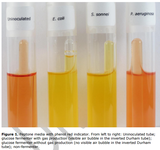 Figure: Image source: ASMCUE
Why some fermenters make no gas
Producing acid and producing gas are separate abilities. An organism can ferment a sugar to acid and make no gas at all. This is not a weakness of the test; it is a real, useful identification feature.
Organisms that ferment glucose but produce no gas are called anaerogenic, and the anaerogenic enterics are a short, high-yield list:
- Shigella species: acid, no gas. This helps separate them from most other Enterobacteriaceae, which are aerogenic.
- Salmonella Typhi: acid, no gas. Its anaerogenic glucose fermentation is one of the classic features that separates it from the non-typhoidal salmonellae, which do produce gas. (This is the same no-gas feature you read on TSI and KIA.)
So a Gram-negative rod that ferments glucose to acid with no gas should raise Shigella or S. Typhi immediately. Gas production, or its absence, is doing real diagnostic work here, not just filling a second column.
Sugar fermentation patterns of some common organisms
| Organism | Glucose | Lactose | Sucrose | Maltose | Mannitol | Gas from glucose |
|---|---|---|---|---|---|---|
| Escherichia coli | + | + | variable | + | + | + |
| Shigella species | + | − | − | variable | variable | − (anaerogenic) |
| Salmonella Typhi | + | − | − | + | + | − (anaerogenic) |
| Klebsiella pneumoniae | + | + | + | + | + | + (often abundant) |
| Proteus vulgaris | + | − | + | + | − | + |
| Proteus mirabilis | + | − | + | − | − | + |
| Staphylococcus aureus | + | + | + | + | + (anaerobic) | − |
Two separations worth noting from this table: maltose splits Proteus vulgaris (positive) from P. mirabilis (negative), and the anaerogenic glucose fermenters (Shigella, S. Typhi) stand out from everything else. The full enteric identification uses these patterns as part of a larger battery (TSI, IMViC, urease); this test contributes the individual sugar reactions that battery reads.
How to remember
Yellow is acid, bubble is gas, and they're separate questions. Phenol red goes yellow when the organism makes acid from the sugar. A bubble in the Durham tube means it also made gas. An organism can do the first without the second. Always read both, and don't assume acid implies gas.
No gas points to Shigella or Typhi. Among glucose-fermenting enterics, the anaerogenic ones, acid but no gas, are a short list led by Shigella and Salmonella Typhi. A yellow glucose tube with an empty Durham tube should make you think of those two. It's the same no-gas feature you read on TSI and KIA.
The panel is a fingerprint; one sugar is just one letter. A single sugar result rarely identifies anything. The pattern across a panel, glucose, lactose, sucrose, maltose, mannitol, is what fingerprints a species. This is exactly what API 20E automates: a row of sugars read as a code.
Read it before it reverts. Once the sugar runs out, the organism eats the peptone and makes alkaline amines that can turn a yellow tube back to red. Read at 18-24 hours. A late read can hide a true positive.
Key exam facts in one table
| Question | Answer | The reason behind it |
|---|---|---|
| What does the test detect? | Acid (and gas) production from a specific sugar | Fermentation lowers pH; gas collects in the Durham tube |
| Indicator (common) | Phenol red: red (pH 7.4) → yellow (acid) | Detects the pH drop |
| Other indicators | Bromocresol purple, bromothymol blue, Andrade's | Each has its own acid color (see Table 1) |
| What detects gas? | Inverted Durham tube | Gas displaces liquid, trapping a bubble |
| Acid only (A) | Yellow broth, no bubble | Ferments sugar, anaerogenic |
| Acid + gas (A/G) | Yellow broth, bubble | Ferments sugar and makes gas |
| Negative | Red broth, no bubble | Does not ferment this sugar |
| Anaerogenic enterics | Shigella, Salmonella Typhi | Acid but no gas from glucose; a key ID feature |
| Maltose separation | P. vulgaris + / P. mirabilis − | Splits the two Proteus species |
| Neisseria panel | gonorrhoeae (glu), meningitidis (glu+mal), lactamica (glu+mal+lac) | Owned by the RCUT and Neisseria articles; linked |
| Why read early (18-24h)? | Peptone breakdown can reverse an acid result | Alkaline amines re-raise the pH |
| Why a single carbohydrate per tube? | To attribute the reaction to that specific sugar | A panel uses one sugar per tube |
| Relationship to OF test | OF distinguishes oxidative vs fermentative; this reads the sugar/gas pattern | See the OF Test article |
| Relationship to API 20E | API 20E automates a sugar panel as a code | The panel-as-fingerprint principle |
| Which sugars need gentle autoclaving? | Lactose, maltose, sucrose, arabinose, salicin, trehalose, xylose | They break down at full autoclave; 3 min only |
| Panel used in | Enteric ID (with TSI, IMViC, urease), API 20E, Enterotube | Sugar reactions are part of a larger battery |
Where students get confused
Thinking acid always comes with gas. They are separate abilities. An organism can ferment a sugar to acid and produce no gas at all (anaerogenic). Read the broth color and the Durham tube as two independent results. A yellow tube with an empty Durham tube is acid-only, which is itself informative.
Reading the tube too late and getting a false negative. Once the sugar is exhausted, the organism starts metabolizing the peptone, releasing alkaline amines that push the pH back up and can turn a yellow (positive) tube back to red. Read at 18-24 hours. A tube read at 72 hours may have reverted. Longer incubation is only for confirming a genuine negative, not for reading positives.
Confusing this test with the OF test. This test reads the sugar and gas pattern. It does not distinguish aerobic oxidation from true anaerobic fermentation, because an oxidizer can still acidify an open tube. The OF test, with its sealed and open tubes, is what makes that distinction. If the question is "fermenter or oxidizer," that is an OF question, not a carbohydrate-fermentation-tube question.
Over-autoclaving heat-labile sugars. Lactose, maltose, sucrose, and several others break down under full autoclaving, which can create free glucose and cause false-positive reactions. These sugars are autoclaved for only 3 minutes, or filter-sterilized and added to a pre-sterilized base. Autoclaving them like glucose invalidates the result.
Forgetting that a bubble is gas, not turbidity. Growth clouds the broth (turbidity); that is not the gas result. Gas is specifically the bubble trapped in the inverted Durham tube. Turbidity with no Durham-tube bubble is growth without gas.
Trying to identify a species from one sugar. One sugar is one data point. The identification comes from the pattern across a panel. A lone glucose-positive result says almost nothing; glucose+/lactose−/sucrose−/maltose+/no-gas starts to point somewhere. Read sugars as a set.
Mixing up the indicator colors. Phenol red goes red → yellow with acid, but bromocresol purple goes purple → yellow, and bromothymol blue goes green → yellow, and Andrade's goes pale → pink/red. The acid color depends on the indicator. Check which indicator the medium uses before calling a result.
References and further readings
- Procop GW, Church DL, Hall GS, Janda WM, Koneman EW, Schreckenberger PC, Woods GL. Koneman's Color Atlas and Textbook of Diagnostic Microbiology. 7th ed. Philadelphia: Wolters Kluwer; 2017.
- Tille PM. Bailey and Scott's Diagnostic Microbiology. 15th ed. St. Louis: Elsevier; 2022.
- MacFaddin JF. Biochemical Tests for Identification of Medical Bacteria. 3rd ed. Philadelphia: Lippincott Williams & Wilkins; 2000.
- Carbohydrate fermentation protocol. American Society for Microbiology (Microbe Library / ASMCUE teaching resource).
Frequently Asked Questions
Which bacteria ferment glucose but produce no gas?
Why should the carbohydrate fermentation tube be read at 18 to 24 hours?
Why are some sugars autoclaved for only 3 minutes?
How does the carbohydrate fermentation test differ from the OF test?
What is the difference between acid and gas production in the carbohydrate fermentation test?
How are sugar fermentation patterns used to identify bacteria?

Tankeshwar Acharya, MSc (Medical Microbiology)
Tankeshwar Acharya is an Assistant Professor in the Department of Microbiology at Patan Academy of Health Sciences (PAHS), Nepal, where he has been teaching and practicing clinical microbiology for over 14 years. He is the founder of Microbe Online, one of the leading free microbiology education resources on the web, covering bacteriology, mycology, parasitology, immunology, and clinical laboratory diagnostics written from direct experience in both the classroom and the diagnostic laboratory.