
Baird-Parker Agar: Composition, Principle, Uses, and Colony Characteristics of S. aureus

On this page
Following a wedding reception, 47 guests develop nausea, vomiting, and abdominal cramps within 1–6 hours of eating. The clinical picture suggests staphylococcal food poisoning — pre-formed enterotoxin, not infection. Food samples from the buffet are sent to the public health laboratory. The investigators need to know not just whether Staphylococcus aureus was present, but how many organisms per gram of food — because regulatory thresholds exist for what constitutes a public health risk.
Baird-Parker agar was designed precisely for this quantitative detection: a selective, differential medium that allows S. aureus colonies to be counted and confirmed from a mixed food matrix, where the organism must be distinguished from the heavy background of other staphylococci, micrococci, and environmental contaminants typically present in food samples.
Baird-Parker Agar was developed by Baird Parker in 1962 and is a modification of the Tellurite-glycine formulation of Zebovitz et al. It is a moderately selective medium for the isolation and differentiation of coagulase-positive staphylococci, especially Staphylococcus aureus. This agar is primarily used in the processing of food, cosmetics, and environmental samples rather than clinical samples.
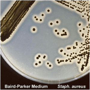 Figure: Colonies of Staphylococcus aureus in Baird Parker Agar Medium
Principle
Baird-Parker agar differentiates Staphylococcus aureus from other organisms through two independent reactions, each producing a visible zone around the colony:
Reaction 1 — Tellurite reduction → black colony colour: Potassium tellurite in the medium is toxic to most bacteria, providing selectivity against non-staphylococcal flora. Staphylococcus aureus and coagulase-positive staphylococci resist this toxicity and additionally possess tellurite reductase — an enzyme that reduces tellurite ions (TeO₃²⁻) to elemental tellurium (Te⁰). Elemental tellurium deposits within the bacterial cell, producing the characteristic black, shiny colony appearance. Most coagulase-negative staphylococci also reduce tellurite but to a lesser degree, producing grey rather than shiny-black colonies.
Lithium chloride provides additional selectivity by inhibiting Gram-negative organisms and most Gram-positive bacteria other than staphylococci.
Reaction 2 — Egg yolk reactions → two distinct halos: The egg yolk supplement added to Baird-Parker agar contains two substrates that S. aureus acts upon through two different enzymes, producing two distinct zones observable at different incubation timepoints:
| Time | Enzyme | Reaction | Zone appearance |
|---|---|---|---|
| 18–24 hours | Lecithinase (phospholipase C) | Hydrolyses lecithin in egg yolk → diglyceride + phosphorylcholine | Clear zone immediately surrounding the colony — lecithin is completely broken down, removing the turbidity from the egg yolk in that area |
| 48 hours | Lipase | Hydrolyses triglycerides and other lipids in egg yolk → fatty acids + glycerol | Opaque (pearlescent) zone develops outside the clear zone — lipolysis products form a milky precipitate in the agar |
The full characteristic colony appearance of S. aureus on Baird-Parker agar at 48 hours is therefore:
Black, shiny colony + inner clear zone (lecithinase) + outer opaque zone (lipase)
Factual note: The opaque zone described in many references as "lipolytic activity" is specifically lipase activity on egg yolk lipids — distinct from the lecithinase-produced clear zone. Both enzymes are virulence factors of S. aureus: lecithinase (phospholipase C) and lipase contribute to membrane disruption and tissue invasion. The two-zone reaction is therefore both a diagnostic characteristic and a virulence indicator.
Sodium pyruvate — protecting injured cells: Sodium pyruvate is added at high concentration (10 g/L) specifically to protect sublethally injured staphylococci present in processed or heat-treated foods. Injured cells have damaged membranes and impaired metabolism — pyruvate provides a readily metabolisable carbon source that enables their recovery on the medium. Without pyruvate, food processing-stressed S. aureus may fail to grow even if present in the sample.
Composition of Baird-Parker Agar
| Ingredient | Amount (g/L) | Function |
|---|---|---|
| Casein peptone | 10.0 | Nitrogen, amino acids, carbon |
| Meat extract | 5.0 | Nitrogen, vitamins, minerals |
| Yeast extract | 1.0 | Vitamins, growth factors |
| Glycine | 12.0 | Selective agent — inhibits many bacteria at this concentration; staphylococci are glycine-tolerant |
| Sodium pyruvate | 10.0 | Protects sublethally injured staphylococci from food processing; stimulates S. aureus growth |
| Lithium chloride | 5.0 | Selective agent — inhibits Gram-negative organisms and most non-staphylococcal Gram-positives |
| Agar | 15.0 | Solidifying agent |
Supplements added after autoclaving:
- Egg yolk tellurite emulsion (50 mL/L): provides lecithin and lipid substrates for the two-zone reaction
- Potassium tellurite solution (3 mL of 3.5% solution): tellurite reduction → black colony colour; selective against most non-staphylococcal bacteria
Final pH: 6.8 ± 0.2 at 25°C
Uses of Baird-Parker Agar
1. Quantitative detection of S. aureus in food safety testing Baird-Parker agar is the ISO and FDA reference method for S. aureus enumeration in foods. A measured, diluted food homogenate is spread-plated onto Baird-Parker plates, incubated for 48 hours, and typical black colonies with clear and opaque zones are counted and reported as CFU/g. This is the primary application globally.
Regulatory significance: In many countries, S. aureus counts >10⁵ CFU/g in ready-to-eat foods constitute a public health action threshold, because enterotoxin production typically occurs at or above this level.
2. S. aureus detection in environmental and cosmetic samples Surfaces, equipment swabs, and cosmetic products are tested for S. aureus contamination using Baird-Parker agar in pharmaceutical and cosmetic manufacturing quality control.
3. S. aureus isolation from clinical specimens (secondary use) While MSA is more commonly used in clinical laboratories for selective S. aureus isolation, Baird-Parker agar can be used when the egg yolk reaction confirmation is desired alongside colony isolation from wound swabs, skin specimens, and nasal swabs.
4. Research applications Baird-Parker agar is used in research settings for studying S. aureus virulence factors (lecithinase, lipase, tellurite resistance), antibiotic resistance, and biofilm formation.
Baird-Parker vs MSA — primary difference: Baird-Parker is the food microbiology standard; MSA is the clinical microbiology standard. In food laboratories, Baird-Parker is preferred because its two-zone reaction provides a visual confirmation of coagulase-positive staphylococci directly on the plate. In clinical laboratories, MSA's simple yellow/pink colour differentiation is sufficient and more economical.
Preparation of the media
- Suspend desired quantity (as per the manufacturer’s instruction) of the medium in 950 ml purified water.
- Then, heat with frequent agitation and boil for one minute to completely dissolve the medium.
- After that autoclave at 121°C for 15 minutes.
- After cooling to 45- 50°C, add 50 mL of egg yolk tellurite supplement and 3 ml sterile 3.5% potassium tellurite solution or 50 ml egg yolk tellurite emulsion.
- Finally, mix thoroughly before dispensing.
Colony Characteristics on Baird-Parker Agar
At 18–24 hours:
| Organism | Colony colour | Egg yolk zone | Interpretation |
|---|---|---|---|
| Staphylococcus aureus | Black, shiny, convex, 1–1.5 mm | Clear zone (lecithinase positive) | Presumptive positive — confirm with coagulase test |
| Coagulase-negative staphylococci (CONS) | Grey to black, less shiny | Absent or narrow/indistinct | Not S. aureus; confirm by coagulase test |
| Micrococcus spp. | Variable — may produce small grey colonies | Absent | Usually inhibited; if growing, coagulase-negative |
| Gram-negative bacteria | No growth or severely inhibited | — | Inhibited by lithium chloride + glycine + tellurite |
At 48 hours (additional reading required): After 48 hours, S. aureus colonies develop the full three-part appearance:
- Black, shiny colony centre
- Inner clear zone (2–5 mm) produced by lecithinase activity — egg yolk cleared completely
- Outer opaque/pearlescent zone produced by lipase activity — milky precipitate at the periphery
The complete 48-hour appearance is the most specific indicator of S. aureus. Both zones must be present for high confidence. A black colony with only a clear zone and no opaque zone may represent other lecithinase-positive staphylococci. A colony with an opaque zone but no black centre is unlikely to be S. aureus.
Confirmation requirement: Colony morphology is presumptive only. All typical colonies must be confirmed by coagulase test — the gold standard for S. aureus identification. In food microbiology, a tube coagulase test is performed on picked colonies from Baird-Parker agar; a positive result confirms S. aureus.
Counting colonies in food microbiology: Count all black colonies with clear and/or opaque zones at 48 hours. Recount after selecting 5 typical and 5 atypical colonies for confirmatory coagulase testing. Adjust the total count based on the proportion of coagulase-confirmed colonies. Report as CFU/g.
How to Remember
Two reactions, two zones, one organism:
The complete Baird-Parker reaction tells a sequential story:
- Organism grows → reduces tellurite → black colony (this happens at 18–24 hrs)
- Lecithinase attacks egg yolk lecithin → inner clear zone (18–24 hrs)
- Lipase attacks egg yolk lipids → outer opaque zone (48 hrs)
Reading the plate at 24 hours shows black + clear zone. Reading at 48 hours adds the outer opaque zone. Always read at 48 hours for full confirmation.
Sodium pyruvate is the food safety innovation: Plain MSA misses sublethally injured S. aureus from heat-treated or processed foods. The high sodium pyruvate concentration in Baird-Parker allows injured organisms to recover and grow. This single difference makes Baird-Parker the food safety standard while MSA remains the clinical standard.
The two enzymes and their products:
- Lecithinase = breaks lecithin (a phospholipid) = clear zone (phospholipid is transparent; when broken down, the turbid egg yolk clears)
- Lipase = breaks triglycerides (fat) = opaque zone (fatty acid products form a milky precipitate)
Both enzymes are S. aureus virulence factors — the same enzymes that damage host membranes in a clinical infection produce the two-zone reaction on the plate. The diagnostic reaction directly reflects pathogenic mechanism.
References
- Stiles, M. E., & Ng, L. K. (1981). Use of Baird-Parker’s Medium to Enumerate Staphylococcus aureus in Meats 1. Journal of food protection, 44(8), 583–587. https://doi.org/10.4315/0362-028X-44.8.583
- Ingham, S. C., Becker, K. L., & Fanslau, M. A. (2003). Comparison of the Baird-Parker agar and 3M Petrifilm Staph Express Count plate methods for enumeration of Staphylococcus aureus in naturally and artificially contaminated foods. Journal of food protection, 66(11), 2151–2155. https://doi.org/10.4315/0362-028x-66.11.2151
Frequently Asked Questions
What produces the clear zone and the opaque zone around S. aureus colonies on Baird-Parker agar, and why do they appear at different times?
Why does Baird-Parker agar use sodium pyruvate at a high concentration (10 g/L), and why is this important for food safety testing?
When is Baird-Parker agar preferred over MSA for Staphylococcus aureus detection, and when is MSA preferred?

Tankeshwar Acharya, MSc (Medical Microbiology)
Tankeshwar Acharya is an Assistant Professor in the Department of Microbiology at Patan Academy of Health Sciences (PAHS), Nepal, where he has been teaching and practicing clinical microbiology for over 14 years. He is the founder of Microbe Online, one of the leading free microbiology education resources on the web, covering bacteriology, mycology, parasitology, immunology, and clinical laboratory diagnostics written from direct experience in both the classroom and the diagnostic laboratory.
Comments
No comments yet. Be the first to share your thoughts.
Leave a comment
All comments are reviewed before they appear.