
Anaerobic Infections: Causes, Where Anaerobes Live, Symptoms, Diagnosis
Anaerobic infections explained: what they are, where anaerobic bacteria live in the body, clinical clues, the organisms that cause them, and how the lab diagnoses them.

On this page
A patient has a swelling that has turned into an abscess, and the pus has a foul, putrid smell. A Gram stain shows bacteria, yet the routine aerobic culture grows nothing. To a trained eye, those three findings together, a foul odor, pus, and no aerobic growth, point almost immediately to anaerobes.
Anaerobic infections are common, often hidden, and easy to miss if the specimen is collected or transported the wrong way. This article explains what anaerobic infections are, where these bacteria normally live in the body, the clues that should make you suspect them, the main organisms involved, and how the laboratory confirms them.
An anaerobic infection is an infection caused by anaerobic bacteria: organisms that grow in the absence of oxygen and are often harmed or killed by it. These infections range from a small local abscess to life-threatening disease such as gas gangrene. Most anaerobic infections are endogenous, meaning they come from the person's own normal flora (the harmless anaerobes that normally live in the mouth, gut, skin, and genital tract) when those bacteria are pushed into a normally sterile site. A smaller number are exogenous, coming from outside the body, such as Clostridium spores from soil entering a deep wound.
What makes a bacterium anaerobic (and why oxygen harms it)
Bacteria differ in how they handle oxygen. Four groups matter here:
- Obligate anaerobes: cannot grow when oxygen is present, and oxygen may kill them. They cause the classic anaerobic infections. Examples: Clostridium perfringens, Bacteroides fragilis, Fusobacterium.
- Facultative anaerobes: grow with or without oxygen, using oxygen when it is there. Examples: E. coli, Staphylococcus aureus.
- Aerotolerant anaerobes: do not use oxygen for energy but can survive in its presence. Example: Cutibacterium acnes (formerly Propionibacterium acnes).
- Microaerophiles: need oxygen, but only at low levels; normal air is too much. Example: Campylobacter. (Note: microaerophiles are not anaerobes, a common point of confusion.)
Why oxygen is toxic to obligate anaerobes. When oxygen is around, normal metabolism produces harmful reactive oxygen species, mainly the superoxide radical and hydrogen peroxide. Aerobes and facultative anaerobes defend themselves with protective enzymes: superoxide dismutase (clears superoxide), catalase, and peroxidase (clear hydrogen peroxide). Obligate anaerobes have little or none of these enzymes. Without them, superoxide and hydrogen peroxide build up and inactivate the cell's own enzymes, so the organism is damaged or killed in air. In short, anaerobes are anaerobic because they lack the toolkit to survive oxygen's byproducts.
For the full classification of bacteria by oxygen requirement, see our article on the oxygen requirements of bacteria.
Where anaerobes live in the human body
Anaerobes are not invaders from outside in most infections. They are part of our own normal flora, living in large numbers wherever the body offers a low-oxygen surface. Knowing where they live explains which infections they cause, because an anaerobic infection usually begins when these resident bacteria escape their normal home into nearby tissue.
The main anaerobic reservoirs:
- Mouth and gums: heavy anaerobic flora, especially in the gaps around teeth (gingival crevices). This is why dental abscesses, gum disease, and aspiration pneumonia often involve anaerobes.
- Large intestine (colon): the densest anaerobic population in the body. Bacteroides fragilis and other gut anaerobes dominate here. This is why bowel perforation, appendicitis, and intra-abdominal abscesses are typically anaerobic.
- Skin: Cutibacterium acnes lives in hair follicles and sebaceous glands, which is why it is linked to acne and to infections of implanted devices.
- Female genital tract: anaerobes are a normal part of vaginal flora, which is why pelvic infections and some obstetric infections involve them.
The clinical rule that follows: an anaerobic infection tends to appear next to a mucosal surface that normally carries anaerobes, once a barrier is broken by trauma, surgery, or perforation. A gut anaerobe in the abdomen after a perforated appendix, or an oral anaerobe in the lung after aspiration, is the normal flora turning up where it does not belong.
Characteristics of Anaerobic Infections
Anaerobic infections are usually polymicrobial, meaning several bacteria are involved together, not just one. Alongside the anaerobes, there may be facultative anaerobes or aerobes. In polymicrobial infections, these different types of bacteria can coexist: for example, facultative anaerobes can deplete the amount of oxygen present, making the environment conducive for strict anaerobe for growth.
The site of the infection may be non-sterile body parts or the sterile site, when the normal skin or mucous flora reaches there via local trauma, surgery, or viscus perforation (for example appendicitis if not diagnosed and treated timely, may lead to perforation of appendix thus giving enteric anaerobes access to the peritoneal cavity). Tissue necrosis and impaired clearance of a sterile site (chronic sinusitis, pneumonia) also predispose to anaerobic infections.
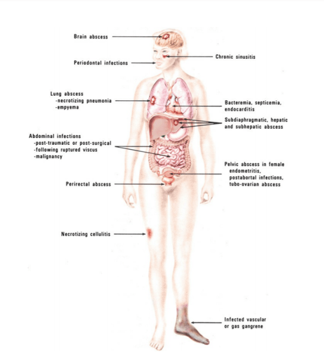 Figure: Common locations of infections involving anaerobic bacteria (Image source: Koneman, Ref-1)
Common anaerobic infections
- Brain abscess
- Dental infections (dental abscesses, gingivitis, and periodontitis)
- Head and neck infections ( suppurative infections of retropharyngeal abscess, peritonsillar abscess, cervical lymphadenitis, deep neck abscesses, and parotitis)
- Abdominal abscesses,
- Aspiration pneumonia,
- Bite infections (animal/human),
- Lung abscesses, and
- Necrotizing infections of soft tissue.
Clinical clues: when to suspect an anaerobic infection
Anaerobes rarely announce themselves by name, but they leave characteristic signs. Any of the following should raise suspicion:
- Foul, putrid smell. Anaerobes produce short-chain fatty acids and other volatile products as they ferment. A rotten or putrid odor from pus or discharge is one of the most reliable bedside clues to anaerobes.
- Gas in the tissues. Some anaerobes, especially Clostridium, produce gas as they metabolize. Gas felt under the skin (crepitus) or seen on imaging suggests an anaerobic process such as gas gangrene.
- Abscess formation. A walled-off collection of pus creates exactly the low-oxygen, dead-tissue environment anaerobes thrive in.
- Necrotic (dead) tissue. Dead tissue has no blood supply and no oxygen, which favors anaerobes and is a hallmark of severe anaerobic infection.
- Infection near a mucosal surface. Infection close to the mouth, bowel, or genital tract, where anaerobes are normal flora, points to those anaerobes as the likely cause.
- Bacteria on Gram stain but no growth on aerobic culture. If the direct Gram smear clearly shows organisms yet the routine aerobic culture is sterile, the organisms are likely anaerobes that need anaerobic culture to grow. This mismatch is one of the strongest laboratory clues.
The bacteria that cause anaerobic infections
Anaerobic infections are caused by a range of Gram-positive and Gram-negative organisms. The table below groups the main ones. Where we have a detailed article on an organism, its name links to it.
| Organism | Gram reaction | Typical anaerobic infection |
|---|---|---|
| Clostridioides difficile | Gram-positive, spore-forming bacilli | Antibiotic-associated and hospital-acquired diarrhea and colitis |
| Clostridium perfringens | Gram-positive, spore-forming bacilli | Gas gangrene (myonecrosis), soft-tissue infection, food poisoning |
| Clostridium tetani | Gram-positive, spore-forming bacilli | Tetanus |
| Clostridium botulinum | Gram-positive, spore-forming bacilli | Botulism (infant, foodborne, wound) |
| Actinomyces israelii | Gram-positive, non-spore-forming bacilli | Actinomycosis |
| Cutibacterium acnes (formerly Propionibacterium acnes) | Gram-positive, non-spore-forming bacilli | Acne; infections of implanted devices |
| Peptostreptococcus and related anaerobic cocci | Gram-positive cocci | Chronic sinusitis, aspiration pneumonia, pelvic and oral infections |
| Bacteroides fragilis | Gram-negative, non-spore-forming bacilli | Intra-abdominal abscess, aspiration pneumonia, brain abscess |
| Fusobacterium spp. | Gram-negative, non-spore-forming bacilli | Peritonsillar abscess, Lemierre's syndrome |
| Prevotella spp. | Gram-negative, non-spore-forming bacilli | Oral, head-and-neck, and perianal abscesses |
| Veillonella spp. | Gram-negative cocci | Usually harmless oral/gut flora; rarely opportunistic infection |
Collecting a good specimen for anaerobic culture
Anaerobic culture succeeds or fails at the collection step. Two problems must be avoided:
- Oxygen exposure kills the target. Anaerobes begin to die as soon as they meet air, so specimens must be protected from oxygen and transported quickly, ideally in an anaerobic transport system. Aspirates (pus drawn into a syringe) and tissue are far better than swabs, which expose bacteria to air and hold very little sample.
- Normal-flora contamination gives a meaningless result. Because anaerobes are heavy normal flora on mucosal surfaces (mouth, gut, vagina), a specimen from or through those surfaces will grow normal-flora anaerobes that have nothing to do with the infection. Such specimens are unacceptable for anaerobic culture.
The best specimen is obtained by tissue biopsy or needle aspiration from a normally sterile, walled-off site. Suitable specimens include:
- Bile
- Biopsy of endometrial tissue obtained with an endometrial suction curette
- Blood
- Bone marrow
- Bronchial washings obtained with a double-lumen plugged catheter
- Cerebrospinal fluid
- Culdocentesis aspirate
- Decubitus ulcer, if obtained from the base of the lesion after thorough debridement of surface debris
- Fluid from normally sterile sites (e.g. joint)
- Material aspirated from abscesses (the best specimens are from loculated or walled-off lesions)
- Percutaneous (direct) lung aspirate or biopsy
- Peritoneal (ascitic) fluid
- Sulfur granules from draining fistula
- Suprapubic bladder aspirate
- Thoracentesis (pleural) fluid
- Tissue obtained at biopsy or autopsy
- Transtracheal aspirate
- Uterine contents, if collected using a protected swab
For the full rules on which microbiological specimens are accepted or rejected, and why, see our article on rejection criteria for microbiological specimens.
Laboratory diagnosis
The culture and isolation of anaerobic organisms are difficult because of their fastidious nature. Proper collection and transport of the sample are of utmost importance to increase their recovery whenever samples are shipped to a reference laboratory.
Gram stain is an essential rapid tool for anaerobic bacteriology. All the specimens submitted for anaerobic culture should be examined by Gram staining prior to culture.
- Gram stain reveals the types and relative numbers of microorganisms and host cells present in the sample.
- Gram stain also serves as a quality control measure for the adequacy of anaerobic techniques.
- After the bacteria’s isolation, the isolate’s clinical relevance is determined by correlating it with the initial Gram stain.
Is there any modification of the Gram staining technique for the anaerobes?
Yes. We use the same standard gram stain procedure and reagents but the safranin counterstain is left on for 3 to 5 minutes. Alternatively, 0.5% aqueous basic fuchsin can be used as the counterstain.
Culturing anaerobes
Growing anaerobes requires an oxygen-free atmosphere, created by one of several systems. Each has its own detailed article:
- GasPak anaerobic system: a sealed jar in which a sachet chemically removes oxygen and generates carbon dioxide. The common workhorse in most labs.
- Robertson's cooked meat medium: a liquid medium whose meat particles lower the oxygen level, good for growing and maintaining anaerobes.
- Commonly used anaerobic media: an overview of the selective and enriched media used to isolate anaerobes.
- Cultivation of aerobic and anaerobic bacteria: the broader methods, including anaerobic jars, glove boxes and chambers, and pre-reduced media.
Other systems include the McIntosh-Fildes jar, anaerobic glove boxes, Coy chambers, and the Anoxomat automated system.
Presumptive identification of anaerobic bacteria can be made using macroscopic examination, examination of Gram-stained smears, cultural characteristics in differential agar media, and results of spot tests. The litmus milk test is one biochemical test used in identifying some anaerobes, notably Clostridium species.
Earlier, microbiologists used to rely on the phenotypic characteristics of the anaerobes for their identification but nowadays, various commercial microsystems (RapID ANA II (Remel), the RapID CB-Plus (Remel), the BBL Crystal ANR ID (BD Microbiology Systems) and the RapID 32A (bioMérieux, Inc.) are available for the identification of anaerobes. Other methods of identification are; the determination of metabolic products by Gas-Liquid Chromatography, the use of molecular and mass spectrometric methods (e.g., MALDI-TOF), etc.
What do positive gram stain and negative culture indicate?
Positive grams stain with a negative culture report gives information regarding the adequacy of sample collection, transport, and culture methods used. This situation may come in the following mentioned conditions:
- Poor transport methods
- Excessive exposure to air during sample processing
- Inadequate types of media or old media, or
- The anaerobic system (jar, pouch, and chamber) failed to achieve an anaerobic atmosphere.
- That microorganisms have been killed by antimicrobial therapy
Treatment
Antibiotic choice for anaerobic infections differs from that for aerobic infections, because many anaerobes (especially Bacteroides fragilis) produce beta-lactamase and resist plain penicillins. The main options, chosen by the site and the organisms involved:
- Metronidazole is the classic anaerobic drug, highly active against most Gram-negative anaerobes such as Bacteroides.
- Beta-lactam and beta-lactamase-inhibitor combinations (such as ampicillin-sulbactam and piperacillin-tazobactam) cover both anaerobes and the aerobes in mixed infections.
- Carbapenems (such as meropenem) are used for serious mixed infections.
- Clindamycin covers many anaerobes, though resistance has grown.
Surgery is often as important as antibiotics. Draining an abscess, removing dead tissue (debridement), or, in severe gas gangrene, amputating, removes the low-oxygen dead tissue the anaerobes depend on. Antibiotics alone often cannot cure an anaerobic infection without this source control.
How to remember
Foul smell, gas, pus, no aerobic growth: think anaerobe. These four clues together are the bedside and bench signature of anaerobic infection. If you remember the quartet, you will suspect anaerobes when it matters.
Anaerobes live where oxygen is low, and infect right next door. Mouth, gut, skin, genital tract. Almost every anaerobic infection is normal flora from one of these sites escaping into nearby tissue after a barrier breaks. Locate the infection and you can guess the resident anaerobe.
No catalase, no SOD, no life in air. Obligate anaerobes lack the enzymes that neutralize oxygen's toxic byproducts, so oxygen's own waste products kill them. That single deficiency is why they are anaerobic.
Specimen quality is the whole game. Air kills anaerobes and normal flora fakes them. Aspirate, not swab; sterile site, not mucosa; fast transport, not a delay. A positive Gram stain with a negative culture usually means the specimen or transport failed, not that there was no infection.
Key exam facts
| Point | Fact | Memory aid |
|---|---|---|
| Definition | Infection by bacteria that grow without oxygen | Anaerobe = no air |
| Most are | Endogenous (from the person's own normal flora) | Your own flora, wrong place |
| Why oxygen is toxic | Anaerobes lack catalase, SOD, peroxidase | No enzymes, no defense |
| Oxygen groups | Obligate anaerobe, facultative, aerotolerant, microaerophile | Campylobacter is microaerophilic, not anaerobic |
| Main body reservoirs | Mouth, colon, skin, genital tract | Anaerobes live in low-oxygen niches |
| Top clinical clues | Foul smell, gas, abscess, necrosis, no aerobic growth | The anaerobe quartet |
| Key Gram-negative rod | Bacteroides fragilis (beta-lactamase producer) | Gut anaerobe, resists penicillin |
| Key gas-forming rod | Clostridium perfringens (gas gangrene) | Spore-forming, gas in tissue |
| Best specimen | Aspirate or tissue from a sterile site, fast anaerobic transport | Aspirate, not swab |
| Positive Gram, negative culture | Usually a collection, transport, or culture failure | The system let air in |
| Drug of choice (Gram-negative anaerobes) | Metronidazole | Metronidazole for anaerobes |
| Treatment also needs | Surgery: drainage, debridement | Source control, not just drugs |
Where students get confused
"Is Campylobacter an anaerobe?" No. Campylobacter is microaerophilic: it needs oxygen, but only a little, less than normal air. Anaerobes grow without oxygen and are often harmed by it. Microaerophiles are a separate group and should not be counted as anaerobes, even though older tables sometimes list them together.
"Where do anaerobes causing infection come from?" Usually the patient's own body. Anaerobes are heavy normal flora in the mouth, colon, skin, and genital tract. Infection happens when a barrier breaks (trauma, surgery, a perforated bowel) and these resident anaerobes reach a normally sterile site. Most anaerobic infections are endogenous, not caught from outside.
"Why does a foul smell mean anaerobes?" Anaerobes ferment tissue and produce volatile short-chain fatty acids and other smelly products. Aerobic infections usually do not smell the same way. A distinctly putrid odor is one of the most useful bedside clues.
"Why did the Gram stain show bacteria but the culture grew nothing?" Almost always a technique failure, not a true negative. Either the specimen met air (killing the anaerobes), transport was too slow, the media were wrong or old, the anaerobic system failed to remove oxygen, or antibiotics had already killed the organisms. It tells you to check your method, not to call the sample sterile.
"Why is surgery needed if there are antibiotics?" Anaerobes live in dead, oxygen-poor tissue and walled-off abscesses that antibiotics penetrate poorly. Draining the pus and removing dead tissue takes away the environment the anaerobes depend on. Without this source control, antibiotics alone often fail.
"Why an aspirate and not a swab?" A swab exposes bacteria to air, holds very little sample, and often picks up surface normal flora. An aspirate or tissue sample protects the anaerobes from oxygen and comes from the true site of infection. Specimen type strongly affects whether anaerobes are recovered.
Frequently Asked Questions
What is an anaerobic infection?
What is an anaerobic infection?
It is an infection caused by anaerobic bacteria, which grow without oxygen and are often harmed by it. These infections range from a small abscess to life-threatening gas gangrene. Most come from the person's own normal flora reaching a normally sterile site.
Where are anaerobic bacteria found in the human body?
Where are anaerobic bacteria found in the human body?
Mainly in low-oxygen sites: the mouth and gums, the large intestine, the skin, and the female genital tract. The colon holds the densest anaerobic population. Infections usually occur when these resident anaerobes escape into nearby tissue after a barrier is broken.
What are examples of anaerobic infections?
What are examples of anaerobic infections?
Dental and gum abscesses, aspiration and lung abscesses, intra-abdominal abscesses after bowel perforation, brain abscesses, gas gangrene, tetanus, botulism, and Clostridioides difficile colitis. Many are polymicrobial.
What are the signs of an anaerobic infection?
What are the signs of an anaerobic infection?
Classic clues are a foul or putrid smell, gas in the tissues, abscess formation, dead (necrotic) tissue, infection near a mucosal surface, and bacteria seen on Gram stain that fail to grow on ordinary aerobic culture.
Why is oxygen toxic to anaerobic bacteria?
Why is oxygen toxic to anaerobic bacteria?
Obligate anaerobes lack the protective enzymes catalase, superoxide dismutase, and peroxidase. Without these, the toxic byproducts of oxygen (superoxide and hydrogen peroxide) build up and damage the cell, so the bacteria are harmed or killed in air.
How are anaerobic infections diagnosed?
How are anaerobic infections diagnosed?
By collecting a good specimen (an aspirate or tissue from a sterile site, protected from air and transported quickly), examining a Gram stain, and culturing it in an oxygen-free system such as a GasPak jar. A Gram stain showing bacteria with no aerobic growth strongly suggests anaerobes.
Why must anaerobic specimens be collected and transported carefully?
Why must anaerobic specimens be collected and transported carefully?
Anaerobes start to die when exposed to air, so delays or oxygen exposure can kill them before culture. Swabs and specimens from mucosal surfaces (which carry normal-flora anaerobes) give poor or misleading results. Aspirates and tissue from sterile sites, transported fast, are best.
How are anaerobic infections treated?
How are anaerobic infections treated?
With antibiotics chosen for anaerobes (metronidazole, beta-lactam/beta-lactamase-inhibitor combinations, carbapenems, or clindamycin) and, just as importantly, with surgery to drain abscesses and remove dead tissue. Removing the low-oxygen dead tissue is often essential for cure.
References
- Procop GW, Church DL, Hall GS, et al. Koneman's Color Atlas and Textbook of Diagnostic Microbiology. 7th ed. Philadelphia: Wolters Kluwer; 2017.
- Tille PM. Bailey & Scott's Diagnostic Microbiology. 15th ed. St. Louis: Elsevier; 2022.
- Leber AL, ed. Clinical Microbiology Procedures Handbook. 4th ed. Washington, DC: ASM Press; 2016. doi:10.1128/9781683670438.CMPH
- Noor A, Rehman Khetarpal S. Anaerobic Infections. In: StatPearls [Internet]. Treasure Island (FL): StatPearls Publishing; 2023. Available from: https://www.ncbi.nlm.nih.gov/books/NBK482349/
- Levinson W, Chin-Hong P, Joyce EA, Nussbaum J, Schwartz B. Review of Medical Microbiology and Immunology. 17th ed. New York: McGraw Hill; 2022.

Tankeshwar Acharya, MSc (Medical Microbiology)
Tankeshwar Acharya is an Assistant Professor in the Department of Microbiology at Patan Academy of Health Sciences (PAHS), Nepal, where he has been teaching and practicing clinical microbiology for over 14 years. He is the founder of Microbe Online, one of the leading free microbiology education resources on the web, covering bacteriology, mycology, parasitology, immunology, and clinical laboratory diagnostics written from direct experience in both the classroom and the diagnostic laboratory.
Comments
No comments yet. Be the first to share your thoughts.
Leave a comment
All comments are reviewed before they appear.