
Haemophilus influenzae: Properties, Virulence Factors, Diseases, and Lab Diagnosis

On this page
In the pre-vaccine era, a child presenting with high fever, stiff neck, and bulging fontanelle in a developing country had a high probability of Haemophilus influenzae type b (Hib) meningitis, the leading cause of bacterial meningitis in children under five years globally before the introduction of Hib conjugate vaccines. In countries with full Hib vaccine coverage, invasive Hib disease has been virtually eliminated. In countries with incomplete coverage, Hib meningitis remains a preventable killer.
The Hib vaccine story is one of the great successes of applied microbiology: understanding the polysaccharide capsule as the key virulence factor led directly to a conjugate vaccine that has saved an estimated 1.5 million children's lives since its introduction.
But H. influenzae is not simply a vaccine-preventable pathogen. Nontypeable strains (NTHi), which have no capsule and are not covered by the Hib vaccine, remain a major cause of otitis media, sinusitis, and lower respiratory tract infections across all age groups and settings, including in fully vaccinated populations.
Most Haemophilus species are normal inhabitants of upper respiratory tract of humans and other animals. The species of Haemophilus that most frequently cause human infections are H. influenzae (respiratory and invasive infections), H. aegyptius (acute conjunctivitis), H. parainfluenzae, H. haemolyticus, H. parahaemolyticus, H. aprophilus, H. paraphrophilus and H. segnis (abscesses and infective endocarditis).
General Properties
Haemophilus influenzae is:
- Gram-negative coccobacillus: pleomorphic; varies from coccobacilli to short rods depending on growth conditions
- Fastidious: requires enriched media; cannot grow on standard nutrient agar or tryptic soy agar
- Requires X factor (hemin/porphyrin) and V factor (NAD): both must be present simultaneously for growth; chocolate agar provides both by heat-lysis of RBCs
- Strictly human pathogen: no animal reservoir; transmitted person-to-person by respiratory droplets
- Aerobic, with enhanced growth in 5–10% CO₂
- Non-motile, non-spore-forming
- Oxidase-positive, catalase-positive
- Characteristic "mouse nest" odor on chocolate agar, due to indole production
- Temperature sensitive: specimens must NOT be refrigerated; cold kills the organism before culture
Two epidemiologically distinct populations:
| Characteristic | Hib (type b encapsulated) | NTHi (nontypeable) |
|---|---|---|
| Capsule | Yes. Type b polyribosyl ribitol phosphate (PRP) | No capsule |
| Serotype | Type b | None (nontypeable) |
| Primary age group | Children <5 years | All ages |
| Key diseases | Meningitis, epiglottitis, septic arthritis, bacteremia | Otitis media, sinusitis, COPD exacerbations, pneumonia |
| Vaccine | Hib conjugate vaccine | No vaccine available |
| Post-vaccine era | Rare in vaccinated populations | Unchanged not affected by Hib vaccine |
| Invasive potential | High | Low (mucosal infections primarily) |
Virulence Factors of Haemophilus influenzae
1. Polysaccharide Capsule (Hib only)
The type b polysaccharide capsule; polyribosyl ribitol phosphate (PRP) is the most important virulence factor of encapsulated H. influenzae:
- Antiphagocytic: inhibits complement-mediated opsonization and neutrophil phagocytosis
- Facilitates bacteremia: enables escape from the mucosal surface into the bloodstream and hematogenous spread to meninges, joints, and epiglottis
- Vaccine immunogen: PRP conjugated to a protein carrier (tetanus toxoid, diphtheria toxoid, or outer membrane protein) is the basis of all Hib conjugate vaccines; conjugation is critical because unconjugated PRP gives poor T-cell-dependent immunity in children under 18 months
Nontypeable H. influenzae lacks a capsule entirely, which is why it cannot cause invasive disease in the same way as Hib, and why there is no vaccine for NTHi.
2. IgA1 Protease
Like N. meningitidis and N. gonorrhoeae, H. influenzae produces IgA1 protease; an enzyme that cleaves secretory IgA1 at the hinge region:
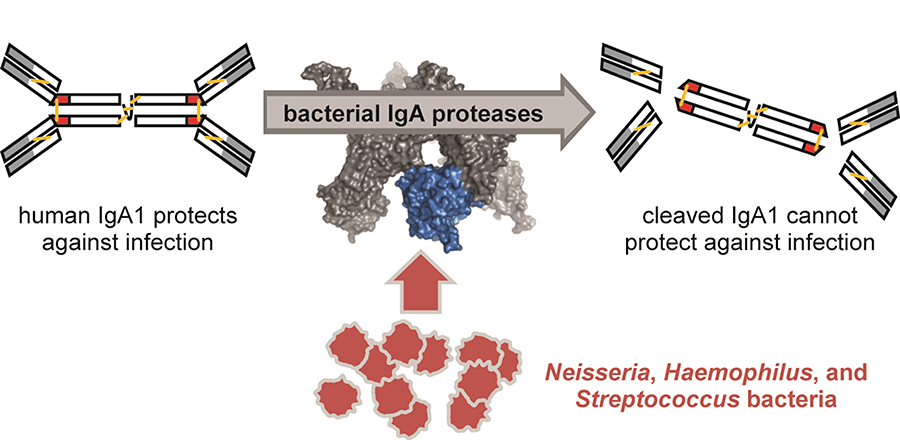 - Destroys the mucosal antibody that would otherwise block attachment to respiratory epithelial cells
- Produced by both Hib and NTHi strains
- Particularly important for NTHi colonization of the nasopharynx and middle ear
3. Adhesins: HMW1, HMW2, and Hia
NTHi strains produce adhesin proteins that mediate attachment to respiratory epithelium:
- HMW1 and HMW2 (high-molecular-weight adhesins): present in ~75% of NTHi strains; mediate attachment to non-ciliated airway epithelial cells; undergo phase variation
- Hia (Haemophilus influenzae adhesin): present in ~25% of NTHi strains without HMW1/2; mediate sialoglycoconjugate-dependent attachment
- These adhesins are critical for the NTHi colonization and infection cycle (nasopharynx → middle ear → sinuses)
4. Phase Variation and Immune Evasion
H. influenzae uses phase variation (on/off switching of surface protein expression) to evade the host immune response:
- Hemoglobin-binding proteins (HgbA, HgbB, HgbC) undergo phase variation
- Adhesins HMW1, HMW2, and OapA undergo phase variation
- Allows adaptation to the changing immune environment of the host during infection
- Contributes to persistence of NTHi in chronic otitis media and COPD exacerbations
5. Lipooligosaccharide (LOS)
Like Neisseria, H. influenzae produces LOS rather than full LPS:
- Endotoxin activity: stimulates TNF-α and IL-1β production causing local inflammation
- Molecular mimicry: LOS structures resemble host glycosphingolipids, reducing immune recognition
- Sialic acid decoration of LOS enhances serum resistance in invasive strains
6. Iron Acquisition
H. influenzae is an obligate human pathogen and must scavenge iron from host proteins in vivo:
- Produces hemoglobin-binding proteins (HgbA, HgbB, HgbC) that extract heme directly from hemoglobin
- Produces transferrin-binding proteins that extract iron from transferrin
- Iron acquisition is essential for survival in the bloodstream and is a potential vaccine/drug target
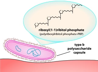 Figure: Capsule of Haemophilus influenzae
Virulence Factor Summary
| Factor | Hib | NTHi | Role |
|---|---|---|---|
| Capsule (PRP) | Yes | No | Antiphagocytic; bacteremia; vaccine target |
| IgA1 protease | Yes | Yes | Destroys mucosal IgA; facilitates colonization |
| HMW adhesins | No | Yes | Epithelial attachment; colonization |
| Phase variation | Yes | Yes | Immune evasion; adaptation |
| LOS | Yes | Yes | Endotoxin; tissue damage; mimicry |
| Iron acquisition | Yes | Yes | Survival in vivo |
Disease
Haemophilus influenzae can cause many kinds of infections. Which can range from mild ear infections to severe diseases, like meningitis. Haemophilus influenzae bacteria most often cause pneumonia, bacteremia, and meningitis mostly in infants and children younger than five years of age.
| Disease | Species | Specimen for culture |
|---|---|---|
| Meningitis | H. influenzae type b | Cerebrospinal fluid (CSF); blood |
| Epiglottis | H. influenzae type b | Blood; laryngeal secretions |
| Otitis media | H. influenzae nontypeable strains | Swab of drainage in ear canal; needle aspiration. If systemic disease is suspected blood/CSF culture is recommended |
| Acute sinusitis | H. influenzae usually nontypeable strains; in rare cases H. parainfluenzae is also involved | Sinus aspirates, surgical specimens |
| Acute Pharyngitis/ laryngotracheobronchitis | H. influenzae type b | Posterior pharyngeal swab; laryngeal secretions |
| Bronchitis | H. influenzae nontypeable strains | Sputum; transtracheal aspirates; bronchial washings |
| Pneumonia | H. influenzae type b; in case of elderly patients nontypeable strains may involve | Sputum; tracheal aspirates; bronchial washings |
| Endocarditis | H. aphrophilus; H. paraphrophilus; H. parainfluenzae; rarely, H. influenzae | Blood |
| Genital tract infection and postpartum bacteremia | H. influenzae nontypeable; H. parainfluenzae | Urethral and endocervical specimens; blood. |
| Conjunctivitis | H. influenzae biogroup aegyptius | Conjunctival swab |
| Chancroid | H. ducreyi | Swab obtained from genital ulcers; aspirates from buboes; endocervical swabs |
| Brazilian purpuric fever | H. influenzae biogroup aegyptius | Blood; conjunctival swabs; skin lesions |
Note: In spite of its name, Haemophilus influenzae does not cause influenza (the “flu”)
There are six identifiable types of Haemophilus influenzae bacteria (a through f) and other non-identifiable types (called nontypeable). Haemophilus influenzae type b, or Hib is associated with most of the clinical cases for which vaccine is also available.
Sample
Specimens used for the laboratory diagnosis of Haemophilus influenzae may be
- Specimens from respiratory tract: Sputum, lung aspirate, pleural fluid
- Body fluids e.g., Blood/ cerebrospinal fluid
- Exudates from the joint, middle ear, other sites
Sample Collection
- Sputum: Collect > 1.0 ml expectorated sputum in a sterile screw-capped container.
- Lung aspirate/ pleural fluid: Collect > 1.0 ml by percutaneous needle aspiration in a sterile screw-capped tube.
- Blood: Clean the venipuncture site with 70% alcohol and iodine, allow it to evaporate and collect blood aseptically in a culture broth with an anticoagulant. In the case of adults, collect 5-10 ml of blood in culture bottle, for children < 12-year old, collect 1.5-2.0 ml of blood. Mix the blood and broth by rotating gently to avoid clotting.
- Cerebrospinal fluid (CSF): Clean the skin over L3-L4 inter-space with 70% alcohol and iodine. Collect > 1.0 ml CSF in a sterile screw-capped tube. Keep the CSF in an incubator at 35-37 degree centigrade, if it is not processed immediately.
- Exudates from joints/middle ear: Collect > 1.0 ml by aspiration in a sterile screw-capped tube or add directly to a culture broth used for blood culture.
Note: DO NOT REFRIGERATE THE SAMPLE
Sample Transport
H. influenzae is a fastidious bacteria. Care must be taken during the transport of the specimen. Specimens must be transported promptly to the laboratory preferably within 1-2 hours.
Blood can only be transported after collecting in a culture broth containing appropriate anticoagulant. The inoculated medium can be held at room temperature (20°C– 25°C) for 4 – 6 hours before incubation at 37°C. The samples during transportation should be protected from extremes of temperature (less than 18°C, more than 30°C) and direct sunlight.
Direct Examination
- Perform Gram stain
- Gram staining shows Gram-negative pleomorphic thin rods or coccobacilli.
Culture
- Inoculate samples onto chocolate agar. Plain blood agar fails because hemin (X) stays locked in intact red cells and its NAD (V) is degraded by red-cell enzymes; gentle heating to make chocolate agar frees the hemin and spares the NAD.
- Incubate at 37°C in an aerobic atmosphere containing 5-10% CO2 for 24-48 hours.
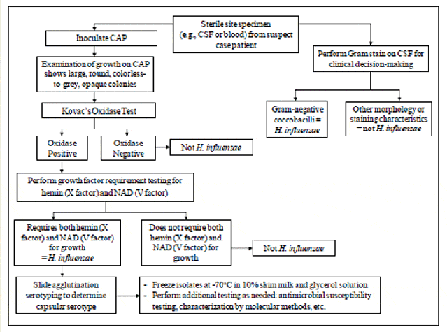 Figure: Flow chart Haemophilus influenzae identification
Colony morphology on chocolate agar:
- large flat, colorless to gray or opaque colonies.
- Colonies are 0.5 – 1mm circular, low convex, smooth, pale grey, and transparent.
- With a characteristic “mouse nest” odor. No hemolysis or discoloration is seen.
- Encapsulated strains appear more mucoid (watery) and non-capsulated strains appear as compact greyish colonies.
Note: Growth is enhanced on chocolate agar and satellitism around Staphylococcus aureus is seen on blood agar.
Biochemical reactions for differentiation
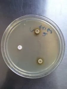 Figure: X and V Factor: Growth is seen around XV Factor only.
Identity is confirmed by the organism's growth-factor requirement. On a factor-free medium with X, V, and XV disks, H. influenzae grows only around the XV disk (it needs both hemin and NAD).
The full procedure, interpretation table across Haemophilus species, and the porphyrin test caveat are covered in the X and V factor test.
Serotyping of Haemophilus influenzae
Commercial antisera against capsular serotypes "a" to "f" are available. Each antiserum contains antibodies against a specific capsular polysaccharide. When mixed with a colony carrying that capsule, visible agglutination occurs, identifying the serotype (type b is the one of major clinical importance).
The same capsule-plus-antiserum principle underlies the Quellung (capsular swelling) reaction, the microscopic method historically used to serotype encapsulated organisms including H. influenzae type b.
Slide agglutination test.
- Apply one drop of normal saline on a slide and make a homogenous suspension with a single suspected colony of H. influenzae.
- Add one drop of specific antiserum and mix thoroughly.
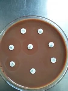 Figure: Anti-microbial sensitivity testing of H. influenzae in Chocolate Agar
Reading
- Observe for agglutination (visible clumping) within 1 minute.
Interpretation.
- A visible clumping within 1 minute is indicative of a positive reaction.
Antimicrobial susceptibility testing
- Perform antimicrobial susceptibility test against a selected group of antimicrobials by a disk-diffusion method.
- Follow the standard techniques as provided in the manual.
Treatment and Antibiotic Resistance
Invasive Hib disease (meningitis, epiglottitis, bacteremia):
- Empirical: Ceftriaxone or cefotaxime IV — covers Hib and other common meningitis pathogens
- Definitive: Ampicillin (if β-lactamase negative); ceftriaxone (if β-lactamase positive or sensitivity unknown)
- Duration: 7–10 days for uncomplicated meningitis; 7–14 days for epiglottitis
Non-invasive NTHi infections (otitis media, sinusitis, COPD exacerbations):
- Amoxicillin: first-line for mild disease
- Amoxicillin-clavulanate: if β-lactamase producing (20–40% of strains in many regions)
- Oral cephalosporins (cefuroxime, cefpodoxime), macrolides, or fluoroquinolones as alternatives
Antibiotic resistance: β-lactamase: Approximately 20–40% of H. influenzae clinical isolates produce TEM-1 or ROB-1 β-lactamase, making them resistant to ampicillin and amoxicillin. β-lactamase testing (nitrocefin disc) should be performed on all H. influenzae isolates from significant specimens.
β-lactamase negative ampicillin resistant (BLNAR) strains: A smaller but clinically important group of strains are ampicillin-resistant through mutations in penicillin-binding proteins (altered PBP3) rather than β-lactamase production. These strains are not detected by the nitrocefin disc test. BLNAR strains require alternative therapy and are an emerging resistance concern in parts of Asia.
Key exam facts in one table
| Fact | What to remember |
|---|---|
| Organism | Fastidious gram-negative coccobacillus; strict human pathogen, no animal reservoir. |
| Growth needs | X factor (hemin) + V factor (NAD); chocolate agar supplies both; needs 5-10% CO2. |
| Cold sensitivity | Never refrigerate specimens (especially CSF); cold kills the organism before culture. |
| Hib | Encapsulated (PRP), invasive (meningitis, epiglottitis), vaccine-preventable. |
| NTHi | No capsule, mucosal disease (otitis media, sinusitis, COPD), no vaccine. |
| Key virulence factor | PRP capsule (antiphagocytic, basis of the conjugate vaccine). |
| Confirmatory ID | X and V factor disk test; H. influenzae grows around XV disk only. |
| Resistance to know | Beta-lactamase (TEM-1/ROB-1, 20-40%); test with nitrocefin. BLNAR (altered PBP3) is nitrocefin-negative. |
| Empirical invasive therapy | Ceftriaxone or cefotaxime IV. |
| Classic sign | Epiglottitis "thumb sign" on lateral neck X-ray; do not use a tongue depressor. |
How to Remember
The two-factor requirement: why chocolate agar:
X factor = hemin (from heme/porphyrin) — used to make cytochromes, catalase, peroxidase V factor = NAD (nicotinamide adenine dinucleotide) — electron carrier
Blood agar contains V factor (released into medium) but X factor is locked inside intact RBCs. Chocolate agar releases both by heat-lysis. That is why H. influenzae grows on chocolate agar but not plain blood agar — unless Staphylococcus aureus lyses the RBCs (satellitism).
Mnemonic for factor requirements: H. parainfluenzae requires V only (p = panch = five = V in Roman numerals). H. ducreyi requires X only (d = das = ten = X in Roman numerals). H. influenzae requires both X and V.
Hib vs NTHi : Hib = capsule → invasive disease (meningitis, epiglottitis) → vaccine prevents this NTHi = no capsule → mucosal disease (otitis media, sinusitis, COPD) → vaccine does not prevent this
The satellite colony: tiny dewdrop colonies growing around Staphylococcus aureus on blood agar suggest Haemophilus influenzae. The Staphylococcus secretes V (NAD); the red cells it lyses nearby release X (hemin). The tiny colonies "orbit" the Staphylococcus like satellites, which is where the name comes from. Full method: satellitism test.
The three most serious Hib diseases (vaccine-preventable):
- Meningitis: leading cause in children <5 years pre-vaccine
- Epiglottitis: "thumb sign" on lateral neck X-ray; medical emergency; do NOT examine throat with tongue depressor
- Septic arthritis / cellulitis: haematogenous spread from bacteremia
CSF rule do NOT refrigerate: CSF for Haemophilus culture must be transported and processed immediately at room temperature or 37°C. The organism is extremely cold-sensitive — refrigerating CSF kills H. influenzae before it can be cultured. Same rule applies to blood culture bottles.
Where Students Get Confused
1. Hib vaccine did not eliminate H. influenzae disease. The vaccine targets only the type b capsule. Nontypeable strains have no capsule, are not covered, and remain a leading cause of otitis media, sinusitis, and COPD exacerbations even in fully vaccinated populations. "Fully vaccinated" does not mean "cannot have H. influenzae."
2. Beta-lactamase testing does not catch every resistant strain. A nitrocefin-negative result rules out beta-lactamase but not ampicillin resistance. BLNAR strains resist ampicillin through altered penicillin-binding proteins (PBP3), not beta-lactamase, so they read nitrocefin-negative yet still fail ampicillin. A negative nitrocefin test is not a green light for ampicillin if the clinical picture is not improving.
3. Refrigerating the specimen destroys the diagnosis. H. influenzae is exceptionally cold-sensitive. Unlike most bacteriology specimens, CSF and blood cultures for suspected H. influenzae must be kept at room temperature or 37°C, never refrigerated. This is a common preanalytical error that produces false-negative cultures.
4. Serotyping tests the isolate's capsule, not the patient's antibody. Slide agglutination uses known antisera to detect the capsular polysaccharide on the colony. It is not a serology test on patient serum. Only encapsulated strains agglutinate; nontypeable strains give no reaction because they have no capsule to detect.
References
- Tille, P. M. (2022). Bailey & Scott's Diagnostic Microbiology (15th ed.). Elsevier.
- Madigan, M. T., et al. (2021). Brock Biology of Microorganisms (16th ed.). Pearson.
- Agrawal, A., & Murphy, T. F. (2011). Haemophilus influenzae infections in the H. influenzae type b conjugate vaccine era. Journal of Clinical Microbiology, 49(11), 3728-3732. https://doi.org/10.1128/JCM.05476-11
- Pittman, M. (1931). Variation and type specificity in the bacterial species Haemophilus influenzae. Journal of Experimental Medicine, 53(4), 471-492.
- Noel, G. J. (2012). A review of levofloxacin for the treatment of bacterial infections. Clinical Medicine Insights: Circulatory, Respiratory and Pulmonary Medicine, 6, 25-38.
- World Health Organization. (2013). WHO position paper on Haemophilus influenzae type b (Hib) vaccination. Weekly Epidemiological Record, 88(39), 413-426.
Frequently Asked Questions
Why is chocolate agar used instead of blood agar for Haemophilus influenzae?
What is the difference between Hib and nontypeable H. influenzae?
Why must specimens for H. influenzae never be refrigerated?
What is a BLNAR strain and why does it matter?
Does the Hib vaccine protect against all Haemophilus influenzae infections?

Tankeshwar Acharya, MSc (Medical Microbiology)
Tankeshwar Acharya is an Assistant Professor in the Department of Microbiology at Patan Academy of Health Sciences (PAHS), Nepal, where he has been teaching and practicing clinical microbiology for over 14 years. He is the founder of Microbe Online, one of the leading free microbiology education resources on the web, covering bacteriology, mycology, parasitology, immunology, and clinical laboratory diagnostics written from direct experience in both the classroom and the diagnostic laboratory.
Comments
No comments yet. Be the first to share your thoughts.
Leave a comment
All comments are reviewed before they appear.