
Urease Test: Principle, Procedure, Interpretation & Urease-Positive Organisms
Urease test principle and procedure: including the PUNCH mnemonic, the rapid-vs-delayed reading windows, and why H. pylori gastric biopsies use this exact reaction.

On this page
The urease test determines whether an organism produces urease, an enzyme that hydrolyzes urea into ammonia and carbon dioxide. The ammonia raises the pH of the medium, which is detected by a color shift in the phenol red indicator: light orange at neutral pH, bright pink (magenta) at alkaline pH.
Why It Matters
Proteus mirabilis is one of the most prolific urease producers in clinical microbiology, and that single enzyme has a direct downstream consequence: in a urinary tract infection, urease-driven ammonia production alkalinizes the urine enough to precipitate magnesium ammonium phosphate crystals, the struvite stones. This is the classic mechanism behind staghorn calculi and recurrent UTIs, and it's the reason urease activity is not just an identification tool. It is a disease mechanism you can trace directly from one enzyme to a kidney stone on imaging.
Urease also shows up in a completely different clinical setting: a rapid urease test (sometimes called the CLO test) performed on a crushed gastric biopsy sample is one of the fastest ways to get presumptive evidence of Helicobacter pylori during an endoscopy: same principle, same color change, completely different patient story (peptic ulcer disease workup instead of a UTI).
Principle
Urea + H₂O → (urease) → 2 NH₃ + CO₂
The ammonia combines with CO₂ and water to form ammonium carbonate, which alkalinizes the medium. Phenol red shifts from light orange to bright pink (magenta) as the pH rises.
 Media: Christensen's urea agar (slant) or Stuart's urea broth. Both contain phenol red as the indicator, but they don't always agree with each other (see below).
Procedure
Christensen's Urea Agar
- Streak the entire slant surface with a heavy inoculum from an 18–24 hour pure culture. Do not stab the butt. It's left unstabbed deliberately, to serve as a color control against the slant.
- Incubate with the cap loosened at 35°C.
- Read at 6 hours and again at 24 hours; do not rely on a single reading.
Stuart's Urea Broth
- Inoculate with a heavy inoculum from an 18–24 hour pure culture and shake gently to suspend.
- Incubate with the cap loosened at 35°C.
- Read at 8, 12, and 24 hours.
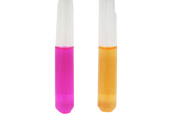 Figure: Urease test using urea broth. Left: Positive, Right: Negative
Where students get confused
- Rapid vs. delayed producers need different reading windows.
Proteus, Morganella morganii, and some Providencia stuartii are rapid urease producers, strong positive within 1 to 6 hours. Klebsiella and Enterobacter are delayed producers, weak positive at 6 hours that intensifies over 24 hours.
If you check only once at 24 hours, you will still catch both, but if you check early and stop, you will miss the delayed group. - Broth and agar can give genuinely different answers for the same organism. This is the trap that actually matters: Stuart's broth has a higher buffering capacity than Christensen's agar, which means delayed-positive organisms like Enterobacter may produce a false negative in broth even though the same organism reads correctly as delayed-positive on agar. If a result looks wrong, check which medium you used before assuming a technical error.
- The unstabbed butt is a control, not a shortcut. On Christensen's agar, comparing the slant color to the unstabbed butt is how you confirm the color shift is real and not just lighting or an old tube.
- A false positive can come from peptone, not urease. In overnight tests run on peptone-containing medium, peptone hydrolysis alone can alkalinize the medium and mimic a positive. This is unrelated to urease activity. Be cautious interpreting overnight positives on peptone-rich media.
- Urea reagent is light-sensitive. It undergoes autohydrolysis if not stored properly, so keep it at 2 to 8°C in the dark, and never reheat prepared medium, since urea decomposes under heat.
Result Interpretation
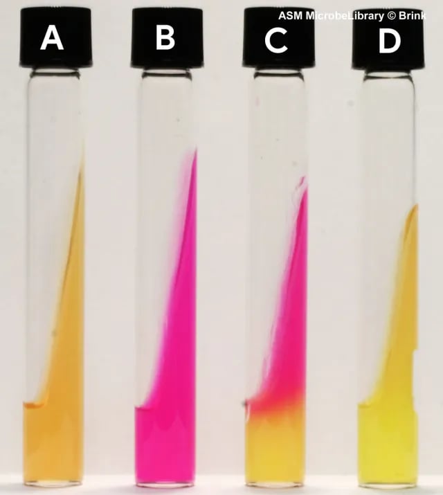 Figure: Urea agar test (a) uninoculated, (b) Proteus mirabilis (rapidly urease positive), (c) Klebsiella pneumoniae (delayed urease positive), (d) Escherichia coli (urease negative). (Benita A. Brink, Adams State College, Alamosa, CO)
| Organism type | Reading window | Result |
|---|---|---|
| Rapid producers (Proteus, M. morganii, some Providencia) | 1–6 hours | Strong positive (bright pink/magenta) |
| Delayed producers (Klebsiella, Enterobacter) | 6–24 hours | Weak, intensifying positive |
| Urease-negative (e.g., E. coli) | Any | Remains light orange, no change |
The PUNCH Mnemonic
Proteus · Ureaplasma · Nocardia · Cryptococcus neoformans · Helicobacter pylori
Note: Brucella and Corynebacterium (some species) are also urease producers that do not have a letter in this mnemonic. Remember them as named exceptions alongside PUNCH.
Diagnostic Applications
- Proteus identification: distinguishes Proteus from other non-lactose-fermenting Enterobacteriaceae.
- H. pylori (gastric biopsy): a crushed tissue sample placed directly into urease broth gives presumptive evidence of infection. This is the basis of the rapid/CLO test used during endoscopy. The related urea breath test uses the same enzymatic principle non-invasively.
- Cryptococcus vs. Candida: Cryptococcus neoformans is rapidly urease-positive; Candida albicans is not, which is useful for presumptive yeast differentiation.
Quality Control
| Organism | Expected result |
|---|---|
| Proteus mirabilis | Urease positive (rapid) |
| Escherichia coli | Urease negative |
Limitations
- Rapid vs. delayed urea splitting means interpretation depends on which organism group you are testing; the reading windows and the peptone and storage pitfalls are covered above.
- Medium choice affects sensitivity: Stuart's broth has higher buffering capacity than Christensen's agar and can mask delayed producers, so agar is more sensitive for that group.
References and further reading
- Brink B. Urease test protocol. Washington, DC: American Society for Microbiology; 2010.
- Leber AL, editor. Clinical Microbiology Procedures Handbook. 4th ed. Washington, DC: ASM Press; 2016. https://doi.org/10.1128/9781683670438.CMPH
Frequently Asked Questions
Why did my Enterobacter test negative in broth but positive on agar?
Stuart's broth has a higher buffering capacity than Christensen's agar, which can mask the weaker, delayed urease activity of organisms like Enterobacter. The agar is more sensitive for delayed producers, if results disagree, trust the agar reading.
My urea agar turned pink overnight even though I expected a negative, what happened?
My urea agar turned pink overnight even though I expected a negative, what happened?
How is the urease test used to diagnose H. pylori?
A crushed gastric biopsy sample is placed directly into urease broth. If H. pylori is present, its urease rapidly hydrolyzes urea and turns the broth pink within minutes to hours, this is the basis of the rapid urease (CLO) test performed during endoscopy.

Tankeshwar Acharya, MSc (Medical Microbiology)
Tankeshwar Acharya is an Assistant Professor in the Department of Microbiology at Patan Academy of Health Sciences (PAHS), Nepal, where he has been teaching and practicing clinical microbiology for over 14 years. He is the founder of Microbe Online, one of the leading free microbiology education resources on the web, covering bacteriology, mycology, parasitology, immunology, and clinical laboratory diagnostics written from direct experience in both the classroom and the diagnostic laboratory.
Comments
No comments yet. Be the first to share your thoughts.
Leave a comment
All comments are reviewed before they appear.