
Tinsdale Agar: Composition, Preparation, Uses, and Colony Characteristics
Tinsdale agar selectively isolates and differentiates Corynebacterium diphtheriae from diphtheroids. Black colonies with brown halos after 48h incubation — learn the principle, preparation, and why both reactions are needed for presumptive identification.
A child presents with a grey pseudomembrane in the throat, a "bull-neck" appearance, and a history of incomplete vaccination. The clinician suspects diphtheria. A throat swab is plated on Löffler's serum medium for rapid growth — and simultaneously on Tinsdale agar for selective isolation and differentiation.
After 48 hours on Tinsdale agar: grey-black colonies surrounded by a distinctly outlined brown halo. This combination — black colony plus brown halo — is the key result. Other organisms growing on the plate (staphylococci, diphtheroids) may reduce tellurite and form black colonies, but they do not produce the surrounding brown halo. The halo is what makes the presumptive identification.
Tinsdale agar base medium was developed by Tinsdale for the selective isolation and differentiation of C. diphtheriae from diphtheroids. Billings later modified the medium improving its differential qualities and reproducibility.
Moore and Parsons used Billings’ modification and confirmed the appearance of a distinct halo around the colonies of C. ulcerans and C. diphtheriae only. They also found modified Tinsdale medium as satisfactory as cystine-tellurite blood agar for the primary isolation of C. diphtheriae from nose and throat cultures. With these findings, they recommended the use of modified Tinsdale medium for routine isolation of C. diphtheriae.
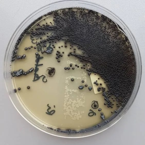 Figure: Corynebacterium diphtheriae in Tinsdale Agar medium; Look for black colonies with brown halo (Image source: Anonymous)
Principle
Tinsdale medium supports the growth of all species of Corynebacterium while inhibiting the growth of normal inhabitants of the upper respiratory tract. Potassium tellurite from the supplement inhibits all gram-negative bacteria and most of the upper respiratory tract normal flora.
L-cystine and sodium thiosulphate form the H₂S indicator system. Black coloration of the colonies results from tellurite reductase activity, resulting in the reduction of tellurium, while the brown halos indicate cystinase activity. Only C. diphtheriae and C. ulcerans give black colonies with a distinctly outlined halo. Other diphtheroids, staphylococci, and some streptococci may also reduce tellurite, although halos around the colonies are not generally present.
Peptic digest of animal tissue provides nitrogenous compounds and agar is for the solidification of the medium.
The two reactions, clearly explained:
- Tellurite reduction → black colony: C. diphtheriae (and some other organisms) reduce potassium tellurite to metallic tellurium, which deposits within and around the colony producing a grey-black colour. This reaction alone is NOT specific — staphylococci, some streptococci, and other diphtheroids also reduce tellurite.
- Cystinase activity → brown halo: C. diphtheriae and C. ulcerans produce cystinase, which reacts with L-cystine and sodium thiosulphate in the medium to produce H₂S. This H₂S reacts with iron salts in the medium to form iron sulphide — the brown/black precipitate that forms the halo around the colony. This reaction IS specific — diphtheroids and most other respiratory commensal organisms do not produce cystinase, so they do not produce halos even if they reduce tellurite.
The diagnostic rule: Black colony + brown halo = presumptive C. diphtheriae or C. ulcerans. Black colony alone = not specific. The halo is everything.
Composition of modified Tinsdale’s Base medium
pH 7.9 ± 0.2 @ 25°C
| Ingredients | Gm/Liter |
|---|---|
| Peptic digest of animal tissue | 20 g |
| Sodium chloride | 5 g |
| L-Cystine | 0.24 g |
| Sodium thiosulfate | 0.24 g |
| Agar | 15 g |
Tinsdale Supplement (for 1 Liter)
- Sterile bovine serum: 100 mL
- Tellurite solution (1%): 30 mL
Preparation of Tinsdale Medium
The Tinsdale’s agar base medium without serum and tellurite is stable indefinitely if stored in closed screw-capped tubes or bottles but after the addition of supplement (serum and tellurite), the medium is stable only for 2/3 days if stored in the refrigerator.
Troubleshooting Tinsdale Agar
| Problem | Cause | Action |
|---|---|---|
| No halo around black colonies | Serum not added after autoclaving; medium overheated with serum | Ensure serum is added aseptically to cooled base (50°C); never autoclave after serum addition |
| Halos present but faint | Insufficient incubation time | Read at 48h minimum; re-incubate up to 72h |
| No growth at all | Medium too inhibitory; tellurite concentration too high | Use correctly prepared modified Tinsdale medium; check potassium tellurite concentration |
| Medium too dark after preparation | Overheating during preparation | Do not overheat base; add serum at exactly 50°C |
| All colonies show halos | Contaminated serum; incorrect tellurite concentration | Check serum sterility; verify potassium tellurite and cystine concentrations |
Procedure
- Streak the Tinsdale agar plate to obtain well-isolated colonies. Note: It is recommended that the agar be stabbed at intervals because browning of the medium can be detected early (within 10-12 hours incubation) in the stab areas.
- Incubate the plates at 35°C in an aerobic incubator. Note: Growth of Corynebacterium diphtheriae may be inhibited if Tinsdale Agar is incubated in carbon dioxide-enriched air e.g. in aCO2 incubator.
- Examine the colonial morphology after 24 hours and 48 hours of incubation.
Results and Interpretation
A brown halo around the black colony is considered presumptive evidence of C. diphtheriae. This can sometimes be seen after 10–12 hours of incubation, although 48 hours may be required for the appearance of typical dark-brown halos. The only related species other than C. diphtheriae that produces this halo is C. ulcerans. Isolates with this colonial morphology are first identified biochemically as C. diphtheriae, and subsequent testing involves demonstrating the production of diphtheria toxin.
Other bacteria, such as coagulase-positive staphylococci, grow well on this medium but do not have a brown halo. Bacteria, such as species of Proteus that produce a heavy, diffuse blackening of the medium, can be distinguished by their Gram stain reaction and biochemical characteristics.
Uses of Tinsdale Agar
1. Primary isolation and presumptive identification of Corynebacterium diphtheriae from throat and nasal swabs
Tinsdale agar is specifically indicated when diphtheria is clinically suspected — in patients with pharyngeal pseudomembrane, bull-neck appearance, hoarseness, or stridor, particularly in unvaccinated or incompletely vaccinated individuals. It is used alongside Löffler's serum medium as part of the standard C. diphtheriae isolation protocol:
- Löffler's serum medium — inoculated simultaneously for rapid growth and Albert stain preparation (results in 12–18 hours)
- Tinsdale agar — for selective isolation and differentiation from respiratory commensals (results at 48 hours)
2. Differentiation of C. diphtheriae from diphtheroids (non-pathogenic corynebacteria)
The normal upper respiratory tract harbours multiple Corynebacterium species and diphtheroids that may grow on general-purpose media alongside C. diphtheriae. Tinsdale agar differentiates them: C. diphtheriae and C. ulcerans produce the characteristic black colony + brown halo; diphtheroids (C. xerosis, C. pseudodiphtheriticum, C. striatum) may produce black colonies from tellurite reduction but do NOT produce a surrounding halo. This distinction allows the laboratory to focus Albert staining and further testing on halo-producing colonies only.
3. Supporting biotype differentiation (in combination with tellurite blood agar)
When biotype identification is required (gravis, mitis, intermedius), Tinsdale agar is used alongside cystine-tellurite blood agar (Hoyle's medium). Different biotypes of C. diphtheriae show characteristic colony morphologies on tellurite blood agar:
- Gravis — flat, grey, irregular "daisy-head" colonies
- Mitis — black, dome-shaped, entire edge colonies
- Intermedius — small, flat, intermediate morphology
Tinsdale agar alone does not reliably differentiate biotypes — this requires tellurite blood agar.
4. Not recommended for routine non-diphtheria throat cultures
Tinsdale agar is specifically indicated for suspected diphtheria. It is not used as a routine throat culture medium — for routine throat culture (to detect S. pyogenes, for example), blood agar remains the standard. Using Tinsdale agar routinely would be inappropriate and costly.
Important limitation: A positive Tinsdale result (black colony + brown halo) is presumptive only. Definitive diagnosis requires:
- Species confirmation by biochemical tests or MALDI-TOF
- Toxigenicity testing by Elek test or PCR for the tox gene — non-toxigenic C. diphtheriae exists and does not cause diphtheria
For the full C. diphtheriae laboratory diagnosis pathway, see: Corynebacterium diphtheriae: Properties, Pathogenesis, and Laboratory Diagnosis
Tinsdale Agar vs Other C. diphtheriae Media
Three media are used in the isolation of C. diphtheriae. They serve complementary roles:
| Medium | Purpose | C. diphtheriae Appearance | Key Advantage |
|---|---|---|---|
| Löffler's serum medium | Rapid initial growth; stimulates metachromatic granule production | Grey-white colonies; granules visible on Albert stain | Fastest growth (visible in 6–8h); excellent for Albert stain |
| Tinsdale agar | Selective isolation and differentiation from diphtheroids | Black colonies with brown halo after 48h | Distinguishes C. diphtheriae from diphtheroids by halo |
| Tellurite blood agar (Hoyle's) | Selective isolation; biotype differentiation | Black colonies; biotype differences in morphology | Distinguishes biotypes (gravis = daisy-head, mitis = black dome) |
Standard protocol: Inoculate both Löffler's serum medium (for rapid morphology/Albert stain at 12–18h) AND Tinsdale agar (for selective isolation and presumptive identification at 48h). Tellurite blood agar may be added when biotype identification is needed.
Key Exam Facts in One Table
| Feature | Detail |
|---|---|
| Type | Selective and differential medium |
| Selective agent | Potassium tellurite — inhibits most respiratory commensals |
| Differential reactions | 1) Tellurite reduction → black colony; 2) Cystinase activity → brown halo |
| C. diphtheriae result | Black colony + brown halo after 48h |
| C. ulcerans result | Black colony + brown halo (same as C. diphtheriae) |
| Diphtheroids result | Black colony (tellurite reduction) but NO halo |
| Key diagnostic rule | Halo presence is specific; black colour alone is not |
| Incubation | 35–37°C for 48h minimum; up to 72h if negative at 48h |
| When to add serum | After autoclaving base, cooled to 50°C — never autoclave with serum |
| Shelf life (base without serum) | Indefinite if stored in sealed screw-cap containers |
| Companion media | Löffler's serum medium + Albert stain; tellurite blood agar |
| Confirmatory test | Elek test or PCR for tox gene (Tinsdale = presumptive only) |
References and further reading
- Forbes BA, Sahm DF, Weissfeld AS. Bailey & Scott's Diagnostic Microbiology. 14th ed. Elsevier; 2023.
- Koneman EW, Allen SD, Janda WM, Schreckenberger PC, Winn WC. Koneman's Color Atlas and Textbook of Diagnostic Microbiology. 6th ed. Lippincott Williams & Wilkins; 2006.
- Murray PR, Rosenthal KS, Pfaller MA. Medical Microbiology. 9th ed. Elsevier; 2020.
- Moore MS, Parsons EI. A study of modified Tinsdale's medium for the primary isolation of Corynebacterium diphtheriae. J Infect Dis. 1958;102(1):88–93.
- World Health Organization. Laboratory Manual for the Diagnosis of Diphtheria. WHO/EUR/93-5. Copenhagen: WHO Regional Office for Europe; 1993.
- Garcia LS. Clinical Microbiology Procedures Handbook. 4th ed. ASM Press; 2016.
Frequently Asked Questions
What are the two reactions that make Tinsdale agar diagnostic for C. diphtheriae?
Why is Tinsdale agar result considered presumptive rather than confirmatory for diphtheria?

Tankeshwar Acharya, MSc (Medical Microbiology)
Tankeshwar Acharya is an Assistant Professor in the Department of Microbiology at Patan Academy of Health Sciences (PAHS), Nepal, where he has been teaching and practicing clinical microbiology for over 14 years. He is the founder of Microbe Online, one of the leading free microbiology education resources on the web, covering bacteriology, mycology, parasitology, immunology, and clinical laboratory diagnostics written from direct experience in both the classroom and the diagnostic laboratory.