

An older man has repeated urinary infections that never fully clear. A scan shows a large stone filling the collecting system of one kidney, shaped like the antlers of a deer. The urine has an unusual ammonia-like smell and is alkaline. All of this points to one organism: Proteus, a bacterium whose single most important trait, the enzyme urease, both drives the infection and builds the stone.
Proteus is a common cause of urinary tract infection, but what makes it distinctive is what it does beyond infecting: it changes the chemistry of urine and turns it into stone. This page is about how Proteus is identified in the laboratory, starting with its dramatic swarming growth, and how its urease connects a simple UTI to a kidney full of stone.
Overview
Proteus is a Gram-negative rod of the family Enterobacteriaceae. It is found widely in the environment and lives as normal flora in the human intestine. It is an opportunistic pathogen: harmless in the gut, but a common cause of trouble when it reaches the urinary tract. Proteus is a frequent cause of urinary tract infection and is strongly associated with infection-related kidney stones. It also causes wound and ear infections, respiratory infections, and hospital-acquired infections.
The name fits the organism. Proteus is named after a sea-god in Greek mythology who could change his shape at will. Its discoverer chose the name because the organism keeps changing form as it spreads across a culture plate in waves, its striking swarming motility, described below.
What Proteus looks like, and what that tells you
A few features identify Proteus, and two of them are unusual enough to be near-diagnostic.
It swarms. On blood agar, Proteus does not form separate colonies. Instead it spreads across the whole plate in a series of thin, concentric waves, like ripples. This swarming is the first visual clue and is unusual for the family. It happens because the organism switches into long, hyper-flagellated "swarmer" cells that move as a group. Most bacteria you plate give tidy separate colonies; a plate covered in a thin filmy veil that grows in rings suggests Proteus.
It is a strong, fast urease producer. This is the single most important Proteus trait. It splits urea rapidly and powerfully, far faster than the weak urease of organisms like Klebsiella. This one enzyme explains both its role in kidney stones and how you identify it. Read more about urease test in this article.
It does not ferment lactose, so it is pale on MacConkey agar. This groups it with the other non-lactose fermenters and separates it from E. coli.
It is oxidase-negative (placing it in the Enterobacteriaceae) and deaminates phenylalanine to phenylpyruvic acid, a reaction most Enterobacteriaceae cannot do, which makes the PPA test a useful Proteus marker.
It has an ammonia-like smell. Because it splits urea into ammonia, Proteus plates often have a distinctive smell that experienced lab workers recognize.
Hold these together: a swarming, pale, oxidase-negative, strongly urease-positive organism with an ammonia smell is Proteus until proven otherwise.
Virulence factors and how Proteus causes disease
Proteus has a small set of virulence factors, and each one does something you can see in the disease.
Fimbriae (pili) for attachment. Proteus uses fimbriae to grip the lining of the urinary tract, so urine flow does not flush it out. As with other uropathogens, attachment is the necessary first step in a UTI.
Swarming motility for spread. The same swarming that is so obvious on a plate helps Proteus move across surfaces in the body, including up the urinary tract and along catheters. Its motility helps it ascend from the bladder toward the kidney.
Urease: the key virulence factor. This is what makes Proteus special and dangerous. Urease splits urea (abundant in urine) into ammonia. The ammonia raises the pH of the urine, making it alkaline. In alkaline urine, minerals that normally stay dissolved come out of solution as solids: magnesium ammonium phosphate (struvite) and calcium phosphate. These build into stones.
Why the stones matter, and why they feed the infection. As the stones grow, they trap bacteria inside them, where antibiotics cannot reach. So the infection cannot be cleared while the stone remains, and the ongoing infection keeps making more stone. It becomes a cycle: infection makes stone, stone shelters infection. Large stones can fill the whole collecting system of the kidney and take its branching shape; these are called staghorn calculi because they look like a deer's antlers. This is why a Proteus UTI is not just an infection to treat but a stone problem to solve.
Putting it together
The sequence explains the illness. Proteus attaches to the urinary tract with fimbriae, ascends using its swarming motility, and then its urease splits urea into ammonia. The alkaline urine that results precipitates struvite stones, which shelter the bacteria from antibiotics and drive recurrent, hard-to-clear infection. Remove the urease from the story and Proteus would be an ordinary UTI organism; the urease is what turns it into a stone-forming one.
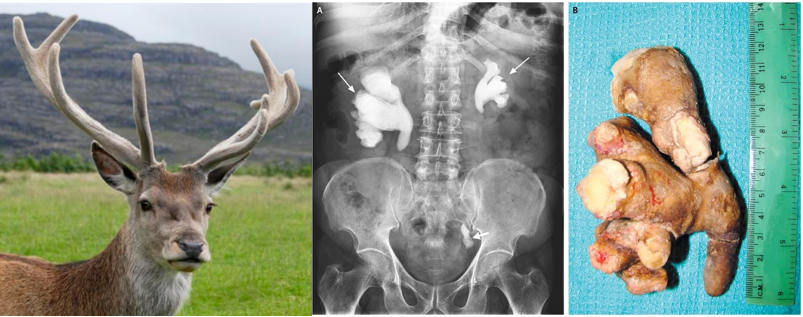 Figure: a staghorn calculus, a branching kidney stone shaped like a deer's antlers.
The urease-driven stone mechanism above is the reason Proteus is so strongly linked to struvite (magnesium ammonium phosphate) stones. Recurrent infection with a urease-producing organism such as Proteus is the classic cause of staghorn calculi, large branching stones that fill the kidney's collecting system.
Antigens and the Weil-Felix connection
Proteus has heat-stable O (somatic) and H (flagellar) antigens, and several serotypes are recognized on this basis.
One historical curiosity is worth knowing because it still appears in exams. Certain strains of Proteus vulgaris (called OX-19, OX-2, and OX-K) carry O antigens that happen to be shared with Rickettsia, the organisms that cause typhus and spotted fevers. Because of this shared antigen, these Proteus strains are used as a stand-in to detect antibodies against Rickettsia in the Weil-Felix test. The patient's serum is tested against these Proteus antigens; agglutination suggests a rickettsial infection. The full principle and its limitations are in the Weil-Felix test article.
Identifying Proteus in the laboratory
Specimen. Midstream urine for UTI, pus aspirate for abscesses, depending on the site. Send to the laboratory promptly.
On the plate.
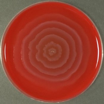 Figure: Swarming in Blood Agar
Proteus grows on all common laboratory media, and its appearance differs by medium. The medium-specific look, especially whether it swarms, is one of the most useful early clues, so it is worth having in one place:
| Medium | Appearance of Proteus |
|---|---|
| Blood agar | Swarming: a thin, filmy veil of growth spreading in concentric waves across the whole plate, rather than separate colonies. The signature Proteus pattern. Colonies may be weakly hemolytic. |
| MacConkey agar | No swarming (bile salts inhibit it); pale, non-lactose-fermenting (colorless) colonies. The stopped swarming plus pale colony is itself a useful clue. |
| Chocolate agar | Grows well as greyish, moist colonies; may show some spreading but the dramatic swarming is less pronounced than on blood agar. No hemolysis is read here because the blood is already lysed. |
| CLED agar | No swarming (CLED is specifically used for urine to prevent it); translucent, pale, non-lactose colonies. Preferred for urine cultures for exactly this reason. |
| Nutrient agar | Swarming growth with the characteristic ammonia-like smell from urea splitting. |
The unifying clues across media are the swarming on non-inhibitory media and the ammonia smell; the specific colony form then varies by medium as above. On the media that suppress swarming (MacConkey, CLED), Proteus shows plain pale non-lactose colonies.
Presumptive identification. An organism that swarms on blood agar, smells of ammonia, and is oxidase-negative can be presumptively called Proteus. A quick spot indole test then splits the two common species.
Differentiating P. mirabilis from P. vulgaris
The two common species are separated by one test: indole. This is the single most useful distinction, and it is worth understanding rather than memorizing which is which.
| Feature | P. mirabilis | P. vulgaris |
|---|---|---|
| Indole | Negative | Positive |
| MacConkey | Pale (non-lactose) | Pale (non-lactose) |
| Swarming | Yes | Yes |
| Urease | Positive (strong) | Positive (strong) |
| H₂S | Positive (strong) | Positive (variable) |
Note: These two organisms can be differentiated on the basis of indole test. *Proteus mirabilis* is indole minus (negative). The letter m ties the more common species to the negative result.
All three of these reactions can be read from a single sulfide indole motility (SIM) tube: both species are motile and H₂S-positive (strongly in P. mirabilis, more variably in P. vulgaris), and the indole reaction splits them cleanly, positive for P. vulgaris and negative for P. mirabilis. SIM is a common way to capture the H₂S, indole, and motility pattern of Proteus in one stab.
The most useful confirmatory test for the genus is the phenylpyruvic acid (PPA) test, positive for Proteus and negative for most other Enterobacteriaceae. One caveat worth knowing: Proteus, Morganella, and Providencia (the three genera of the tribe Proteeae) are all PPA-positive, so PPA identifies the group rather than Proteus alone. Within that group, the strong rapid urease and swarming point to Proteus.
Why swarming is a laboratory problem, and how to stop it
Swarming is a useful clue, but it is also a nuisance. When Proteus is mixed with other organisms on a plate, its veil of growth spreads over everything and covers the colonies you are trying to isolate. Several methods reduce swarming:
- Increasing the agar concentration (up to about 6% instead of the usual 1 to 2%), which makes the surface harder to move across.
- Adding inhibitors such as chloral hydrate, sodium azide, or boric acid to the medium.
- Using CLED agar for urine samples, which does not support swarming, instead of blood agar and MacConkey.
The Dienes phenomenon
Proteus mirabilis can tell "self" from "non-self" on a plate. When two different strains of P. mirabilis swarm toward each other, a visible line with sparse growth, the Dienes line, forms where they meet, because the two strains do not merge. When two identical strains meet, they merge smoothly with no line. This is used to tell whether two isolates are the same strain, which is useful in tracking outbreaks (for example, checking whether infections in different patients came from one source).
How to remember
Urease is the whole story. One enzyme explains almost everything about Proteus: it splits urea into ammonia, which makes urine alkaline, which precipitates struvite stones, which shelter the bacteria from antibiotics. Picture the chain: urea to ammonia to alkaline urine to stone. Remove urease and Proteus is just another UTI organism.
Swarming looks like ripples. Picture dropping a stone in water and the rings spreading out. Proteus grows across blood agar in the same concentric waves. No separate colonies, just a spreading veil. And it smells of ammonia, because of the urea splitting.
mirabilis is minus. The two common species differ by indole: *P. mirabilis* is indole-minus (negative), P. vulgaris is positive. The shared letter m links the species to the sign.
Weil-Felix borrows Proteus to catch Rickettsia. The odd one to remember: a Proteus antigen is used to detect antibodies against a completely different organism, Rickettsia, because they happen to share an antigen. The test grows Proteus but diagnoses typhus.
Staghorn = struvite = urea splitter. A branching kidney stone shaped like antlers means a struvite stone, which means a urease-producing organism, which usually means Proteus.
Key exam facts
| Fact | Detail |
|---|---|
| Gram reaction / shape | Gram-negative rod, family Enterobacteriaceae |
| Motility | Swarming motility on blood agar (concentric waves); does not swarm on MacConkey |
| MacConkey | Pale (non-lactose fermenter) |
| Oxidase | Negative |
| Urease | Strongly and rapidly positive (key trait) |
| PPA (phenylalanine deaminase) | Positive (marker vs most Enterobacteriaceae) |
| H₂S | Positive (strong in P. mirabilis, variable in P. vulgaris) |
| Species split | Indole: P. mirabilis negative, P. vulgaris positive |
| Main disease | Urinary tract infection; wound, ear, respiratory, hospital infections |
| Stone link | Urease → ammonia → alkaline urine → struvite (magnesium ammonium phosphate) → staghorn calculi |
| Weil-Felix | P. vulgaris OX-19, OX-2, OX-K antigens cross-react with Rickettsia; used to detect rickettsial antibodies |
| Dienes phenomenon | Different P. mirabilis strains form a Dienes line where they meet; identical strains merge |
| Swarming control | Higher agar %, chloral hydrate / sodium azide / boric acid, or CLED agar for urine |
Where students get confused
Swarming counted as contamination. A beginner may read the spreading veil as a contaminated or overgrown plate. It is a characteristic Proteus growth pattern. On MacConkey the swarming stops and normal pale colonies appear.
mirabilis vs vulgaris. The two are nearly identical; the one reliable splitter is indole (mirabilis negative, vulgaris positive). Remember mirabilis is minus.
Urease seen as just a test result. Urease is not only how you identify Proteus; it is why Proteus forms stones.
Weil-Felix thought to diagnose Proteus. The test uses Proteus antigens but diagnoses Rickettsia infection (typhus, spotted fever). This reversal is a classic exam trap.
Strong vs weak urease. Proteus urease is strong and fast. Other organisms (Klebsiella, some others) are weakly urease-positive. A rapid, strong positive points to Proteus; do not treat all urease-positives as equal.
Staghorn stones assumed to be calcium. The staghorn stones linked to infection are struvite (magnesium ammonium phosphate), formed because urease makes the urine alkaline. They are not the common calcium oxalate stones of metabolic origin.
References
- Tille, P. M. (2022). Bailey & Scott's Diagnostic Microbiology (15th ed.). Elsevier.
- Procop, G. W., et al. (2017). Koneman's Color Atlas and Textbook of Diagnostic Microbiology (7th ed.). Wolters Kluwer.
- Madigan, M. T., Bender, K. S., Buckley, D. H., Sattley, W. M., & Stahl, D. A. (2021). Brock Biology of Microorganisms (16th ed.). Pearson.
- Schaffer, J. N., & Pearson, M. M. (2015). Proteus mirabilis and urinary tract infections. Microbiology Spectrum, 3(5). https://doi.org/10.1128/microbiolspec.UTI-0017-2013

Comments
No comments yet. Be the first to share your thoughts.
Leave a comment
All comments are reviewed before they appear.