
Phage Typing Method: Principle, Procedure, Results
How phage typing uses a bacterium's pattern of susceptibility to a phage panel to trace outbreaks, and why a "positive" result means the phage won.
A hospital records several severe Staphylococcus aureus wound infections across different wards over a few weeks. Infection control needs an answer fast: is this one outbreak strain moving between patients, or several unrelated infections that happened to cluster in time?
Culturing and identifying the species answers "what is it," but every case is already known to be S. aureus. What's needed is a way to tell whether these particular isolates are the same strain. Phage typing answers exactly that question by exposing each isolate to the same standardized panel of phages and reading which ones cause lysis. Two isolates with the same lysis pattern, the same phage type, are far more likely to share a common source; two isolates with different patterns very likely don't.
This is the same logic still used today for Salmonella Typhimurium DT104 and Salmonella Enteritidis PT4 in foodborne outbreak investigations, and it's why the "pattern," not any single result, is what phage typing is actually reporting.
Bacteriophages, also known as phages, are viruses that attack bacteria. For the full history and range of phage applications, see the Bacteriophage Hub. Depending on the method of replication, phages can be broadly classified as virulent phages (replicate via the lytic pathway) and temperate phages (replicate via both lytic and lysogenic pathways).
In phage typing, a panel of lytic phages is inoculated on a lawn inoculum of the bacteria under investigation. Phages which are able to set up a lytic infection in that isolate produce a clear zone. As the ability to be infected (and lysed) by different phages varies between different strains of bacteria, the pattern of lysis forms the basis of phage typing.
Microbiologists are using phage typing for several decades to determine the relatedness of species and also for various epidemiological purposes (surveillance, outbreak investigations etc.) This phenotypical method is being replaced by various molecular typing methods Phage-typing methods are gradually being superseded by genotypic techniques such as clustered regularly interspaced short palindromic repeats (CRISPR) typing, whole-genome sequencing etc.
Principle
Bacterial strains are grown on a suitable culture medium and then subjected to attack by a series of different known phages. Some phages will kill the bacteria and lyse their colony, which can be visualized and measured but others won’t be able to kill a given bacteria. Depending on which groups of phages can lyse or fail to lyse bacterial strain, the bacteria are given a number, also called phage-type.
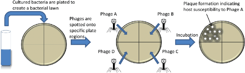 Figure: Bacteriophage typing method (Image source: Ref-2)
Phage typing has been used for decades for subtyping of Salmonella Typhimurium to determine the epidemiological relation among isolates. The system distinguishes more than 300 definitive phage types (DT) of Salmonella Typhimurium based on their patterns of lysis to a unique collection of Salmonella phages e.g., S. Typhimurium DT104. Phage typing is also done for other species of Salmonella e.g. Salmonella Enteritidis PT4. Similarly, Staphylococci are typed to determine whether the isolates belonged to the more virulent phage types so that the appropriate infection control method could be instituted.
Salient features of phage typing methods
- Phage typing is a rapid and low-cost approach for the epidemiological surveillance and outbreak investigation (identification of the source of infection).
- Phage-typing is the most widely recognized typing method for Staphylococcus aureus and is also still used widely for sub-dividing serotypes of Pseudomonas aeruginosa and Salmonella / Shigella spp. Phage typing still remains as the gold standard method for epidemiological surveillance of S. Typhimurium.
Procedure
- Label each plate with the name/number of test bacterium.
- Place a sterile cotton swab in the bacterial suspension and remove the excess fluid by pressing and rotating the cotton against the inside of the tube above the fluid level.
- Streak the swab in three directions over the surface of the agar medium to obtain uniform growth. A final sweep is made around the rim of the agar. This is done to make a lawn culture of bacterium.
- Allow the plates to dry for five minutes.
- Divide the plate in four quarters (using a pencil, by drawing a line in the backside of the plate) and name each quarter with the name of the bacteriophage which you are going to inoculate in that region.
- Once the agar media has dried completely, spot-inoculate 10 µl phages (according to the labelling) by dropping just a tiny amount of the phage suspension from the pipette tip.
- Repeat the above procedure with a fresh pipette tip and spot-inoculate this phage on its specifically labelled region.
- Allow the phage inocula to dry completely.
- Incubate at 37°C for 1-2 days (or 30°C if incubation is more than 2 days).
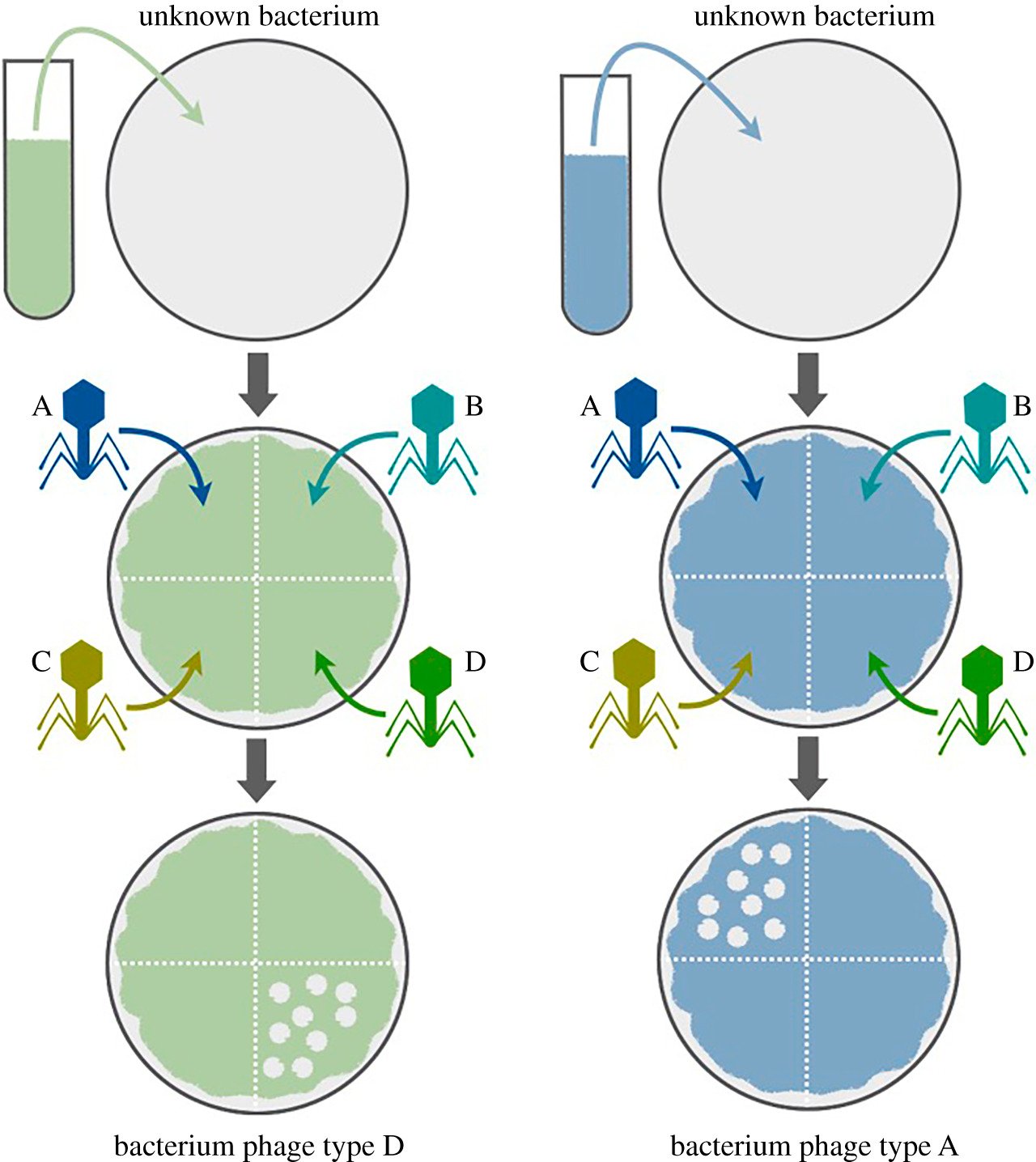 Figure: Phage Typing Procedure (Image source: Ref-3)
Reporting
Examine the plates for evidence of lysis (a giant plaque) in the area where phage was inoculated and tabulate the results. Record positive for lysis (= sensitivity of a bacterial strain to a particular phage).
This is the same reading logic used in antibiotic susceptibility testing by disk diffusion: a clear zone means something killed the bacteria there, susceptibility to a phage in this case rather than to an antibiotic. It's also worth distinguishing this "giant plaque" from the discrete, countable plaques used in a plaque assay. Here, a concentrated spot of phage either produces a zone of lysis or it doesn't; the result is qualitative (sensitive or resistant to that phage), not a count of individual plaques.
Limitations
- Phage typing requires different phages so phage typing is beyond the scope of local diagnostic laboratories. It is generally performed only at reference laboratories.
- Phage typing requires substantial technical expertise to perform. Careful control of environmental conditions and other variables is technically demanding.
- Maintenance of typing phages by the reference laboratory is time consuming and expensive approach.
- phage-types can change following lysogenic conversion, loss of prophages, or gain or loss of R plasmids, and this variability is coupled with the continuous need to maintain the typing set of bacteriophages in a viable state by regular serial passage. This instability is the same phenomenon covered in Bacteriophage Structure and Life Cycle: a strain's phage type is not a fixed identity, it can shift if the strain gains or loses a prophage through lysogenic conversion, or gains or loses an R plasmid.
How to Remember
- Reading the result: a clear zone always means something got killed there, whether the killer is a phage (phage typing) or an antibiotic (disk diffusion susceptibility testing). Positive for lysis = that isolate is sensitive to that particular phage.
- "Giant plaque" isn't a real plaque count: phage typing asks a yes/no question per phage (did it lyse this isolate or not), while a plaque assay asks a how-many question (how many individual phage particles are in a sample). Same visual idea, zone of clearing, different job.
- Why a phage type isn't permanent: think of a phage type as a fingerprint written on something that can still be edited. Gain or lose a prophage through lysogenic conversion, or gain or lose an R plasmid, and the same strain's phage type can shift.
Key exam facts in one table
| Concept | Detail | Why it matters |
|---|---|---|
| Principle | A strain's susceptibility pattern to a standardized panel of phages defines its phage type | Same underlying idea as antibiogram-based typing, just with phages instead of antibiotics as the discriminating agent |
| Classic examples | Salmonella Typhimurium DT104; Salmonella Enteritidis PT4; Staphylococcus aureus phage types | DT104 and PT4 are still commonly tested names in food/outbreak microbiology questions |
| Positive result | A clear zone (lysis) at a phage's spot = that isolate is sensitive to that phage | Mirrors disk diffusion susceptibility logic: clear zone = the challenger won |
| Main limitations | Requires reference-lab expertise and phage maintenance; technically demanding; phage types can change over time | Explains why phage typing is largely restricted to reference laboratories, not routine diagnostic labs |
| Why phage types shift | Lysogenic conversion, prophage loss, R plasmid gain or loss | Ties directly to the phage life cycle content covered in Structure and Life Cycle |
| Current status | Being progressively replaced by CRISPR typing and whole-genome sequencing | A common "which method is now preferred" exam angle |
Where Students Get Confused
- Assuming phage typing counts plaques the way a plaque assay does. It doesn't. Phage typing is a per-phage yes/no susceptibility call across a panel; a plaque assay is a quantitative titer. The "giant plaque" language in the reporting step describes a zone of confluent lysis from a concentrated spot, not an individually countable plaque.
- Treating phage type as a fixed, permanent strain identity. It can change if the strain gains or loses a prophage or an R plasmid, which is exactly why molecular methods like whole-genome sequencing have largely superseded it for definitive strain identification.
- Assuming phage typing is still the routine, first-line method everywhere. It remains historically important and is still used in some reference labs and for some organisms, but it has been progressively replaced by CRISPR typing and whole-genome sequencing for most epidemiological work.
References and further reading
- Mohammed, M. Phage typing or CRISPR typing for epidemiological surveillance of Salmonella Typhimurium?. BMC Res Notes 10, 578 (2017). https://doi.org/10.1186/s13104-017-2878-0
- Van der Merwe, Ruben & Helden, Paul & Warren, R & Sampson, Samantha & Gey van Pittius, Nico. (2014). Phage-based detection of bacterial pathogens. The Analyst. 139. 10.1039/c4an00208c.
- Kirchhelle Claas The forgotten typers: The rise and fall of Weimar bacteriophage-typing (1921–1935)0Notes Rec. http://doi.org/10.1098/rsnr.2019.0020
Frequently Asked Questions
What is phage typing used for?
What does a positive phage typing result mean?
Is a "giant plaque" in phage typing the same as a plaque in a plaque assay?
Why is phage typing mostly performed at reference laboratories rather than routine diagnostic labs?
Can a bacterial strain's phage type change over time?

Tankeshwar Acharya, MSc (Medical Microbiology)
Tankeshwar Acharya is an Assistant Professor in the Department of Microbiology at Patan Academy of Health Sciences (PAHS), Nepal, where he has been teaching and practicing clinical microbiology for over 14 years. He is the founder of Microbe Online, one of the leading free microbiology education resources on the web, covering bacteriology, mycology, parasitology, immunology, and clinical laboratory diagnostics written from direct experience in both the classroom and the diagnostic laboratory.