
Methyl Red (MR) Test: Principle, Procedure, Results & MR-Positive Organisms
MR test principle, procedure, and the mixed-acid vs. butanediol fermentation logic that tells you which organisms will turn red — without memorizing two separate lists.
The Methyl Red (MR) test determines whether an organism performs mixed acid fermentation and produces enough stable acid end products to drop the broth's pH to 4.4 or below. It's the second test in the IMViC panel, and it's always run alongside its mirror-image partner, the Voges-Proskauer test — same broth, opposite logic.
Why It Matters
Every Enterobacteriaceae member ferments glucose to pyruvic acid the same way. What happens next to that pyruvate is where the family splits into two camps, and that split is exactly what MR and VP detect:
- Mixed acid pathway → large amounts of stable acid (lactic, acetic, succinic) → pH drops below 4.4 → MR positive
- 2,3-Butanediol pathway → acid is converted into a neutral compound (acetoin) → pH stays near neutral → MR negative, VP positive
This single fork is why E. coli is MR+/VP– and Klebsiella/Enterobacter are MR–/VP+ — and why, historically, this reciprocal pattern was used in public health labs to distinguish fecal coliforms (E. coli, implying fecal contamination of water) from environmental coliforms that don't carry the same contamination significance. The two tests were never meant to be memorized as separate organism lists — they're reading the same metabolic decision from two different angles.
Principle
In mixed acid fermentation, the organism produces enough acetic, lactic, and succinic acid to drop the medium's pH below 4.4. Methyl red (a pH indicator) is red at pH ≤ 4.4 and yellow at pH ≥ 5.8, with an intermediate orange in between.
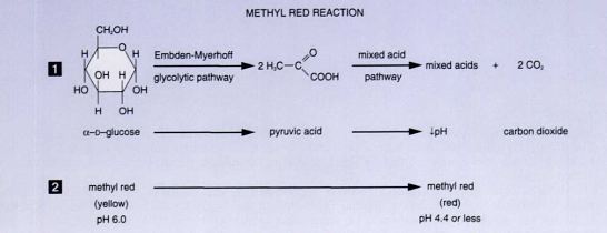 Figure: Methyl Red (MR) Test Reaction
Because methyl red only flips color at a notably low pH, the organism has to genuinely overpower the broth's buffering capacity — a weak acid producer simply won't trigger it. That's the whole point: this test only flags strong, stable acid producers.
Why MR and VP are opposite sides of one fork
MR and VP are run from the same MR-VP broth because they read the two possible fates of pyruvate, and most Enterobacteriaceae commit to one or the other. After glucose is fermented to pyruvate, an organism takes one of two roads. The mixed-acid road produces stable acids (acetic, lactic, formic, succinic) and drives the pH below 4.4, which MR detects. The butanediol road produces neutral acetoin and 2,3-butanediol, spares the pH, and consumes acids that were already made, which VP detects.
Because the two roads are largely mutually exclusive, MR and VP usually give opposite results in the same organism. E. coli takes the mixed-acid road: MR positive, VP negative. Klebsiella and Enterobacter take the butanediol road: MR negative, VP positive. This is why the tests share a broth and why a result on one usually predicts the other. The exceptions, Hafnia alvei and Proteus mirabilis, which can be positive for both, are worth remembering precisely because they break the reciprocal rule.
Procedure
MR-VP broth is shared between this test and the VP test — only the reagent added at the end differs.
- Bring the MR-VP broth to room temperature.
- Inoculate two tubes of MR-VP broth with a pure culture of the test organism.
- Incubate at 35°C for at least 48 hours.
- Add 5 drops of methyl red indicator to the first tube. (The second tube is reserved for the VP test, using Barritt's reagent instead.)
- Read the color.
Where students actually get confused
- 48 hours is a minimum, not a suggestion. Reading the tube too early is the single most common reason for a false-negative or unreliable MR result — the organism may not have produced enough acid yet to drop the pH past the threshold.
- Orange is not positive. An intermediate orange color means some acid was produced, but not enough to count. Only a stable, distinct red counts as positive — if you're unsure, this usually means under-incubation, not a "weak positive."
- Over-inoculation skews the result. Too heavy an inoculum (above ~10⁹ viable cells/mL) inhibits growth and can produce a misleading reaction — use a standard light inoculum, not "more is better."
- MR and VP should normally be reciprocal — but not always. Hafnia alvei and Proteus mirabilis are the classic exceptions: both can give a positive MR and a positive VP (the VP reaction is often delayed). If your "reciprocal" expectation breaks, check whether you're looking at one of these two before assuming a technical error.
Quality Control
Examine each new lot of broth for contamination, dehydration, or deterioration before use. Run one known-positive and one known-negative control organism with every new lot.
| Organism | Expected result |
|---|---|
| Escherichia coli ATCC 25922 | MR positive (red) |
| Klebsiella pneumoniae ATCC 13883 | MR negative (yellow) |
Result Interpretation
| Color | Interpretation |
|---|---|
| Stable red | MR positive — pH ≤ 4.4, strong mixed-acid fermentation |
| Yellow | MR negative — pH stayed near neutral |
| Orange | Not a valid positive — usually under-incubation |
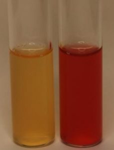 Figure: Methyl Red Test Left: Negative Right: Positive
MR-positive organisms
E. coli, Shigella species, Salmonella species, Citrobacter species, Proteus species, Yersinia species
MR-negative organisms
Enterobacter species, Hafnia species, Serratia marcescens, Klebsiella pneumoniae
Instead of memorizing both lists separately: if it's a strong acid producer (mixed-acid pathway), it's almost always also VP-negative. Learn the pathway, and both lists fall out of it — with Hafnia alvei and P. mirabilis as the two named exceptions worth remembering by name.
Limitations
- Avoid over-inoculation — bacterial growth is inhibited above ~10⁹ viable cells/mL, which can distort the reaction.
- Incubating for less than 48 hours risks a false-negative or unreliable intermediate color.
How to remember
MR is the acid test, and E. coli is its poster child. Mixed-acid fermenters hold a low pH against the buffer, and methyl red only turns red at a genuinely low pH (4.4). So MR positive means the organism made a lot of stable acid and kept it. E. coli, Shigella, Salmonella, Proteus, Yersinia: MR positive. The Klebsiella-Enterobacter group takes the neutral road and stays yellow.
MR and VP are a see-saw. Same broth, opposite answers. If you know one, you usually know the other. When both come back positive, suspect the two rule-breakers, Hafnia alvei and Proteus mirabilis.
Orange is not a pass. An intermediate orange is not a weak positive. It means the acid did not reach the threshold. Reincubate and retest rather than call it. Read no earlier than 48 hours, because an under-incubated tube can mislead in both directions.
References and further readings
- Leber AL, editor. Clinical Microbiology Procedures Handbook. 4th ed. Washington, DC: ASM Press; 2016. doi:10.1128/9781555818814
- Procop GW, Church DL, Hall GS, Janda WM, Koneman EW, Schreckenberger PC, Woods GL. Koneman's Color Atlas and Textbook of Diagnostic Microbiology. 7th ed. Philadelphia: Wolters Kluwer; 2017.
- MacFaddin JF. Biochemical Tests for Identification of Medical Bacteria. 3rd ed. Philadelphia: Lippincott Williams & Wilkins; 2000.
- Tille PM. Bailey and Scott's Diagnostic Microbiology. 15th ed. St. Louis: Elsevier; 2022.
Frequently Asked Questions
Why is my MR test giving an orange color instead of a clear red or yellow?
Can an organism be both MR-positive and VP-positive?
Why do the methyl red and Voges-Proskauer tests use the same broth?
Why must the methyl red test be incubated for at least 48 hours?
Why is over-inoculation a problem in the methyl red test?

Tankeshwar Acharya, MSc (Medical Microbiology)
Tankeshwar Acharya is an Assistant Professor in the Department of Microbiology at Patan Academy of Health Sciences (PAHS), Nepal, where he has been teaching and practicing clinical microbiology for over 14 years. He is the founder of Microbe Online, one of the leading free microbiology education resources on the web, covering bacteriology, mycology, parasitology, immunology, and clinical laboratory diagnostics written from direct experience in both the classroom and the diagnostic laboratory.