
Clostridium perfringens: Toxins, Gas Gangrene, Diagnosis
How Clostridium perfringens alpha-toxin destroys muscle, why gas gangrene spreads so fast, the A–E toxin types, and how to read Nagler, target hemolysis, and stormy clot.

On this page
A man is recovering at home after surgery to repair a deep leg wound from a farm accident two days ago. He returns because the pain has become severe, far worse than his healing wound should cause. The skin over the muscle is tense and has a bronze discoloration, and in one area you feel fine crackling under your fingers, gas in the tissue. A Gram stain of fluid from the wound shows thick, blunt-ended gram-positive rods and very few inflammatory cells. That last detail is the clue. Most infections pull in a flood of neutrophils. This one destroys them before they arrive. This is clostridial myonecrosis, and the organism behind it is Clostridium perfringens.
Introduction
Clostridium perfringens is a gram-positive, anaerobic, spore-forming rod with a characteristic boxcar shape. It is the main cause of clostridial myonecrosis (gas gangrene) and a common cause of food poisoning, and it also causes necrotizing enteritis and gangrenous cholecystitis. Almost everything it does traces back to the toxins it releases, especially alpha-toxin. That single idea organizes the whole organism: the toxins are the disease.
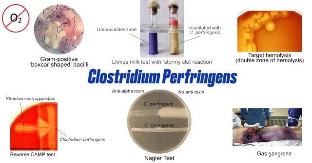 Figure: Common properties of Clostridium perfringens
Properties
- Gram staining: gram-positive, thick, straight rods with blunt ends, occurring singly or in pairs. Older cultures can stain gram-variable. The blunt, thick shape is described as boxcar-shaped.
- It is capsulated and non-motile. The capsule and the lack of motility (unusual for a clostridium) are both useful identification clues.
- Spores are subterminal and bulging in theory, but in practice C. perfringens rarely sporulates in tissue or on routine media. This is a useful clue: gram-positive rods without visible spores in a wound smear point to C. perfringens, while spore-bearing rods suggest other clostridia.
- It is invasive as well as toxigenic.
Transmission
C. perfringens lives in soil and as normal flora in the large intestine of humans and animals. Human infection follows two routes: spores or organisms entering a wound (leading to gas gangrene), and ingestion of heavily contaminated food (leading to food poisoning or, rarely, necrotizing enteritis). The wound route is detailed in the pathogenesis section.
Virulence factors and pathogenesis of Clostridium perfringens
C. perfringens is not very invasive on its own. Its power comes from a large set of secreted toxins and enzymes. It produces at least 12 toxins, grouped as four major toxins (alpha, beta, epsilon, iota) and eight minor toxins, plus enterotoxin and neuraminidase. The major toxins define the five classic strains (toxinotypes A to E). The one that matters most in human gas gangrene is alpha-toxin.
Alpha-toxin: the central weapon
What it is: a phospholipase C (lecithinase) with sphingomyelinase activity, produced by all five strains.
Why it matters: alpha-toxin attacks the phospholipids in host cell membranes. Because every cell membrane contains these phospholipids, the toxin damages many cell types at once. It lyses red cells (hemolysis), platelets (thrombocytopenia), and muscle cells, and it damages the lining of blood vessels. The membrane damage in vessel walls causes leak and thrombosis, which cuts off blood supply to the muscle. Loss of blood supply deepens the anaerobic environment, which lets the organism grow even faster. This is why myonecrosis spreads so quickly: the toxin creates the very low-oxygen conditions the organism prefers.
Alpha-toxin is also the basis of two lab tests. Its lecithinase activity is what you see as opalescence in the Nagler reaction, and its incomplete hemolysis forms the outer zone of target (double-zone) hemolysis on blood agar.
Theta-toxin (perfringolysin O)
What it is: an oxygen-labile, pore-forming cytolysin.
Why it matters: theta-toxin damages blood vessels and drives leukostasis and thrombosis, reducing perfusion and worsening tissue hypoxia. It also destroys leukocytes and stimulates cytokine release, which contributes to shock. Together with alpha-toxin, theta-toxin explains the striking finding in the hook: very few inflammatory cells in the wound smear, because the neutrophils are killed at the vessel wall before they reach the tissue. Theta-toxin produces the inner zone of complete hemolysis on blood agar.
Enterotoxin (CPE)
What it is: C. perfringens enterotoxin, produced mainly by type A strains during sporulation in the gut.
Why it matters: CPE binds tight-junction proteins in the intestinal epithelium and forms pores in the mucosal cell membrane. This disrupts the epithelial barrier, causes fluid loss, and produces the watery diarrhea and cramping of C. perfringens food poisoning. The toxin is made when the organism sporulates in the intestine, which is why food poisoning follows ingestion of large numbers of organisms rather than preformed toxin.
Beta, epsilon, and iota toxins
Beta-toxin: a lethal, necrotizing, pore-forming toxin from type B and type C strains. It is central to necrotizing enteritis (enteritis necroticans) because it is normally destroyed by intestinal proteases (trypsin), so disease appears when trypsin is low, in protein-poor diets, or when trypsin inhibitors are eaten (for example, sweet potato in the Papua New Guinea "pig bel" outbreaks).
Epsilon-toxin: a potent pore-forming toxin from type B and D strains. It causes enterotoxemia in sheep and goats and is studied for a possible role in human disease. It is regarded as a potential select agent because of its potency.
Iota-toxin: a binary toxin from type E strains, known for lethality, dermonecrosis, and cytotoxicity.
Spreading and immune-evasion enzymes (minor toxins)
These do not kill tissue directly. They help the organism spread and survive host defenses.
| Enzyme (toxin) | Activity | What it does in the body |
|---|---|---|
| Kappa (collagenase) | Breaks down collagen | Lets the organism infiltrate and spread through muscle and connective tissue |
| Mu (hyaluronidase) | Degrades hyaluronic acid | Acts as a spreading factor, opening tissue planes for toxin to advance |
| Lambda (protease) | Degrades IgG, C3, fibrinogen, fibronectin | Protects the organism from innate and adaptive immune attack |
| Nu (DNase) | Degrades DNA | Role in pathogenesis not well defined |
| Neuraminidase | Removes sialic acid from red cells | Makes red cells panagglutinable, raises blood viscosity, promotes capillary thrombosis |
Putting it together: how gas gangrene develops
- Entry. Spores or vegetative organisms enter through a deep wound with dead, poorly perfused tissue (trauma, surgery, ischemia). Low oxygen in devitalized tissue lets the anaerobe grow.
- Toxin release and local damage. Alpha-toxin and theta-toxin destroy muscle cells, red cells, platelets, and the leukocytes that would normally fight back. This is why the smear shows organisms but little pus.
- Vascular collapse and spread. Damage to vessel walls causes thrombosis and cuts off blood supply. Collagenase and hyaluronidase open tissue planes, so the infection advances through muscle. Fermentation produces gas, felt as crepitus and seen on imaging.
- Systemic toxicity. Toxins enter the bloodstream, causing hemolysis, shock, and organ damage. Without rapid surgical debridement, the process can be fatal within hours.
Toxin types (toxinotypes) of C. perfringens
The five classic strains are defined by which of the four major toxins they produce.
| Strain (type) | Major toxins produced | Main disease |
|---|---|---|
| Type A | Alpha | Gas gangrene (myonecrosis) in humans and animals; food poisoning (enterotoxin) |
| Type B | Alpha, beta, epsilon | Severe enteritis in young calves, foals, lambs, piglets |
| Type C | Alpha, beta | Necrotizing enteritis (enteritis necroticans, "pig bel") in humans |
| Type D | Alpha, epsilon | Enterotoxemia in sheep and goats, rarely cattle |
| Type E | Alpha, iota | Enterotoxemia in calves and lambs |
Note: a revised typing scheme (types A to G) was introduced in 2018. It adds type F (the enterotoxin-producing food-poisoning strains formerly grouped under type A) and type G. Most current textbooks and exams still use the A to E scheme shown here, so learn A to E first and treat A to G as an update to be aware of.
Clinical manifestations
Many C. perfringens wound infections are polymicrobial and involve other clostridia. Various manifestations include:
Clostridial Wound Infections
- Simple wound contamination: It involves the wound surface contamination, without invasion of underlying tissue, as occurs in the absence of devitalized tissue.
- Crepitant cellulitis (anaerobic cellulitis) is seen in diabetic patients. It involves the fascial plane with minimal toxin release, without muscle invasion.
- Anaerobic myositis (gas gangrene): Clostridial myonecrosis (gas gangrene) is a serious infection that involves rapid invasion and liquefactive necrosis of muscle with the gas formation and clinical signs of toxicity. Pockets of gas are seen within the muscles and subcutaneous tissue and as the liquefaction of muscle continues a thin, blackish fluid exudes from the skin. The other clostridia most often involved in gas gangrene are C. sordellii, C. septicum, and occasionally C. histolyticum.
Clostridial Enteric infection
- Food poisoning: enterotoxin-producing C. perfringens (classic type A) is a major cause of food-borne disease. It follows eating raw or improperly stored meat and other foods that carry large numbers of the organism (roughly 10⁸ viable cells). Heat-resistant spores survive cooking and germinate as the food cools. The enterotoxin (CPE) is then made when the organisms sporulate in the gut, as covered in the pathogenesis section.
- Necrotizing enteritis (enteritis necroticans): is a life-threatening infection caused by Clostridium perfringens type C and type A and characterized by ischemic necrosis of the jejunum and gas in the tissue plane. The disease was called “Darmbrand” (meaning “fire bowels”) in Germany and “Pig bel” in Papua New Guinea.
Other Clostridial infections are puerperal infections, anaerobic pleuropulmonary infections, and clostridial bacteremia. In rare cases, C. perfringens also causes meningitis, meningoencephalitis, and subdural empyema.
Laboratory diagnosis
Based on the clinical diagnosis of gas gangrene, treatment should be started as early as possible. Laboratory diagnosis has a role only for (1) confirmation of the clinical diagnosis, (2) species identification.
Laboratories can diagnose C. perfringens food poisoning by detecting the bacterial toxin in feces using enzyme immunoassay or by quantitative anaerobic cultures. Serotyping of the isolates is done to determine if the same serotype of C. perfringens is present in the epidemiologically implicated outbreaks.
Specimen
- Ideal specimens in the case of gas gangrene are necrotic tissues, muscle fragments, and exudates from deeper part of the wound where the infection appears to be more active. Other specimens depending on the type of infections include suspected food (to investigate food poisoning), feces, etc.
- Blood culture may be positive for C. perfringens and C. septicum. However, C. perfringens bacteremia can occur even in the absence of gas gangrene
- Swabs rubbed over the wound surface or soaked in exudates are not satisfactory.
- Specimens should be put into Robertson’s cooked meat broth and transported immediately to the laboratory.
Direct Microscopy
Gram-stained smears of aspirated material from myonecrosis show a necrotic background, few or no inflammatory cells, and gram-positive bacilli with the morphology of C. perfringens or other clostridia. Gram stained films provide clues about the species of clostridia present.
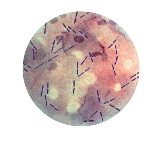 Figure: Boxcar shaped C. perfringens
- Thick, stubby, boxcar-shaped, gram-positive bacilli without spore are suggestive of C. perfringens. The cells of C. perfringens are usually 0.8 to 1.5 μm in diameter × 2 to 4 μm long and have blunt ends. They are often described as boxcar-shaped.
- Spore bearing gram-positive bacilli suggest other clostridia species
Citron bodies (boat or leaf-shaped) pleomorphic irregularly stained bacilli with spores suggest C.septicum. Large rods with oval sub-terminal spores- suggest C. novyi
Culture
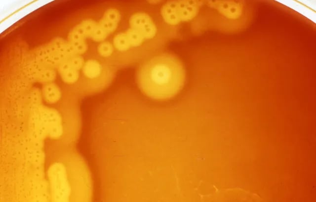 Figure: Target hemolysis (Double zone hemolysis) Image source: UPEI University
Culture plates are incubated anaerobically at 35–37°C for up to 2 days. Clostridium perfringens do not produce spores in media routinely used in the clinical laboratory.
Blood agar: After overnight incubation on blood agar, colonies are usually 1 to 3 mm in diameter, but may reach a diameter of 4 to 15 mm after prolonged incubation. Colonies are usually flat, somewhat rhizoid, and raised centrally. Some colonies tend to spread, but they do not swarm.
Target hemolysis (double zone hemolysis)
On anaerobic blood agar, C. perfringens produces target (double-zone) hemolysis: an inner narrow zone of complete hemolysis from theta-toxin, surrounded by a wider zone of incomplete hemolysis from alpha-toxin.
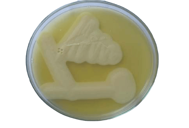 Figure: Lecithinase activity of C. perfringens in Egg Yolk Agar (Image source: Palaniappan Srinivasan)
Nagler’s reaction
Principle: alpha-toxin is a lecithinase (see pathogenesis above). On egg yolk agar, its lecithinase activity produces an opalescent zone around the streak line. Expected result for C. perfringens: opalescence that is abolished on the half of the plate spread with anti-alpha-toxin (this specific inhibition confirms the toxin). The test is also positive for other lecithinase-producing clostridia such as C. bifermentans, C. baratti, and C. sordellii.
Principle: alpha-toxin is a lecithinase (see pathogenesis above). On egg yolk agar, its lecithinase activity produces an opalescent zone around the growth. Expected result for C. perfringens: opalescence that is abolished on the half of the plate spread with anti-alpha-toxin, and this specific inhibition confirms the toxin. Other lecithinase-producing clostridia such as C. bifermentans, C. baratti, and C. sordellii can also give opalescence, so the anti-toxin inhibition step is what makes the test specific. For the full procedure, controls, and how to read borderline plates, see the Nagler reaction (lecithinase test) article.
Reverse CAMP test
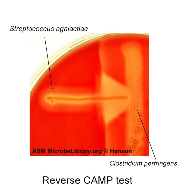 Figure: Reverse CAMP test
Suspected C. perfringens isolate is streaked over the center of anaerobe blood agar plate and Streptococcus agalactiae is streaked perpendicular to it. Presence of arrowhead of synergistic hemolysis with the tip of the arrow pointing from the Streptococcus toward the C. perfringens, is a positive reverse CAMP test.
Rapid growth at 45°C
C. perfringens can grow when Robertson’s cooked meat (RCM) broth is incubated at 45°C for 4-6 hours. This differentiates it from other organisms in the specimen.
Litmus milk test
In litmus milk, C. perfringens ferments lactose to acid, which clots the milk protein, and produces vigorous gas that tears the clot apart. This is the stormy clot reaction.
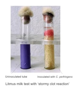 Figure: Stormy clot reactions by Clostridium perfringens
Other properties of Clostridium perfringens
- Non-motile
- Catalase negative
- No growth on chocolate agar in 5% CO₂ (it is an anaerobe)
Treatment of Clostridium perfringens infection
Gas gangrene is a surgical emergency first. The single most important step is prompt, aggressive surgical debridement of all dead tissue, because the toxins are produced in the necrotic, poorly perfused tissue that antibiotics cannot reach well. Antibiotics support surgery, they do not replace it.
The antibiotic of choice is penicillin, usually combined with a protein-synthesis inhibitor (clindamycin) that suppresses toxin production. Clindamycin is added because stopping toxin synthesis matters as much as killing the organism in a toxin-driven disease. For penicillin allergy, alternatives include metronidazole or a carbapenem, guided by local practice.
Hyperbaric oxygen is used in some centers as an adjunct to raise tissue oxygen and limit the spread of this anaerobe, but it must never delay surgery.
Food poisoning is self-limiting and needs only supportive care with fluids. Antibiotics are not indicated.
How to Remember
| Device | The memory hook |
|---|---|
| Boxcar shape | Blunt-ended, thick rods lined up like railway boxcars. Boxcars have no visible "cargo" (no spores) in clinical smears, matching the finding that C. perfringens rarely shows spores in tissue. |
| Target hemolysis = two toxins, two zones | Inner complete zone = theta (think "tight inner target"), outer incomplete zone = alpha (the "aura" around it). Two toxins make the two rings. |
| Alpha-toxin = lecithinase | Alpha is "A," the All-strains toxin and the Architect of gas gangrene. It attacks membrane lecithin, which is why Nagler (lecithinase) is positive. |
| "No pus" clue | Alpha and theta kill leukocytes before they arrive, so the smear shows bugs but little pus. Bacteria without neutrophils in a necrotic wound = think clostridial myonecrosis. |
| Stormy clot | In litmus milk, C. perfringens ferments lactose to acid (clots the milk) and produces vigorous gas that tears the clot apart, a "stormy" clot. Storm = gas-driven violence, the same gas that causes crepitus in tissue. |
| Beta-toxin and pig bel | Beta is destroyed by trypsin, so disease needs low trypsin. Low-protein diet or trypsin inhibitors (sweet potato) → "pig bel." B for beta, B for the bowel disease. |
Key exam facts in one table
| Feature | Clostridium perfringens |
|---|---|
| Morphology | Gram-positive, boxcar-shaped rod, blunt ends, non-motile, capsulated |
| Spores | Subterminal, bulging; rarely seen in tissue or on routine media |
| Oxygen | Anaerobe; tolerates brief air exposure better than most clostridia and grows relatively fast for an anaerobe |
| Catalase | Negative |
| Central toxin | Alpha-toxin (phospholipase C / lecithinase), made by all strains |
| Second key toxin | Theta-toxin (perfringolysin O), oxygen-labile pore-former |
| Food-poisoning toxin | Enterotoxin (CPE), made during sporulation in the gut, classic type A |
| Blood agar | Target (double-zone) hemolysis: theta (inner complete) + alpha (outer incomplete) |
| Nagler reaction | Positive: lecithinase opalescence on egg yolk agar, inhibited by anti-alpha-toxin |
| Reverse CAMP | Positive with Streptococcus agalactiae (arrowhead of synergistic hemolysis) |
| Litmus milk | Stormy clot reaction (acid clot torn by gas) |
| Heat/rapid growth | Grows in cooked meat broth at 45°C in 4–6 hours (fast generation time) |
| Toxinotypes | A–E by major toxins (revised A–G scheme since 2018); type A = human gas gangrene and food poisoning; type C = enteritis necroticans |
| Main diseases | Myonecrosis (gas gangrene), food poisoning, necrotizing enteritis, bacteremia |
Where Students Get Confused
| Confusion | The clarification |
|---|---|
| Alpha vs. theta on blood agar | Both hemolyze, but they make different zones. Theta = inner complete zone. Alpha = outer incomplete zone. Together = target hemolysis. |
| "Does it sporulate or not?" | It can form subterminal spores, but in tissue and on routine media it usually does not. So absent spores in a wound smear support C. perfringens, not against it. |
| Food poisoning: preformed toxin or not? | Not preformed like S. aureus or B. cereus emetic. You must swallow large numbers of live organisms; the enterotoxin is made when they sporulate in the gut. |
| Why so little pus in gas gangrene? | Alpha- and theta-toxin destroy leukocytes and block perfusion, so neutrophils never reach the tissue. Few inflammatory cells is expected, not reassuring. |
| Type A vs. type C disease | Type A = gas gangrene and food poisoning. Type C = necrotizing enteritis (pig bel). Different toxins, different diseases. |
| A–E vs. A–G confusion | Older scheme A–E is still the exam standard. The 2018 A–G scheme moved food-poisoning strains from "type A" to "type F." Learn A–E, know A–G exists. |
| Nagler positive = C. perfringens? | Not exclusively. Any lecithinase producer (C. bifermentans, C. sordellii, C. baratti) can be positive. Specific inhibition by anti-alpha-toxin is what confirms it. |
References and further readings
- Tille, P. M. (2022). Bailey & Scott's Diagnostic Microbiology (15th ed.). Elsevier.
- Procop, G. W., & Koneman, E. W. (2017). Koneman's Color Atlas and Textbook of Diagnostic Microbiology (7th ed.). Wolters Kluwer.
- Carroll, K. C., Pfaller, M. A., et al. (2020). Murray's Medical Microbiology (9th ed.). Elsevier.
- Savva, C. G., Clark, A. R., Naylor, C. E., et al. (2019). The pore structure of Clostridium perfringens epsilon toxin. Nature Communications, 10, 2641.
- Sakurai, J., Nagahama, M., Oda, M., Tsuge, H., & Kobayashi, K. (2009). Clostridium perfringens iota-toxin: structure and function. Toxins, 1(2), 208–228.
- Rood, J. I., Adams, V., Lacey, J., et al. (2018). Expansion of the Clostridium perfringens toxin-based typing scheme. Anaerobe, 53, 5–10. (Basis of the A–G revision.)

Tankeshwar Acharya, MSc (Medical Microbiology)
Tankeshwar Acharya is an Assistant Professor in the Department of Microbiology at Patan Academy of Health Sciences (PAHS), Nepal, where he has been teaching and practicing clinical microbiology for over 14 years. He is the founder of Microbe Online, one of the leading free microbiology education resources on the web, covering bacteriology, mycology, parasitology, immunology, and clinical laboratory diagnostics written from direct experience in both the classroom and the diagnostic laboratory.
Comments
No comments yet. Be the first to share your thoughts.
Leave a comment
All comments are reviewed before they appear.