
CLED Agar: Composition, Principle, Uses, and Colony Characteristics
CLED (Cystine-Lactose-Electrolyte-Deficient) agar is the preferred single medium for urine culture. Learn its composition, why it inhibits Proteus swarming, colony colors of key uropathogens, and how it compares to MacConkey and blood agar.

On this page
A 28-year-old woman presents with dysuria, frequency, and suprapubic pain. Her urine dipstick shows nitrites and leukocytes. The clinician sends a midstream urine sample for culture. In the laboratory, the technician has two options: plate it onto two separate media (blood agar plus MacConkey agar), or use a single plate of CLED agar.
The CLED plate costs less, takes up half the bench space, and in 18–24 hours produces colonies that are color-coded by lactose fermentation (yellow for fermenters like E. coli, blue-green for non-fermenters like Pseudomonas) without any concern that Proteus will swarm across the plate and overgrow everything else.
That combination of economy, safety, and clarity is why CLED agar became the standard medium for urine culture in many diagnostic microbiology laboratories worldwide.
CLED (Cystine-Lactose-Electrolyte-Deficient) agar is a non-selective, differential culture medium used for the isolation and enumeration of bacteria in urine specimens from suspected cases of urinary tract infection (UTI). Each word in the name describes a functional component of the medium:
- Cystine: added as a growth supplement to support the growth of cystine-dependent "dwarf colony" coliforms that would otherwise be missed
- Lactose: the fermentable carbohydrate that drives color differentiation between uropathogens
- Electrolyte-Deficient: deliberately low in electrolytes (sodium chloride and other salts) to prevent Proteus species from swarming
- Agar: the solidifying agent
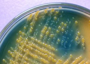 Figure: Lactose fermenting (yellow colonies) and Lactose Non-fermenting colonies in CLED
CLED is preferred over a combination of blood agar (BA) and MacConkey agar for routine urine culture because it supports the growth of all potential urinary pathogens and a number of contaminants such as diphtheroids, lactobacilli, and micrococci. It also differentiates between lactose fermenting and non-lactose fermenting colonies and inhibits the swarming of Proteus spp. With all these benefits, CLED agar can be used as a sole medium, reducing the cost without compromising the quality.
Principle
The major constituents of CLED agar are lactose, pancreatic digest of gelatin, pancreatic digest of casein, and beef extract. Lactose is the energy source for organisms able to ferment it. Enzymatic digest of casein, enzymatic digest of gelatin, and beef extract provide the nitrogen, vitamins, and carbon. L-Cystine is added as a growth supplement for cystine-dependent coliforms. It permits the growth of “dwarf colony” coliforms.
Bromothymol blue is a pH indicator to differentiate lactose fermenters from non-fermenters. Lactose fermenters lower the pH of the medium by producing acid, changing the indicator from blue-green to yellow. Electrolytes are reduced to restrict the swarming of Proteus species. Agar is used as a solidifying agent.
Composition of CLED agar
Final pH 7.3 +/- 0.2 at 25°C
| Ingredients | gm/L |
|---|---|
| Lactose | 10.0 |
| Pancreatic digest of gelatin | 4.0 |
| Pancreatic digest of casein | 4.0 |
| Beef extract | 3.0 |
| L-Cystine | 0.128 |
| Bromothymol Blue | 0.02 |
| Agar | 15.0 |
Note: CLED agar is electrolyte-deficient, not electrolyte-free. Formulations contain no added sodium chloride, but residual electrolytes are present from the peptones and beef extract. It is the low concentration, not total absence, that prevents Proteus swarming. Exact formulations vary between manufacturers, so check the package insert.
Why Electrolyte-Deficient? The Proteus Swarming Problem
Proteus mirabilis is a common cause of complicated urinary tract infections, particularly in catheterized patients and those with urinary tract abnormalities. It has an unusual property: on standard media containing electrolytes, it differentiates from normal short rods into hyperflagellated, elongated swarmer cells that spread rapidly in concentric waves across the entire plate surface.
The result on a standard blood agar or nutrient agar plate is a thin, spreading film of Proteus that overgrows and obscures all other colonies, making it impossible to identify co-infecting organisms or perform accurate colony counting.
CLED agar addresses this in a straightforward way: electrolytes (primarily sodium chloride) are a required trigger for the swarming differentiation. By keeping the electrolyte concentration very low, the medium prevents the morphological switch from vegetative cells to swarmer cells. Proteus still grows on CLED agar, producing its characteristic translucent blue colonies, but it stays confined to discrete colonies rather than spreading.
Key exam point: CLED agar inhibits Proteus swarming by electrolyte deficiency. MacConkey agar also inhibits swarming, but by a different mechanism. Bile salts at the concentration used in MacConkey inhibit the surface translocation required for swarming, rather than acting through electrolyte depletion. On blood agar, Proteus will swarm freely, which is one reason CLED is preferred over blood agar for urine culture.
Preparation of CLED Agar
- Weigh 36 grams of dehydrated CLED agar powder and suspend in one liter of distilled or deionized water. Follow the manufacturer's instructions, since formulations vary slightly between manufacturers (Oxoid, HiMedia, Mast, BD).
- Mix well and heat with frequent agitation, boiling for one minute until the powder is completely dissolved. The medium will appear green at this stage due to the bromothymol blue indicator.
- Sterilize by autoclaving at 121°C for 15 minutes.
- Cool to 50–55°C before pouring to avoid condensation on plates.
- Dispense approximately 20 mL into sterile Petri plates under aseptic conditions.
- Allow to solidify on a level surface, label with medium name and date of preparation.
- Store inverted at 2–8°C. Allow plates to reach room temperature before use.
Important: Do not over-incubate CLED plates beyond 24 hours. If lactose-fermenting organisms predominate, continued acid production can turn the entire medium yellow, masking non-lactose fermenters present in lower numbers.
Inoculation and Incubation
- Allow the plate to reach room temperature before use.
- Inoculate with a calibrated bacteriological loop (1 µL or 10 µL) so that the colony count can be converted to colony-forming units per milliliter. This calibrated inoculation is what makes CLED a quantitative urine-culture medium, not just a presence-or-absence one.
- Streak the loopful across the plate using a standard urine-culture streaking pattern for semi-quantitative counting.
- Incubate aerobically at 35 to 37°C for 18 to 24 hours. Carbon dioxide is not required.
- Read at 18 to 24 hours. Do not incubate beyond 24 hours: dominant lactose fermenters can acidify the whole plate to yellow, masking blue non-fermenters present in smaller numbers.
Colony Characteristics
Lactose fermenting colonies appear yellow but have distinguishable colony morphology based on size, consistency, etc. Non-lactose fermenting colonies appear blue. Colonies of Gram-positive cocci appear yellow but smaller in size as compared to Gram-negative ones.
The bromothymol blue indicator in CLED agar gives a clear color signal: lactose fermenters produce acid, lowering the pH and turning colonies yellow. Non-lactose fermenters produce alkaline or neutral reactions, giving blue or blue-green colonies. This color logic applies across all organisms and makes reading CLED plates fast and intuitive.
| Organism | Colony color | Colony characteristics | Lactose fermentation |
|---|---|---|---|
| Escherichia coli | Yellow | Opaque, slightly deeper yellow center, 2–3 mm | Yes |
| Klebsiella pneumoniae | Yellow to whitish-yellow | Extremely mucoid, large, dome-shaped | Yes |
| Enterococcus faecalis | Yellow | Small (0.5 mm), entire margin | Yes (slowly) |
| Staphylococcus aureus | Deep yellow | Uniform, opaque, 1–2 mm; yellow reflects both acid production and staphyloxanthin pigment | Weak/variable |
| Coagulase-negative Staphylococci | Pale yellow | More opaque than Enterococcus, smaller than S. aureus | Variable |
| Proteus mirabilis | Translucent blue | Discrete colonies (no swarming), 1–2 mm | No |
| Pseudomonas aeruginosa | Green to blue-green | Matted surface, rough irregular periphery; may produce pyocyanin (blue-green diffusible pigment) | No |
| Candida albicans | White to cream | Raised, opaque, yeast-like; may show foot-like projections at margins on prolonged incubation | No |
| Streptococcus agalactiae (GBS) | Yellow | Tiny colonies; may be missed if mixed with larger coliforms | Yes |
Reading tip: On a typical UTI culture plate, a pure growth of large yellow mucoid colonies strongly suggests Klebsiella. Pure yellow non-mucoid colonies with a deeper center suggest E. coli. Blue-green colonies with a matted surface and characteristic sweet/grape-like odor suggest Pseudomonas. Translucent blue discrete colonies in a patient with a urinary catheter suggest Proteus.
Quality Control of CLED Agar
QC of CLED agar should be done by performing sterility testing and performance testing.
- Sterility testing: Incubate 3-5 plates of uninoculated medium at 35°C to 37°C for 18-24 hrs. Any growth in the medium should be regarded positive and the whole lot should be discarded.
- Performance testing should be performed by inoculating one or more standard strains onto the prepared medium and incubating at 35 ± 2°C in an aerobic atmosphere for 18-24 hours. Observe for the growth, pigmentation, colony size, and inhibition of Proteus swarming/spreading after overnight incubation.
| Organism | Desired characteristics |
|---|---|
| Escherichia coli ATCC 25922 | Luxuriant Growth; colonies are yellow, medium yellow |
| Proteus vulgaris ATCC 8427 | Good growth; colonies are colorless to blue; swarming is inhibited; however, slight spreading is acceptable |
| Enterococcus faecalis ATCC 29212 | Growth; small colonies, colorless to yellow |
| Staphylococcus aureus ATCC 25923 | Good growth; colonies small, yellow to yellowish |
| Uninoculated plates | Green to blue-green |
Advantages of CLED Agar
- Good discrimination of gram-negative bacteria on the basis of lactose fermentation and colony appearance;
- Inhibits swarming of Proteus spp (Proteus mirabilis and Proteus vulgaris are frequently involved in urinary tract infection);
- Relatively low cost (compared with the combined use of blood agar and MacConkey agar for urine culture).
Note: MacConkey medium, which contains bile salts, also prevents the swarming of Proteus species.
Urine Culture: Quantitative Interpretation on CLED Agar
A critical and often under-taught aspect of CLED agar use is that it is designed for quantitative culture, not just presence or absence of growth. A calibrated bacteriological loop (1 µL or 10 µL) is used to inoculate the plate, which allows colony counts to be converted to colony-forming units per milliliter (CFU/mL) of urine.
Standard interpretation thresholds:
| Colony count (CFU/mL) | Interpretation |
|---|---|
| ≥ 10⁵ (100,000) | Significant bacteriuria, likely true infection in symptomatic patient |
| 10⁴ – 10⁵ | Borderline, repeat culture or interpret with clinical context |
| < 10⁴ | Likely contamination in most clinical contexts |
Important caveats:
- A count of ≥ 10² CFU/mL may be significant in catheter specimens or in symptomatic men (lower contamination risk)
- Suprapubic aspirate is a sterile-site specimen, so any growth is considered significant regardless of count
- Single organism growth supports infection; three or more species suggests contamination in most cases
This quantitative approach is one of CLED agar's key practical advantages over non-calibrated plating: the same plate used to identify the organism also provides the count needed for clinical interpretation.
Choosing Your Urine Culture Medium
The three media commonly used for urine culture each have distinct strengths. The table below clarifies when each is most appropriate, and why many laboratories have moved to CLED as a single-medium approach.
| Feature | CLED Agar | MacConkey Agar | Blood Agar |
|---|---|---|---|
| Gram-positive growth | Good | Inhibited (selective for GN) | Good |
| Gram-negative growth | Good | Good | Good |
| Lactose differentiation | Yes (yellow vs. blue-green) | Yes (pink vs. colorless) | No |
| Proteus swarming | Inhibited (electrolyte deficiency) | Inhibited (bile salts) | Not inhibited, will swarm |
| Candida growth | Good | Poor | Good |
| Hemolysis visible | No | No | Yes |
| Quantitative culture | Yes (with calibrated loop) | Yes | Yes |
| Cost as sole medium | Low (single plate) | Moderate (needs BA companion) | Moderate (needs MAC companion) |
| Best for | Routine urine culture (sole medium) | GN organism isolation and differentiation | Broad-spectrum, hemolysis assessment |
Practical note for resource-limited laboratories: CLED agar as a sole medium has been validated in multiple studies as equivalent to the BA + MacConkey combination for routine urine culture. In settings where cost or plate availability is a constraint, CLED alone is an evidence-based choice.
How to Remember
Use the name as your framework. CLED, for Cystine-Lactose-Electrolyte-Deficient, is not just an abbreviation. It is a three-part description of how the medium works:
- C = Cystine supports dwarf coliforms (without it, some E. coli strains produce tiny colonies easily missed)
- L = Lactose drives the yellow/blue color split, the most useful visual cue on the plate
- ED = Electrolyte-Deficient stops Proteus from swarming
Color logic, a simple rule:
Yellow colony = acid from lactose = lactose fermenter (most common uropathogens: E. coli, Klebsiella, enterococci) Blue/green colony = no acid = non-fermenter (Proteus, Pseudomonas) White/cream = yeast (Candida)
A clinical anchor to make it stick: Think of CLED agar as the "urine culture plate that solved three problems at once": it grew everything, told you what fermented lactose by color, and stopped Proteus from ruining the plate. Before CLED, laboratories needed two plates (blood agar + MacConkey) and still had the Proteus swarming problem on the blood agar. CLED was the single-medium solution.
Limitations
- Poor discrimination of some Gram-positive organisms. While enterococci and staphylococci grow on CLED, their colonies can be small and overlooked if mixed with larger Gram-negative colonies. Blood agar gives better colony morphology for Gram-positive organisms.
- No hemolysis visible. CLED does not contain blood, so hemolytic organisms (e.g., beta-hemolytic streptococci, S. aureus) cannot be identified by hemolysis pattern. Blood agar is required for this.
- Over-incubation problem. Beyond 24 hours, acid accumulation from dominant lactose fermenters can turn the entire plate yellow, masking non-fermenters present in smaller numbers.
- Not suitable for fastidious organisms. Haemophilus and Neisseria will not grow on CLED agar. For urethral/genital specimens where gonorrhea is suspected, chocolate agar is required alongside CLED.
- Cannot differentiate within lactose fermenters by color alone. Both E. coli and Klebsiella produce yellow colonies; colony morphology (mucoid vs. non-mucoid) and further biochemical testing are required for species-level identification.
Where Students Get Confused
- CLED is differential but not selective, and that is the whole design. Students meet MacConkey first, learn that a color-coded medium suppresses Gram-positives, then assume CLED does the same. It does not. CLED has no bile salts, no crystal violet, no inhibitory dye. It grows enterococci, staphylococci, and Candida alongside the coliforms. That is precisely why one CLED plate can replace a blood agar plus MacConkey pair for urine culture: nothing is being held back.
- Electrolyte-deficient does not mean electrolyte-free. The medium contains no added sodium chloride, but the peptones and beef extract carry residual electrolytes. Proteus swarming is triggered above a concentration threshold, so lowering the concentration is enough. The organism still grows perfectly well and forms normal discrete colonies. It simply cannot make the switch to elongated swarmer cells.
- Yellow does not always mean lactose fermenter. E. coli turns the medium yellow by fermenting lactose to acid. Staphylococcus aureus also appears deep yellow, but that color owes as much to its own staphyloxanthin pigment as to acid production. Reading color alone across the Gram divide will mislead. Color plus colony size plus Gram stain is the reliable combination.
- An uninoculated plate is green, not blue. Bromothymol blue is yellow below pH 6.0, green around neutrality, and blue above pH 7.6. At CLED's final pH of 7.3, the plate sits in the green-to-blue-green range. Students expecting a blue plate sometimes think the batch has failed. Non-fermenting colonies then appear blue against that green background.
- CLED and MacConkey both stop Proteus, but not by the same route. CLED withholds the electrolytes that trigger swarmer-cell differentiation. MacConkey uses bile salts, which suppress the surface migration swarming depends on. Same visible outcome, different mechanism. Exam questions frequently target this distinction.
- Colony count thresholds are not one number. The familiar 10⁵ CFU/mL figure applies to voided midstream urine from a symptomatic patient. It does not apply to catheter specimens, where far lower counts matter, and it certainly does not apply to suprapubic aspirate, where any growth is significant because the specimen comes from a normally sterile site. Applying 10⁵ universally will cause real infections to be reported as insignificant.
References and further readings
- Tille PM. Bailey & Scott's Diagnostic Microbiology. 15th ed. St. Louis: Elsevier; 2022.
- Cheesbrough M. District Laboratory Practice in Tropical Countries, Part 2. 2nd ed. Cambridge: Cambridge University Press; 2006.
- Sandys GH. A new method of preventing swarming of Proteus spp. with a description of a new medium suitable for use in routine laboratory practice. J Med Lab Technol. 1960;17:224–233.
- Mackey JP, Sandys GH. Laboratory diagnosis of infections of the urinary tract in general practice by means of a dip-inoculum transport medium. BMJ. 1966;2(5504):1286–1288.
- Clinical and Laboratory Standards Institute. M35: Abbreviated Identification of Bacteria and Yeast. CLSI.
Frequently Asked Questions
What does CLED stand for?
Is CLED agar selective or differential?
Why is CLED agar preferred for urine culture?
It does in one plate what blood agar and MacConkey do in two. It grows all common uropathogens including Gram-positives and Candida, differentiates lactose fermenters by color, prevents Proteus swarming from obscuring the plate, and supports quantitative colony counting with a calibrated loop. The result is lower cost and less bench space without losing diagnostic information.
How does CLED agar prevent Proteus swarming?
Proteus swarming requires differentiation from short vegetative rods into elongated, hyperflagellated swarmer cells, and that switch depends on adequate electrolyte concentration. CLED omits added sodium chloride, keeping electrolytes below the triggering threshold. Proteus grows normally and produces translucent blue colonies, but stays as discrete colonies rather than spreading in waves.
What color are lactose fermenters on CLED agar?
Yellow. Acid from lactose fermentation lowers the pH and shifts bromothymol blue from green toward yellow. E. coli gives opaque yellow colonies with a deeper yellow center; Klebsiella gives large, markedly mucoid yellow colonies. Non-fermenters such as Proteus and Pseudomonas remain blue or blue-green.
Why should CLED plates not be incubated beyond 24 hours?
Can Candida grow on CLED agar?
Yes. Candida albicans produces white to cream, raised, opaque yeast-like colonies, and may show foot-like projections at the colony margin with prolonged incubation. This is an advantage over MacConkey, where Candida grows poorly, and it matters in catheterized and diabetic patients where candiduria is common.
What colony count is significant in urine culture?
What is the difference between CLED agar and MacConkey agar?
MacConkey is selective and differential; it contains bile salts and crystal violet that inhibit Gram-positive organisms, and lactose with neutral red for differentiation. CLED is differential only, with no inhibitors, so it grows Gram-positives and yeasts as well. Both suppress Proteus swarming, MacConkey through bile salts and CLED through electrolyte deficiency. For urine culture, CLED's ability to recover Gram-positive uropathogens on the same plate is the main advantage.
Why can not CLED agar be used for all specimen types?
Why can not CLED agar be used for all specimen types?
It contains no blood, so hemolysis cannot be assessed, and it does not support fastidious organisms. Haemophilus and Neisseria will not grow on it, so genital specimens where gonorrhea is suspected need chocolate agar or a selective medium such as Thayer-Martin alongside. CLED is designed for urine, and it is excellent there.

Tankeshwar Acharya, MSc (Medical Microbiology)
Tankeshwar Acharya is an Assistant Professor in the Department of Microbiology at Patan Academy of Health Sciences (PAHS), Nepal, where he has been teaching and practicing clinical microbiology for over 14 years. He is the founder of Microbe Online, one of the leading free microbiology education resources on the web, covering bacteriology, mycology, parasitology, immunology, and clinical laboratory diagnostics written from direct experience in both the classroom and the diagnostic laboratory.
Comments
No comments yet. Be the first to share your thoughts.
Leave a comment
All comments are reviewed before they appear.