

A man develops a slow, hard, painless swelling along his lower jaw over several weeks. He had a tooth removed a couple of months earlier. The lump does not behave like an ordinary abscess: antibiotics for a few days do not clear it, it does not stay inside one tissue layer, and eventually the skin over it opens and drains pus that contains tiny hard yellow grains.
Those grains are the clue. They are tangled colonies of a branching bacterium that normally lives quietly in his mouth and only turned invasive after the tooth extraction broke the lining that kept it in place. This is cervicofacial actinomycosis, or "lumpy jaw," caused by Actinomyces.
Introduction
Actinomyces is a group of gram-positive, branching, filamentous rods. Their key properties are:
- Anaerobic to microaerophilic. The main human pathogen, Actinomyces israelii, needs anaerobic conditions to grow, which is why it is slow and hard to culture. (The genus as a whole is best described as aerotolerant anaerobes; some species prefer air with carbon dioxide, but the disease-causing ones favor low oxygen.)
- Non-spore-forming
- Non-acid-fast (an important contrast with Nocardia, which is partially acid-fast)
- Non-motile
One idea organizes the whole topic. Actinomyces is normally a harmless resident of the mouth, gut, and female genital tract. It causes disease only when the surface lining that contains it is broken, and it then produces a slow, chronic infection that ignores normal tissue boundaries and drains to the surface through sinus tracts. Almost every feature of actinomycosis follows from this.
Mode of infection
Actinomycosis is an endogenous infection. This is a defining point: the organism is already living on the patient's own mucosal surfaces, so infection does not come from outside and does not spread from person to person. Disease begins when a break in the mucosa lets the organism reach the deeper tissue, for example after dental extraction, jaw trauma, aspiration, bowel surgery or perforation, or a long-retained intrauterine device.
Actinomycosis is also almost always polymicrobial. Actinomyces rarely acts alone. It is helped by other mouth and gut bacteria that grow with it, and these companion organisms lower the local oxygen level and damage tissue, which creates the anaerobic conditions Actinomyces needs. This synergy is part of why the infection is chronic and hard to treat.
How Actinomyces causes disease
Actinomyces is a weak pathogen on its own. Its disease depends on three things working together.
A breach in the mucosa. As long as the lining of the mouth, gut, or genital tract is intact, Actinomyces stays a harmless commensal. Disease starts only when that barrier is broken and the organism reaches deeper, poorly oxygenated tissue.
Help from other bacteria. Actinomycosis is a mixed infection. The companion bacteria consume oxygen and damage tissue, producing the low-oxygen environment that lets the anaerobic Actinomyces grow. This is why the disease behaves as a synergistic, polymicrobial process rather than a single-organism infection.
The sulfur granule. As it grows, Actinomyces forms dense, tangled masses of filaments called sulfur granules. These are not made of sulfur; they are named for their yellow color. The granule is more than a curiosity: it behaves like a protected community (a biofilm) that shields the bacteria inside from phagocytes and antibodies. This protection is a major reason the infection is so chronic, resists short antibiotic courses, and needs prolonged treatment.
Putting it together: the barrier breaks, companion bacteria create a low-oxygen pocket, Actinomyces grows and forms protected granules, and the immune system cannot clear it. The infection then advances slowly and steadily, crossing normal tissue planes (unlike many infections that stay within one compartment) and eventually tunneling to the surface as draining sinus tracts that discharge sulfur granules. The ability to ignore tissue boundaries and to form draining sinuses is the clinical signature of actinomycosis.
Infections
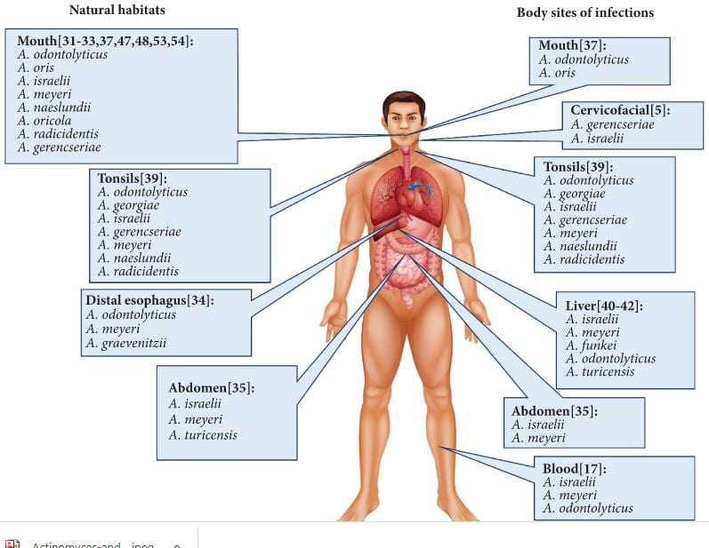 Many Actinomyces species are part of the oral microflora of humans. Some species of Actinomyces are found in supragingival and subgingival plaques of adult patients suffering from periodontitis and gingivitis. These same organisms also play a significant role in the development of root-surface caries. Actinomyces also cause classical actinomycosis, wound infections, abscesses, and genital tract infections.
Actinomycosis is a chronic infection that is both suppurative (pus-forming) and granulomatous. Its typical lesion is a hard, non-tender swelling that develops slowly, crosses tissue planes, and eventually drains pus through sinus tracts. The pus contains hard yellow sulfur granules, which are masses of the branching organism. Over time the lesion causes fibrosis and tissue destruction.
Actinomyces israelii is the most common cause of human actinomycosis, along with A. gerencseriae and Propionibacterium propionicum (formerly Arachnia propionica). Actinomycosis can affect any organ and characteristically manifests as:
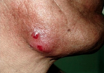 1. Cervicofacial actinomycosis ("lumpy jaw"): the most common form, associated with poor dental hygiene, dental extractions or manipulation, and head-and-neck infections. It presents as the slow, hard jaw swelling described above.
2. CNS actinomycosis: It results after hematogenous dissemination or by direct extension of actinomycotic infections in the head/neck areas. Patients usually present with chronic meningitis or meningoencephalitis complicated by the development of subdural empyema, epidural and spinal abscesses, and cranial osteomyelitis. When the CNS is involved (which is uncommon), the usual presentation is one or more brain abscesses, most often in the temporal or frontal lobes.
3. Thoracic actinomycosis: It involves the chest and mediastinal region.
4. Abdominal actinomycosis: Actinomycosis of the Ileocecal region.
5. Pelvic actinomycosis: occurs in women who have had an intrauterine device (IUD) in place for a long time. The long-retained IUD is the classic risk factor, and Actinomyces-like organisms are sometimes seen on cervical smears (Pap tests) in IUD users. This is a high-yield association for exams.
1. Cervicofacial actinomycosis ("lumpy jaw"): the most common form, associated with poor dental hygiene, dental extractions or manipulation, and head-and-neck infections. It presents as the slow, hard jaw swelling described above.
2. CNS actinomycosis: It results after hematogenous dissemination or by direct extension of actinomycotic infections in the head/neck areas. Patients usually present with chronic meningitis or meningoencephalitis complicated by the development of subdural empyema, epidural and spinal abscesses, and cranial osteomyelitis. When the CNS is involved (which is uncommon), the usual presentation is one or more brain abscesses, most often in the temporal or frontal lobes.
3. Thoracic actinomycosis: It involves the chest and mediastinal region.
4. Abdominal actinomycosis: Actinomycosis of the Ileocecal region.
5. Pelvic actinomycosis: occurs in women who have had an intrauterine device (IUD) in place for a long time. The long-retained IUD is the classic risk factor, and Actinomyces-like organisms are sometimes seen on cervical smears (Pap tests) in IUD users. This is a high-yield association for exams.
Laboratory Diagnosis
Sample
Based on the affected site, the specimens collected include discharge from the sinuses or fistula, rarely bronchoalveolar lavage, sputum or tissue sections.
Diagnosis in the laboratory is made by
- seeing gram-positive branching rods, especially in the presence of sulfur granules, and
- seeing growth when pus or tissue specimens are cultured under anaerobic conditions. Organisms can be identified by immunofluorescence.
Direct Microscopy
Pus discharge is thoroughly washed in saline in a test tube and the sediment is collected that contains gritty, white or yellowish sulfur granules, of <5 mm in size. Granules are crushed between two slides and smears are made.
Gram-staining (Brown-Brenn modification)
It shows a central mass of gram-positive filamentous bacilli, radiating peripherally with hyaline, club-shaped ends. Clubs are composed of complexes formed due to interaction of bacteria derived polysaccharide and protein with host cell salts and polypeptides.
Granules of actinomycosis are hard and not emulsifiable which differentiates them from granules produced other conditions. Actinomyces species can also be detected directly from the sample by methods such as:
- Fluorescent antibody techniques using fluorescent-tagged species-specific monoclonal antibodies.
- Fluorescent in situ hybridization (FISH) using species-specific probes.
Unlike Nocardia, which is partially acid-fast, Actinomyces is not acid-fast. This single stain is one of the fastest ways to separate the two branching gram-positive organisms. The full side-by-side comparison is given in the article on Nocardia.
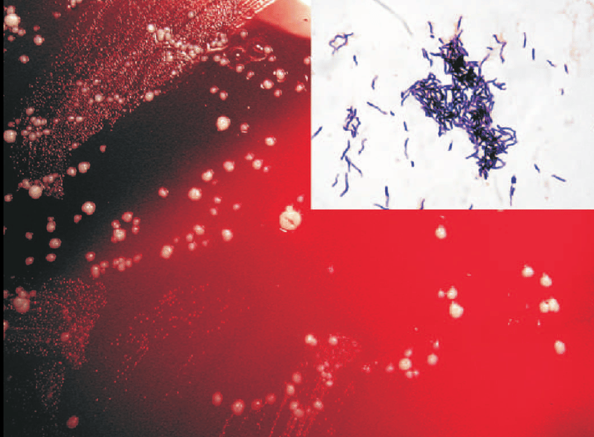 Figure: Actinomyces in culture and Gram-stain
Histopathological staining such as hematoxylin-eosin and Gomori’s stained tissue sections may reveal granules composed of eosinophilic clubs surrounding basophilic filaments and foamy macrophages (sun-rays appearance).
Culture
Pus containing sulfur granules is washed and cultured anaerobically at 35 to 37°C. Culture is slow and often takes up to two to three weeks, so the laboratory must be told that actinomycosis is suspected and cultures must be held long enough. Suitable media include:
- Thioglycollate broth: Growth of A. israelii resembles fluffy balls at the bottom of the tube, this can be differentiated from other species (A. bovis produces uniform turbidity).
- Brain heart infusion (BHI) agar: forms small "spider colonies" (branching filaments radiating from a center) at about 48 hours, which enlarge over about 10 days into heaped, rough colonies with a "molar tooth" appearance.
Species identification
It is done when the culture isolate is subjected to:
- Biochemical reactions
- Gas-liquid chromatography (GLC) for detection of the products of glucose metabolism.
- Molecular methods, such as PCR-RFLP are also available for speciation.
Treatment and prevention
The drug of choice is penicillin, and there is no significant penicillin resistance. Two features of treatment are worth understanding. First, treatment is prolonged, often given for months, because the sulfur granules and dense fibrosis protect the bacteria from both the immune system and short antibiotic courses. Second, surgery to drain abscesses and remove dead tissue is often needed alongside antibiotics. In IUD-associated pelvic disease, removing the device is part of management.
This is also the treatment half of the Actinomyces-versus-Nocardia split: Actinomyces responds to penicillin, while Nocardia is treated with a sulfonamide (co-trimoxazole).
There is no vaccine. The main preventive measure is good oral hygiene and dental care, which reduces the Actinomyces burden and the chance of invasive disease after mucosal injury.
How to Remember
| Device | The memory hook |
|---|---|
| Lumpy jaw | Actinomyces = the "lumpy jaw" bug. Slow, hard, painless jaw swelling after a dental problem. |
| Sulfur granules | Yellow grains in the draining pus. Not actually sulfur, just yellow. Tangled colonies of the branching organism. |
| From the inside, not outside | Actinomyces is endogenous: it already lives in your mouth and gut. Disease starts when the lining breaks, not from an outside source. (Contrast Nocardia, which comes from the environment.) |
| Crosses tissue planes | Most infections respect anatomical boundaries. Actinomycosis ignores them and tunnels to the surface as sinus tracts. Boundary-crossing plus sinuses = think Actinomyces. |
| IUD and the pelvis | Long-retained IUD → pelvic actinomycosis. Remember the "I" link. |
| Penicillin, and long | Actinomyces takes Penicillin (contrast Nocardia = Sulfonamide), and it must be given for a long time because the granules shield the bug. |
| Molar tooth colony | Older colonies on agar look like a molar tooth. A dental bug with a dental-looking colony. |
Key exam facts
| Feature | Actinomyces |
|---|---|
| Gram stain | Gram-positive, branching, filamentous, non-spore-forming |
| Acid-fast | Negative (contrast: Nocardia partially acid-fast) |
| Oxygen | Anaerobic to microaerophilic (A. israelii needs anaerobic conditions) |
| Motility | Non-motile |
| Habitat | Normal flora of human mouth, gut, and female genital tract |
| Infection type | Endogenous; no person-to-person spread |
| Nature | Chronic, polymicrobial, crosses tissue planes, forms draining sinuses |
| Hallmark finding | Sulfur granules in pus (biofilm-like protected colonies) |
| Main species | A. israelii, A. gerencseriae, Propionibacterium propionicum |
| Cervicofacial form | "Lumpy jaw," after dental problems (most common) |
| Pelvic form | Long-retained IUD association |
| Culture | Slow (up to 2–3 weeks) anaerobic; "spider" then "molar tooth" colonies |
| Drug of choice | Penicillin (prolonged), plus surgical drainage |
Where Students Get Confused
| Confusion | The clarification |
|---|---|
| Actinomyces vs Nocardia | Actinomyces = anaerobic, not acid-fast, normal mouth flora, penicillin. Nocardia = aerobic, partially acid-fast, environmental, sulfonamide. The full table is on the Nocardia page. |
| Is it a fungus? | No. The name and the branching filaments look fungal, but Actinomyces is a true bacterium. It is treated with antibacterials, not antifungals. |
| Are sulfur granules made of sulfur? | No. They are named only for their yellow color. They are dense masses of the branching organism that act like a protective biofilm. |
| Where does the infection come from? | From the patient's own flora (endogenous). It is not caught from the environment or from other people. Disease needs a break in the mucosa. |
| Why does it need such long treatment? | The sulfur granules and surrounding fibrosis shield the bacteria from the immune system and from short antibiotic courses, so penicillin must be continued for months, often with surgical drainage. |
| Why does it cross tissue planes? | This boundary-ignoring spread, along with draining sinuses, is a clinical signature of actinomycosis and helps separate it from ordinary abscesses that stay within one compartment. |
References and Further Readings
- Tille, P. M. (2022). Bailey & Scott's Diagnostic Microbiology (15th ed.). Elsevier.
- Procop, G. W., & Koneman, E. W. (2017). Koneman's Color Atlas and Textbook of Diagnostic Microbiology (7th ed.). Wolters Kluwer.
- Carroll, K. C., Pfaller, M. A., et al. (2020). Murray's Medical Microbiology (9th ed.). Elsevier.
- Wong, V. K., Turmezei, T. D., & Weston, V. C. (2011). Actinomycosis. BMJ, 343, d6099.

Comments
No comments yet. Be the first to share your thoughts.
Leave a comment
All comments are reviewed before they appear.