
Trichrome Staining for Fecal Smears: Principle, Procedure, and Results for Intestinal Protozoa
Learn the Wheatley trichrome staining technique for intestinal protozoa — reagents, step-by-step procedure, colour results for Entamoeba, Giardia, and Balantidium, and troubleshooting common staining problems.
A laboratory technician examines a direct wet mount from a patient with chronic diarrhoea — she sees what might be amoebic cysts, but the internal structure is not clear enough to confidently distinguish Entamoeba histolytica from E. coli. One is a significant pathogen requiring treatment; the other is a non-pathogenic commensal. Treating the wrong one or missing the right one has real consequences. She makes a permanent stained smear using trichrome stain.
Twenty-four hours later, under oil immersion, the internal nuclear detail is unmistakable: 1–4 nuclei with finely beaded peripheral chromatin and a small central karyosome — E. histolytica. The patient receives treatment.
This is why trichrome staining exists: to answer the questions that wet mount raises but cannot definitively resolve. It is the standard permanent stain for intestinal protozoa worldwide.
Stained fecal films are the single most productive means of stool examination for the detection of intestinal protozoan parasites and eggs of helminths. Because the refractive index of protozoan cysts and some helminth eggs are near that of water, staining procedures are required for optimal detection and for detailed study of their internal structures. Stained smears facilitate the detection of small protozoa missed by wet mount examination and afford a permanent record of encountered protozoa.
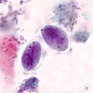 Figure: Cyst of Giardia lamblia
Permanent stains enhance the identification of Entamoeba histolytica and the detection of Giardia, two of the more commonly encountered and important protozoa. Two types of permanent stains are commonly applicable to visualize intestinal protozoa in fecal smears:
- The iron hematoxylin stain and
- The modified (Wheatley’s) Gomori’s Stains
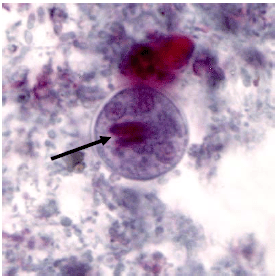 Figure: Cyst of Entamoeba histolytica
Why Trichrome? When Wet Mount Is Insufficient
The direct wet mount is fast but has significant limitations for protozoa:
- Internal cyst structure (nuclei, chromatoid bodies, glycogen vacuoles) is poorly resolved without staining
- E. histolytica and E. dispar cannot be distinguished morphologically — only the presence of ingested RBCs gives a clue
- E. histolytica and E. coli have overlapping cyst sizes; nuclear number (up to 4 vs up to 8) is critical for differentiation — not visible reliably on wet mount
- Small protozoa (Dientamoeba fragilis, Blastocystis hominis) are easily missed or misidentified
Trichrome staining is indicated when:
- Protozoan cysts suspected on wet mount but species cannot be confirmed
- E. histolytica vs E. coli vs E. dispar distinction is needed
- Immunocompromised patient with unexplained diarrhoea (multiple organisms possible)
- Formal quality assurance in a reference laboratory (permanent record required)
Trichrome staining does NOT stain: Cryptosporidium, Cyclospora, Cystoisospora oocysts; these require modified acid-fast staining.
Principle
The Wheatley trichrome technique for the fecal specimen is a modification of the original Gomari technique for tissue staining. It is a simple yet rapid technique that produces uniformly well-stained smears of the intestinal protozoa, human cells, yeast, and artifact material. Wheatly trichrome staining technique contains chromotrope 2R and light green SF as the primary staining agents. Trichrome stain is widely useful in clinical parasitology laboratories. It is easy to perform, and good results are obtained with both fresh and polyvinyl alcohol (PVA)-preserved fecal material.
| Reagents | Amount |
|---|---|
| Chromotrope 2R | 0.6 g |
| Light green SF | 0.3 g |
| Phosphotungstic acid | 0.7 g |
| Acetic acid (glacial) | 1.0 mL |
| Distilled water | 100 mL |
Reagents
- Add 1.0 mL of acetic acid to the dry components.
- Allow the mixture to sit for 15-30 minutes at room temperature before adding 100 mL of distilled water.
- The stain should appear purple.
- When properly stored in glass or plastic bottles at room temperature, the shelf life is 2 years.
Iodine-Alcohol Solution (70% ethanol plus iodine)
- Add sufficient iodine crystals to 70% alcohol to make a dark, concentrated stock.
- At the time of use, dilute the desired amount to the stock solution with 70% alcohol until a port-wine working solution is obtained.
- The exact concentration is not critical.
Quality Control
It is recommended to use a control slide of a known protozoan such as Giardia spp. from a PVA preserved specimen with each staining run. Properly fixed and correctly stained smears produce a blue-green cytoplasm of protozoan trophozoites with a slight tinge of purple. Nuclei, inclusions (chromatoid bodies, red blood cells, bacteria), and Charcot-Leyden crystals appear red, often tinged with purple.
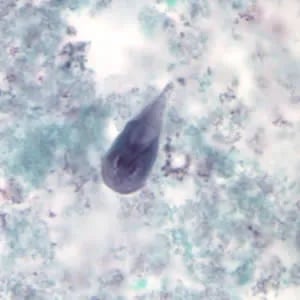 Figure: Trophozoite of Giardia lamblia
Cloudy preparations may result if dehydration is incomplete owing to failure to change alcohol solutions that may have become contaminated with water. Staining quality can be improved by draining slides between transfers from one reagent to another. If staining is of poor quality, the smears may have been too thick or poorly fixed, or residual HgCl2 may not have been removed owing to the use of an alcohol-iodine mixture that was too weak.
Procedure
Wear gloves when handling stool specimens.
Preparation of the Smear
- With a small portion of the fresh stool specimen, prepare two smears on a microscope slide by using an applicator stick or brush
- Immerse smears immediately in Schaudinn’s fixative and allow to fix for a minimum of 30 minutes. Overnight fixation is preferred.
- If the specimen is liquid, mix several drops of fecal material with three or four drops of PVA on a glass slide and let it dry for several hours in a 37°C incubator.
- If the specimen is in PVA, pour some of the mixtures onto a paper towel to absorb out the PVA. Prepare slides of the material from the paper towels as described.
Staining Procedure
- After smears are properly fixed and dried, place slides in the 70% ethyl alcohol for 3 minutes.
- Place slides in alcohol-iodine working solution for 2-5 minutes.
- Wash with two changes of 70% alcohol, one for 5 minutes and one for 2-5 minutes.
- Place in trichrome-staining solution for 10 minutes.
- Place in acidified 90% ethyl alcohol for 5 seconds.
- Dip slides several times in 100% ethanol.
- Place in two changes of 100% ethyl alcohol.
- Remove alcohol with two changes of xylene or toluene for 2-5 minutes each.
- Add mounting medium and overlay with a #1 coverslip.
- Examine under oil immersion for parasitic forms.
Results
The cytoplasm of thoroughly fixed and well-stained cysts and trophozoites are blue-green, tinged with purple. The nuclear chromatin, chromatoid bodies, and ingested red blood cells appear red to red-purple. The background material appears green providing a nice color contrast with the protozoa.
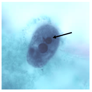 Figure: Trophozoite of Entamoeba histolytica with ingested RBC
Organism-Specific Trichrome Results
| Organism | Stage | Cytoplasm colour | Nuclear features | Distinguishing detail |
|---|---|---|---|---|
| E. histolytica | Trophozoite | Blue-green, granular | Red karyosome; fine peripheral chromatin | Ingested RBCs appear red |
| E. histolytica | Cyst | Blue-green | 1–4 nuclei; fine peripheral chromatin; blunt-ended chromatoid bars (red) | ≤4 nuclei |
| E. coli | Cyst | Blue-green | Up to 8 nuclei; irregular peripheral chromatin; splinter-ended chromatoid bars | >4 nuclei = E. coli |
| Giardia lamblia | Trophozoite | Blue-green; pale | Two nuclei; median bodies visible | "Old man face" appearance |
| Giardia lamblia | Cyst | Blue-green | 4 nuclei (often overlapping); longitudinal fibres | Oval shape |
| Balantidium coli | Trophozoite | Blue-green | Large kidney-shaped macronucleus; small round micronucleus | Largest human intestinal protozoan |
| Dientamoeba fragilis | Trophozoite only (no cyst) | Blue-green | 1–2 nuclei; fragmented central chromatin | No cyst stage — must catch trophozoite |
| Yeast cells | — | Red-purple | — | Common background contaminant; spherical, budding |
| Background fecal debris | — | Green | — | Contrast against protozoa |
Procedure Notes
- If specimens are improperly fixed, protozoan forms may take a dirty red color.
- Incomplete removal of mercuric chloride (Schaudinn’s fixative) may result in the deposit of highly refractive granules that may interfere with the detection of parasitic forms. Be sure to change the 70% ethanol-iodine solution regularly so this does not happen.
- Smears that are too green may indicate inadequate removal of iodine by 70% ethanol. Lengthening the time of washing in 70% alcohol and frequent changes of solution will minimize this problem.
Where Students Actually Get Confused
1. "Trichrome stains all intestinal parasites." Trichrome stains intestinal protozoa well. It does NOT stain coccidian oocysts (Cryptosporidium, Cyclospora, Cystoisospora) — modified acid-fast stain is required for these. Helminth eggs are visible on trichrome but wet mount and concentration methods are superior for eggs.
2. "Any fixative can be used before trichrome staining." Trichrome staining of fecal smears requires fixation in Schaudinn's fixative (or PVA preservative for preserved specimens) before the staining procedure. Using formalin as the fixative before trichrome gives poor staining quality — formalin-fixed specimens require a different staining protocol (iron-haematoxylin).
3. "E. histolytica and E. coli can be distinguished solely by cyst size." Size overlap is significant. The critical distinguishing features are: (a) number of nuclei — E. histolytica cysts have 1–4 nuclei; E. coli cysts have 5–8 (occasionally up to 8); (b) chromatoid bar ends — E. histolytica = rounded/blunt; E. coli = splintered/pointed.
4. "Cloudy background on trichrome means the stain is wrong." Cloudy preparations result from incomplete dehydration — the alcohol solutions have become contaminated with water. The procedure note specifically addresses this: drain slides between transfers and change alcohol solutions regularly.
5. "The slide can be stored permanently after mounting." Yes — this is one of trichrome's advantages over wet mount. Properly mounted trichrome-stained slides are stable for years under correct storage and constitute the official permanent record. Wet mount preparations are temporary and cannot be stored.
Key Exam Facts in One Table
| Fact | Detail | Memory hook |
|---|---|---|
| Stain type | Permanent stain (not temporary like wet mount) | Stable slide = permanent record |
| Primary stains | Chromotrope 2R (0.6 g) + Light green SF (0.3 g) | Two dyes; three colours |
| Cytoplasm of protozoa | Blue-green | Background is green |
| Nuclei and inclusions | Red to red-purple | Chromatoid bars, RBCs = red |
| Fixative required | Schaudinn's fixative (or PVA preserved) | NOT formalin |
| E. histolytica cyst nuclei | 1–4 | ≤4 nuclei |
| E. coli cyst nuclei | Up to 8 | >4 = E. coli |
| E. histolytica chromatoid bars | Blunt, rounded ends | "Smooth ends = histolytica" |
| E. coli chromatoid bars | Splintered, pointed ends | "Splintered = coli" |
| Does NOT detect | Cryptosporidium, Cyclospora, Cystoisospora | Need modified acid-fast instead |
| Oil immersion objective | 100× required for nuclear detail | Can't skip oil immersion |
| Shelf life of stain | 2 years (stored in glass at room temperature) |
Self-Check Questions
- A wet mount shows protozoan cysts but you cannot confidently identify the species. You run a trichrome stain. Under oil immersion you count 6 nuclei in a cyst with splintered chromatoid bars. What organism is this and is it pathogenic?
- A trichrome stain shows no Cryptosporidium oocysts. The clinician suspects Cryptosporidium in an HIV patient. Is a negative trichrome sufficient to exclude this diagnosis?
- Why must Schaudinn's fixative (not formalin) be used before trichrome staining?
- Your trichrome slides show a cloudy green background with poor nuclear detail. What is the most likely cause and how do you correct it?
- What is the single most important diagnostic feature that distinguishes E. histolytica trophozoites from E. coli trophozoites on trichrome stain?
Answers
- Entamoeba coli — up to 8 nuclei and splintered chromatoid bar ends are characteristic. E. coli is a non-pathogenic commensal of the human colon and does not require treatment. However, its presence confirms faecal contamination of food or water.
- No — trichrome stain does not stain Cryptosporidium oocysts. They are acid-fast organisms requiring a modified Ziehl-Neelsen or Kinyoun stain. A negative trichrome result has no bearing on Cryptosporidium diagnosis.
- Formalin-fixed specimens require a different staining protocol (iron-haematoxylin); formalin alters smear characteristics such that the trichrome dye sequence produces poor, unreliable staining. Schaudinn's fixative (mercury chloride + acetic acid in alcohol) or PVA is specifically formulated to prepare the smear for trichrome staining.
- Incomplete dehydration — the 70–100% alcohol solutions have become contaminated with water, preventing thorough dehydration of the smear. Correct by replacing all alcohol solutions with fresh preparations, draining slides thoroughly between transfers, and ensuring the 100% ethanol is anhydrous.
- Ingested red blood cells inside the cytoplasm — this is the hallmark of actively invasive E. histolytica. E. coli trophozoites contain bacteria and other food vacuole contents but never RBCs. Note: this feature is absent in non-invasive or cyst-stage infections, so absence of ingested RBCs does not exclude E. histolytica.
References and further readings
- Garcia, L. S. (2016). Diagnostic Medical Parasitology (6th ed.). ASM Press.
- Wheatley, W. B. (1951). A rapid staining procedure for intestinal amoeba and flagellates. American Journal of Clinical Pathology, 21(10), 990–991. https://doi.org/10.1093/ajcp/21.10.990
- Cheesbrough, M. (2006). District Laboratory Practice in Tropical Countries (2nd ed., Part 1). Cambridge University Press.
- World Health Organization. (2012). Bench aids for the diagnosis of intestinal parasites (2nd ed.). WHO. https://apps.who.int/iris/bitstream/handle/10665/37323/9789241544764_eng.pdf
- CDC – DPDx: Stool specimens — staining procedures. Centers for Disease Control and Prevention. https://www.cdc.gov/dpdx/diagnosticprocedures/stool/staining.html
- Stensvold, C. R., & Clark, C. G. (2020). Current status of Blastocystis research — a need to focus on fecal methods. Gut Pathogens, 12, 38. https://doi.org/10.1186/s13099-020-00372-7
Frequently Asked Questions
What does trichrome staining detect and what are the characteristic colours?
How do you distinguish Entamoeba histolytica from Entamoeba coli on trichrome stain?
Does trichrome staining detect Cryptosporidium?

Tankeshwar Acharya, MSc (Medical Microbiology)
Tankeshwar Acharya is an Assistant Professor in the Department of Microbiology at Patan Academy of Health Sciences (PAHS), Nepal, where he has been teaching and practicing clinical microbiology for over 14 years. He is the founder of Microbe Online, one of the leading free microbiology education resources on the web, covering bacteriology, mycology, parasitology, immunology, and clinical laboratory diagnostics written from direct experience in both the classroom and the diagnostic laboratory.