
Size of Bacteria: Dimensions in μm, nm, and mm, with a Comparison Table

On this page
Bacteria are too small to see through the naked eye. This does not necessarily mean that bacteria are of uniform size and shape, in fact, bacteria come in a great many sizes and several shapes.
Most bacterial size range from 0.2 to 2.0 μm in diameter and 2 to 8 μm in length. The ubiquitous Escherichia coli is about 1 μm in diameter and 1-2 μm long. The largest known bacterium, Thiomargarita magnifica is up to 2 cm long and is visible to the naked eye, whereas the smallest bacteria, Mycoplasma, is about the same size as the largest viruses (0.15 to 0.3 μm).
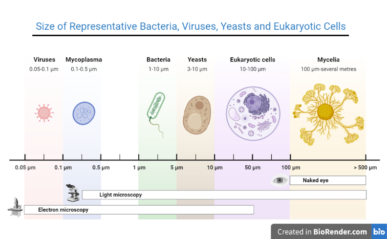 Figure: Sizes of representative bacteria, viruses, yeasts, and human cells. The bacteria range in size from Mycoplasma, the smallest, to Bacillus anthracis, one of the largest.
Units of Measurement in Microbiology
Microbiologists use the metric system to measure organisms. The relevant units are:
| Unit | Symbol | Equivalent | Approximate size of |
|---|---|---|---|
| Millimeter | mm | 10⁻³ m (0.001 m) | Pinhead; smallest visible object with naked eye |
| Micrometer (micron) | μm | 10⁻⁶ m (0.001 mm) | Most bacteria; fungal spores; RBC (7 μm) |
| Nanometer | nm | 10⁻⁹ m (0.001 μm) | Viruses; large molecules; ribosomes |
| Angstrom | Å | 10⁻¹⁰ m (0.1 nm) | Atoms; molecular bonds |
Practical size reference points:
| Object | Approximate size |
|---|---|
| Human hair diameter | ~100 μm |
| Human red blood cell | ~7–8 μm |
| Unaided eye resolution limit | ~200 μm |
| Light microscope resolution limit | ~0.2 μm (200 nm) |
| Electron microscope resolution | ~0.2 nm |
Observation of Bacterial Cells
Experts believe that an unaided eye with normal vision can see objects ≥200 μm, so most bacteria are too small to be seen without a microscope. Humans can see only a handful of giant bacteria without a microscope.
Why bacterial size matters
The size of microorganisms is not just an academic measurement. It has direct practical implications in diagnostic microbiology and infection control:
1. Gram stain detection threshold To be visible on a gram-stained smear under the light microscope, bacteria must be present in a specimen at approximately 10⁴ to 10⁵ organisms per mL. Below this concentration, bacteria are statistically unlikely to appear in the microscopic field examined. This is why a negative gram stain does not rule out infection — early infections with low bacterial counts may not be detectable.
2. Filter sterilization Filtration sterilization uses membrane filters with precisely defined pore sizes to remove microorganisms from heat-sensitive liquids. The choice of pore size depends on the target organism:
- 0.45 μm filters: remove most bacteria (cocci and rods) but may not retain the smallest bacteria (Mycoplasma, some Pseudomonas)
- 0.22 μm filters, the standard for sterile filtration, retain essentially all conventional bacteria. Mycoplasma is the notable exception: lacking a cell wall, it deforms and can pass through. Viruses pass through freely.
- Viruses cannot be removed by standard filtration: they are too small (20–300 nm); viral removal requires ultrafiltration (0.01–0.05 μm) or virus inactivation methods
3. Why Mycoplasma was initially mistaken for a virus
Before molecular methods existed, the filter was the test. An agent that passed a 0.22 μm bacteriological filter was, by definition, a virus, because every known bacterium was retained. Mycoplasma broke that definition. Its cells measure about 0.15 to 0.3 μm, which is already borderline, but the decisive factor is that Mycoplasma has no cell wall. Without a rigid wall, the cell deforms and squeezes through pores that would stop a conventional bacterium of the same nominal diameter.
The classification held until Mycoplasma was grown on artificial culture media. Viruses require living host cells and cannot be cultured on agar. Growth on a cell-free medium settled the question: Mycoplasma is a bacterium, and it remains the smallest one capable of free-living existence.
The wider lesson is that a filter measures deformability as much as diameter, which is why filtration alone was never a reliable way to separate bacteria from viruses.
4. Surface area-to-volume ratio and bacterial metabolism Bacteria must be small because their metabolism depends on diffusion — nutrients enter and waste products exit through the cell surface. As a cell gets larger, volume grows as the cube of radius but surface area grows only as the square. Beyond a certain size, the surface area is insufficient to support the metabolic needs of the interior. This physical constraint is one of the fundamental reasons all bacteria are microscopic.
Size Reference Table For Microorganisms and Cells
Bacteria
Bacteria | Size | Visible by | Notes |
Thiomargarita magnifica (largest bacterium) | Up to 2 cm | Naked eye | Sulfur-oxidizing; found in marine sediments |
Epulopiscium fishelsoni | Up to 600 μm | Naked eye | Gut symbiont of surgeonfish |
Thiomargarita namibiensis | 100–750 μm | Naked eye (barely) | Sulfur bacterium; marine sediments |
Bacillus anthracis | 4–8 × 1–1.5 μm | Light microscope | One of the largest clinically important bacteria |
Clostridium perfringens | 4–6 × 1–1.5 μm | Light microscope | Large gram-positive rod |
Escherichia coli | 1–2 × 0.5–1 μm | Light microscope | Standard reference organism |
Staphylococcus aureus | 0.5–1.5 μm diameter | Light microscope | Typical gram-positive coccus |
Streptococcus pyogenes | 0.6–1.0 μm diameter | Light microscope | Typical gram-positive coccus |
Neisseria gonorrhoeae | 0.6–1.0 μm diameter | Light microscope | Small gram-negative diplococcus |
Treponema pallidum | 0.1–0.2 μm diameter; 6–15 μm long | Dark-field microscopy | Too thin for standard light microscopy |
Leptospira interrogans | 0.1 μm diameter; 6–20 μm long | Dark-field microscopy | Very thin — barely visible even by dark-field |
Mycoplasma pneumoniae (smallest bacterium) | 0.15–0.3 μm | Electron microscopy only | Smallest free-living organism; passes 0.22 μm filters |
Virus
Virus | Size | Visible by |
Smallpox virus (Poxvirus) | ~200 nm (0.2 μm) | Electron microscopy |
HIV | ~100–120 nm | Electron microscopy |
Influenza virus | ~80–120 nm | Electron microscopy |
SARS-CoV-2 | ~100 nm | Electron microscopy |
Hepatitis B virus | ~42 nm | Electron microscopy |
Poliovirus | ~28 nm | Electron microscopy |
Note: Smallpox virus is the largest virus of medical importance. At about 200 nm it sits at the theoretical resolution limit of light microscopy and can be glimpsed as a dot in stained preparations. Poliovirus, at 28 nm, is one of the smallest animal viruses.
Fungi
Yeast/Mold | Size | Visible by | Notes |
Candida albicans (yeast) | 3–8 μm diameter | Light microscope | Pseudohyphae much larger |
Aspergillus fumigatus conidia | 2–3.5 μm diameter | Light microscope | Smaller than A. niger conidia |
Cryptococcus neoformans | 5–7 μm + 1–30 μm capsule | Light microscope | Capsule can be much larger than cell |
Histoplasma capsulatum (yeast form) | 2–4 μm | Light microscope | Intracellular — in macrophages |
Parasites
Parasites | Size | Visible by | Notes |
Plasmodium falciparum ring form | 1–2 μm diameter | Light microscope (100×) | Smallest visible parasite form |
Giardia lamblia trophozoite | 9–21 × 5–15 μm | Light microscope (40×) | Pear-shaped; bilateral symmetry |
Entamoeba histolytica trophozoite | 15–30 μm | Light microscope (40×) | Contains ingested red blood cells |
Microfilaria | 150–320 × 5–8 μm | Light microscope (10×) | Blood film examination |
Human Cells
Organism/object | Size | Visible by |
Red blood cell (No nucleus) | 7–8 μm diameter | Light microscope |
Neutrophil (Multi-lobed nucleus) | 12–15 μm | Light microscope |
Lymphocyte (small) | 7–10 μm | Light microscope |
Macrophage | 15–80 μm | Light microscope |
Hepatocyte (liver cell) | 20–30 μm | Light microscope |
Reference Points
Organism/object | Size | Notes |
Light microscope resolution limit | 0.2 μm | Below this, objects cannot be resolved |
Standard sterilizing filter pore size | 0.22 μm | Retains essentially all bacteria except Mycoplasma; viruses pass through |
Standard 0.45 μm filter | 0.45 μm | Retains most bacteria; Mycoplasma may pass |
Giant Bacteria
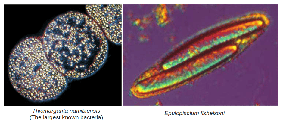 Figure: Thiomargarita namibiensis the largest known bacteria
Thiomargarita magnifica is the world’s biggest single-cell bacteria. It is up to 2 cm long and is visible to the naked eye. It is roughly 50 times larger than other giant sulfur bacteria, which were themselves already exceptional. Thiomargarita namibiensis, meaning "sulfur pearl of Namibia," is another of the largest known prokaryotes. This sulfur chemolithotroph can be 750 μm in diameter and nearly visible to the naked eye. This gram-negative coccoid proteobacterium is about 100 times larger than an average bacterial cell.
Epulopiscium fishelsoni is another very large prokaryote with cells longer than 600 μm (0.6 millimeters). This bacterium is phylogenetically related to the endospore-forming bacterium Clostridium and is found in the gut of the surgeonfish.
Why does bacterial size vary?
Bacterial size is not fixed; it varies depending on several factors:
Growth phase: Bacteria are smallest during the logarithmic (exponential) growth phase when they are dividing rapidly and resources are allocated to division rather than cell enlargement. In the stationary phase, cells may be larger due to incomplete division or storage compound accumulation.
Nutrient availability: Bacteria grown in nutrient-rich media are generally larger than the same species grown in minimal or nutrient-depleted media.
Temperature: Lower growth temperatures slow bacterial metabolism and may produce slightly larger cells.
Species and strain differences: Size is ultimately genetically determined — the range for each species is relatively constant under standard conditions.
Clinical specimens vs culture media: Bacteria directly from clinical specimens may appear smaller or more pleomorphic than the same organism grown on ideal culture media — reflecting the nutrient-poor and immunologically hostile environment of the host.
Cell-size Comparison
- Eukaryotic cells are known with diameters as small as 0.8 μm or as large as several hundred micrometers.
- Cells of yeast, Saccharomyces cerevisiae measures 8 μm in diameter.
- Borrelia is longer than a human blood cell. It is 10 μm long, whereas RBCs are 7 μm in diameter.
- Viruses vary in size, with the smallest known viruses being only about 10 nm in diameter.
Lower Limits of Cell Size
Small cells have a higher surface-to-volume (S/V) ratio. A higher S/V ratio of smaller cells supports a faster rate of nutrient exchange per unit of cell volume compared with larger cells. Thus, smaller cells generally grow faster than larger cells, and a given amount of resources will support a larger population of small cells than large ones. Smaller cells also support faster evolution, since a larger population carries a larger pool of mutations.
Although small size confers a selective advantage, a cell still needs a minimum volume to accommodate biomolecules essential for its growth. A diameter of roughly 0.15 to 0.2 μm is the smallest that can house the essential components of a free-living cell, such as proteins, nucleic acid, and ribosomes.
References and further readings
- Madigan MT, Bender KS, Buckley DH, Sattley WM, Stahl DA. Brock Biology of Microorganisms. 16th ed. Pearson; 2021.
- Tille PM. Bailey & Scott's Diagnostic Microbiology. 15th ed. St. Louis: Elsevier; 2022.
- Murray PR, Rosenthal KS, Pfaller MA. Medical Microbiology. 9th ed. Elsevier; 2020.
- Volland JM, et al. A centimeter-long bacterium with DNA contained in metabolically active, membrane-bound organelles. Science. 2022;376(6600):1453–1458.
Frequently Asked Questions
What is the average size of a bacterium?
What is the smallest and largest known bacterium?
Why can bacteria not be seen with the naked eye?
Why does Mycoplasma pass through bacteriological filters?
How does bacterial size affect gram stain detection?
What is the relationship between bacterial size and surface area-to-volume ratio?
How do bacterial size and viral size compare?
Can bacteria be seen without staining under a light microscope?

Tankeshwar Acharya, MSc (Medical Microbiology)
Tankeshwar Acharya is an Assistant Professor in the Department of Microbiology at Patan Academy of Health Sciences (PAHS), Nepal, where he has been teaching and practicing clinical microbiology for over 14 years. He is the founder of Microbe Online, one of the leading free microbiology education resources on the web, covering bacteriology, mycology, parasitology, immunology, and clinical laboratory diagnostics written from direct experience in both the classroom and the diagnostic laboratory.
Comments
No comments yet. Be the first to share your thoughts.
Leave a comment
All comments are reviewed before they appear.