

An immunocompromised, post-renal transplant patient on tacrolimus, develops unexplained fever and a rising creatinine on day 30 after transplantation. The clinician suspects CMV disease and needs a diagnostic answer fast: delay in treatment with ganciclovir risks graft loss. Conventional cell culture for CMV takes 14–21 days i.e., far too slow to guide management. PCR would be ideal, but the laboratory has limited molecular capacity today.
The solution is shell vial culture: a modified technique that can detect CMV in 16–48 hours by centrifuging the specimen directly onto a cell monolayer, then using early antigen immunofluorescence to detect viral proteins before CPE ever appears. The speed advantage is the clinical point of the entire method.
Shell vial cell culture, a centrifuge-enhanced tissue culture assay, is a modification of conventional cell culture for the rapid detection of viruses in vitro. Rapid shell vial cultures are used to detect many viruses.
Principle of Shell Vial Cell Culture
The technique involves inoculation of the clinical specimen on to cell monolayer grown on a coverslip in the culture tube, followed by low-speed centrifugation and incubation.
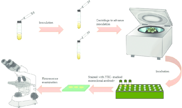 Figure: Shell Vial Cell Culture Technique
It is thought that minor trauma caused by low-speed centrifugation into the susceptible cell surface enhances viral infectivity by enhancing viral entry.
The infected cell monolayer is then stained for the presence of viral antigens by direct fluorescent antibody (DFA) or indirect fluorescent antibody (IFA) staining within hours or days of inoculation. In this way, viruses that normally take days to weeks to produce a cytopathic effect (CPE) can be detected within 1 to 2 days by this culture technique.
Preparation of Shell Vials
Shell vials (15 mm X 45 mm 1-dram vials) are prepared by adding a round coverslip to the bottom of the shell vial, covering them with a normal growth medium, and adding appropriate cells. During incubation, a cell monolayer forms on top of the coverslip. Shell vials should be used 5 to 9 days after the cells have been added.
Shell vials with the already-formed monolayer can be purchased from various suppliers. MRC-5 (Human Fibroblast cells) is used as a cell line in shell vial culture.
Uses of Shell Vial Cell Culture
Rapidity achieved without compromising sensitivity has made shell vial cell culture an important technique in diagnostic virology. This technique can detect most viruses that grow in conventional cell culture and is especially useful for viruses that require relatively long incubation for producing cytopathic effects (CPE).
It is used to identify medically important viruses such as;
- Cytomegalovirus (CMV),
- Varicella-zoster Virus (VZV),
- Herpes Simplex Virus (HSV),
- Adenovirus,
- Influenza A&B virus
- Parainfluenza 1,2,3 virus and
- Respiratory Syncytial Virus (RSV)
It is also used to culture obligate intracellular bacteria such as Chlamydia trachomatis.
Advantage
The advantage of a shell vial is its speed; most viruses are detected within 24 hours.
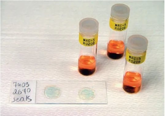 Figure: Shell vial cell culture tubes and stained coverslips
Limitations
- Only one type of virus can be detected per shell vial. For example, a specimen that might contain influenza A and B, or adenovirus, would need to be inoculated to three separate virus-specific conjugates. This limitation can be overcome by using pooled antibodies followed by staining with individual antibody conjugates if positive in pool antibody testing.
How to Remember
Shell vial = centrifuge + early antigen staining, not CPE. The whole point of shell vial culture is that it bypasses waiting for cytopathic effect, which takes days to weeks depending on the virus. Instead, centrifugation forces viral particles into close contact with the cell monolayer (enhancing adsorption that would happen slowly by diffusion alone), and fluorescent antibodies detect viral early antigens expressed in the first hours of replication, long before the cell shows any visible damage. Speed comes from detecting proteins, not damage.
"One virus, one vial" is the limiting rule. Pooled antibodies can screen for multiple viruses in a single vial, but definitive serotyping or identification still typically requires separate vials per virus type. In practice, labs running a respiratory panel might inoculate the same specimen into several shell vials simultaneously (each with a different cell line and a different antibody cocktail) to screen broadly in one run.
CMV and the transplant patient are the prototype clinical scenario. Shell vial was developed largely around CMV detection in immunocompromised hosts where the clinical urgency is real and the virus is slow-growing in conventional culture. If an exam question describes a rapid culture technique for a slow-growing herpesvirus in a transplant patient, shell vial is the answer.
Key Exam Facts
| Feature | Detail |
|---|---|
| Principle | Centrifugation enhances viral adsorption onto cell monolayer; early antigen immunofluorescence detects infection before CPE |
| Time to result | 16–48 hours (vs. days–weeks for conventional shell tube culture) |
| Cell lines used | MRC-5 (human fetal lung diploid), primarily for herpesviruses; others matched to target virus |
| Detection method | Fluorescent antibody staining for viral early antigens |
| Primary clinical application | CMV detection in immunocompromised patients (post-transplant, HIV) |
| Other detectable viruses | HSV-1, HSV-2, VZV, influenza A/B, parainfluenza, RSV, adenovirus, dengue, West Nile, Japanese encephalitis |
| Key advantage over conventional culture | Speed. Detects early antigen before CPE; 16–48 hours vs. weeks |
| Key advantage over PCR | Recovers live virus (enables susceptibility/resistance testing); no molecular infrastructure required |
| Key limitation | One virus per vial (unless pooled antibodies used); sensitivity lower than PCR for some viruses; requires cell culture infrastructure |
| Centrifugation role | Forces virus-cell contact; enhances adsorption rate dramatically; without it, viral attachment relies on slow diffusion |
Where Students Get Confused
"Shell vial culture detects CPE just faster than conventional culture." It doesn't detect CPE at all. Shell vial uses immunofluorescent antibodies to detect viral early antigens that are expressed in the first few hours of replication, before the cell shows any morphological change. Conventional culture waits for CPE (visible cell damage), which takes much longer. Shell vial skips the CPE step entirely.
"Centrifugation is just a standard lab step, the virus would infect the cells anyway." The centrifugation step is the mechanistic heart of the technique. In conventional culture, viruses contact cells by diffusion, which is slow and random. Centrifugation (typically 700g for 40 minutes) forces viral particles physically against the cell monolayer surface, dramatically increasing the number of virus-cell contacts per unit time and concentrating even low-titer specimens against the cells. Without centrifugation, the "shell vial" is just a tube.
"Shell vial is only useful for CMV." CMV was the original application, but shell vial has been validated for a wide range of viruses including HSV, VZV, influenza, parainfluenza, RSV, adenovirus, and arboviruses such as dengue and Japanese encephalitis. The breadth of application depends on having the right cell line and the right fluorescent antibody for the target virus.
"Shell vial has replaced PCR for virus detection." In most well-resourced settings, the opposite has occurred. PCR has largely replaced shell vial for routine diagnostic virology because it is faster, more sensitive, more specific, and doesn't require living virus. Shell vial retains a role where live virus is needed (antiviral susceptibility testing), where molecular infrastructure is limited, or where the target virus isn't covered by available PCR panels.
References and further readings
- Jayakeerthi, R. S., Potula, R. V., Srinivasan, S., & Bhanu, R. V. (2006). Shell vial cell culture assay for the rapid diagnosis of Japanese encephalitis, West Nile and dengue-2 viral encephalitis. Virology Journal, 3, 2. https://doi.org/10.1186/1743-422X-3-2
- Tille, P. M. (2017). Bailey & Scott's Diagnostic Microbiology (14th ed.). Elsevier Mosby.

Comments
No comments yet. Be the first to share your thoughts.
Leave a comment
All comments are reviewed before they appear.