
Saline Wet Mount for Intestinal Parasites: Principle, Procedure, and Results
Learn how to prepare and examine a saline and iodine wet mount for intestinal parasites — trophozoites, cysts, and helminth eggs — with organism-specific results, interpretation tips, and exam mnemonics.
A mother brings her 5-year-old to a district hospital in rural Nepal. The child has had loose stools and abdominal cramping for two weeks. The doctor suspects giardiasis or amoebiasis but wants to confirm before prescribing metronidazole. The laboratory technician takes a pea-sized portion of the fresh stool sample, mixes it with a drop of normal saline on a glass slide, and within five minutes has an answer: motile trophozoites with a characteristic tumbling motion — Giardia lamblia.
This is the saline wet mount — the fastest, cheapest, and most widely available diagnostic test for intestinal parasitic infections. No staining required, no concentration step needed, no special equipment beyond a standard microscope. Its power and its limitation are the same thing: it works best when the parasite burden is high enough to find in a small sample, and when trophozoites are present and motile.
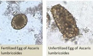 Figure: Egg of Ascaris in wet mount
Gastro-intestinal infestations by parasites (protozoan/helminths) are primarily diagnosed by detecting live motile trophozoites (for protozoans); cyst (inactive dormant stage of protozoa) or eggs (in case of helminths) in the stool. Eggs of helminths are often easier to find and identify because of their size and their distinctive morphological features. These agents are identified based on their morphological/staining (For eggs of helminth; bile stained or not bile stained) characteristics.
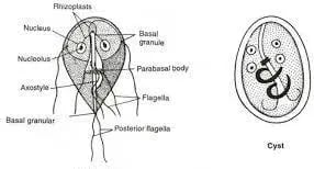 Figure: Trophozoite (left) and Cyst of Giardia lamblia (right)
Saline wet mount is made by mixing a small quantity (about 2 mg) of feces in a drop of saline placed on a clean glass slide. The smear is then examined under a microscope. Saline wet mount is used for the detection of trophozoites and cysts of protozoa, and eggs and larvae of helminths. It is particularly useful for the detection of live motile trophozoites of E. histolytica, Giardia lamblia, and Balantidium coli.
It is usually more efficient for laboratories to do a simple concentration to avoid overlooking parasites that may be present in very small numbers. A modification of the direct smear procedure, the Kato-Katz technique, is especially useful for field surveys aimed to detect schistosome or soil-transmitted nematode (roundworm, whipworm, and hookworm). It also gives an estimation of the intensity of infection.
Why Two Preparations? Saline vs Iodine
The standard wet mount uses two drops side by side on the same slide — one in saline, one in iodine. Each serves a distinct purpose that the other cannot:
| Saline wet mount | Iodine (Lugol's) wet mount | |
|---|---|---|
| Primary purpose | Detect motile trophozoites | Reveal internal cyst structure |
| Best for | E. histolytica, Giardia, B. coli trophozoites; helminth eggs and larvae | Protozoan cysts (glycogen mass, nuclei stained brown-yellow) |
| Organisms killed? | No — motility preserved | Yes — iodine kills; no motility |
| Cyst internal detail | Poor — low contrast | Good — glycogen stains brown, nuclei visible |
| Helminth eggs | Good morphology | Less useful than saline |
The clinical workflow: Always examine the saline preparation first for motility, then switch to the iodine preparation for structural identification of any organisms found.
Uses of Direct Wet Mount
- To assess the worm burden of patient
- To provide a quick diagnosis of the heavily infected specimen
- To check organism motility (primarily protozoan trophozoites)
- To diagnose organisms that might not be seen from permanent stain methods
Reagents and equipment
- Normal saline (0.85% NaCl); Lugol’s Iodine
- Glass slides
- Coverslips
- Pipettes
- Gloves
- Microscope
Procedure
- With a wax pencil or other marker, write the patient’s name or identification number and the date at the left-hand end of a clean microscope slide.
 1. Place a drop of saline in the center of the left half of the slide and place a drop of iodine solution in the center of the right half of the slide (Fig .1). (Note. Iodine wet mount preparations are most useful for protozoa, less so for helminths.)
With an applicator stick or match, pick up a small portion of faeces (approximately 2 mg which is about the size of a match head) and add it to the drop of saline: add a similar portion to the drop of iodine. Mix the faeces with the drops to form suspensions (Fig. 2).
Cover each drop with a coverslip by holding the coverslip at an angle, touching the edge of the drop, and gently lowering the coverslip onto the slide so that air bubbles are not produced (Fig. 3).
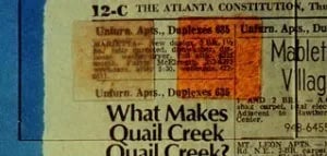 Figure: Suitable Wet Mount Preparation (Img Source: DPDx)
Ideal preparations containing 2 mg of feces are uniform – not so thick that fecal debris can obscure organisms, nor so thin that blank spaces are present. The mount should be just thick enough that newspaper print can be read through the slide.
- If desired the coverslip (s) can be sealed using petroleum jelly and Paraffin oil or other suitable sealing preparations. Sealing the coverslip keeps organisms from moving when using oil immersion objectives and prevents the preparation from drying out.
Examination
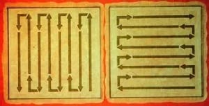 Figure: Scanning the stained smear (Image source: dpdx)
- Examine the specimen with the low power objective (10x) and low light. Begin at one corner of the smear and systematically examine (either up and down or laterally) successive adjacent swaths with the low power microscope. Low power examination includes an entire area of 22 by 22 mm coverslip preparation (both saline and iodine).
- When a parasite-like object comes into view, switch to higher magnification to see the more detailed morphology of the object in question. It should be more closely examined and identified under high power (40x) objective. High dry power examination should include at least one-third of the coverslip area (both saline and iodine).
Results
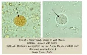 Figure: Wet Mount Smear of Stool showing E. histolytica
Results from the direct smear examination should often be considered presumptive; however, some organisms could be definitely identified (Giardia lamblia cysts and Entamoeba coli cysts, helminth eggs, and larvae, Isospora belli oocysts). These reports should be considered “preliminary”, while the final report would be available after the results of concentration and permanent stained smear were available.
What You Find: Organism-Specific Results
On Saline Wet Mount
| Organism | Stage found | Key identifying feature |
|---|---|---|
| Entamoeba histolytica | Trophozoite | Directional, progressive motility using pseudopodia; ingested RBCs inside cytoplasm (pathognomonic) |
| Entamoeba coli | Trophozoite | Sluggish, non-progressive motility; no ingested RBCs; larger than E. histolytica |
| Giardia lamblia | Trophozoite | "Falling leaf" or tumbling motility; pear-shaped, bilateral symmetry visible |
| Balantidium coli | Trophozoite | Largest human intestinal protozoan; rapid spiral movement; kidney-shaped macronucleus |
| Ascaris lumbricoides | Egg (fertilised) | Oval, bile-stained brown-yellow, mammillated outer coat; unfertilised eggs longer and thinner |
| Hookworm | Egg | Oval, thin-shelled, colourless; segmented embryo inside; identical for Ancylostoma and Necator |
| Trichuris trichiura | Egg | Barrel-shaped with prominent bipolar plugs (like a barrel with corks) — unmistakable |
| Hymenolepis nana | Egg | Round, thin-shelled, with polar filaments between inner and outer membranes |
| Taenia spp. | Proglottid or egg | Eggs spherical, radially striated; species differentiation requires proglottid examination |
| Strongyloides stercoralis | Rhabditiform larva | Larvae (not eggs) in stool; short buccal cavity distinguishes from hookworm larva |
On Iodine Wet Mount
| Organism | Cyst features on iodine |
|---|---|
| Entamoeba histolytica | 1–4 nuclei; chromatoid bars (blunt ends); glycogen mass stains brown |
| Entamoeba coli | Up to 8 nuclei; chromatoid bars have splintered ends |
| Giardia lamblia | Oval cyst, 4 nuclei; median bodies; longitudinal fibres visible |
| Balantidium coli | Large cyst, single large macronucleus |
The single most important result to look for: Entamoeba histolytica trophozoites with ingested red blood cells in the cytoplasm — this distinguishes invasive E. histolytica from non-pathogenic E. dispar (which looks identical morphologically but does not ingest RBCs) and from E. coli (non-pathogenic commensal).
Where Students Actually Get Confused
1. "Iodine wet mount shows motility." Iodine kills trophozoites immediately — no motility is seen. The iodine preparation is for structural identification only. Always examine the saline preparation first if motility is the diagnostic clue you need.
2. "E. histolytica and E. dispar look different on wet mount." They do not. Both are morphologically indistinguishable on saline and iodine wet mount. The only wet mount clue to E. histolytica specifically is the presence of ingested RBCs in the cytoplasm — this indicates active invasion and strongly suggests E. histolytica. Definitive species differentiation requires stool antigen EIA or PCR.
3. "A negative wet mount excludes parasitic infection." Direct wet mount examines roughly 2 mg of stool — a tiny fraction of the total specimen. In low-burden infections, parasites may not be in that 2 mg. The wet mount is a preliminary screening test; results should be considered presumptive. Concentration techniques and permanent stained smears are always recommended when suspicion remains.
4. "Saline and normal saline are different things." Normal saline (0.85% NaCl) is exactly what the procedure uses. The tonicity matches that of the organisms and preserves their morphology. Using tap water causes osmotic lysis of cysts; using hypertonic saline distorts morphology.
5. "The smear can be any thickness." The standard teaching is that the smear should be thick enough to see but thin enough to read newsprint through the slide. Too thick — fecal debris obscures organisms. Too thin — organisms too spread out to find efficiently.
Key Exam Facts in One Table
| Fact | Detail | Memory hook |
|---|---|---|
| Stool volume used | ~2 mg (size of a match head) | Small portion — mix thoroughly |
| Saline concentration | 0.85% NaCl (normal saline) | Isotonic — preserves morphology |
| Saline purpose | Detect motile trophozoites; helminth eggs | Motion first |
| Iodine purpose | Reveal cyst structure (kills motility) | Structure second |
| E. histolytica hallmark | Ingested RBCs in trophozoite cytoplasm | RBC ingestion = invasion = E. histolytica |
| Giardia trophozoite motility | "Falling leaf" / tumbling pattern | Bilateral symmetry + flagella |
| Trichuris egg shape | Barrel-shaped with bipolar plugs | Unmistakable — "barrel with corks" |
| Strongyloides in stool | Rhabditiform larvae (not eggs) | Only helminth to appear as larvae in fresh stool |
| Result status | Preliminary only | Confirm with concentration + permanent stain |
| Processing time for liquid stool | Within 30 minutes | Trophozoites degenerate rapidly |
Limitations
- Once Iodine is added to the preparation, the organism will be killed and motility will be lost.
- Oil immersion examination is not recommended (organism morphology is not that clear)
Self-Check Questions
- You are examining a saline wet mount and see a large amoeboid trophozoite with rapid directional movement and red blood cells visible inside its cytoplasm. What organism is this, and what does the RBC ingestion indicate?
- You add iodine to the right half of the slide and examine it first. You see brown-stained cysts but no motility. A colleague says the specimen is negative for trophozoites — do you agree?
- What is the correct saline concentration for wet mount preparation and why does the concentration matter?
- A wet mount of a liquid stool collected 3 hours ago is negative. The clinician is surprised given the clinical picture. What is the most likely reason for the false-negative result?
- On iodine mount you see a cyst with 8 nuclei. Which organism does this suggest, and is it pathogenic?
Answers
- Entamoeba histolytica trophozoite. Ingested RBCs in the cytoplasm indicate active tissue invasion; this distinguishes E. histolytica from morphologically identical but non-pathogenic E. dispar, which does not ingest RBCs.
- No — iodine kills trophozoites immediately. No motility will be seen on the iodine preparation regardless of whether trophozoites are present. The saline preparation must be examined for motility; the iodine preparation is used only for cyst structure.
- 0.85% NaCl (normal saline). This concentration is isotonic with the organisms and preserves their morphology. Tap water causes osmotic lysis; hypertonic saline distorts and shrinks organisms.
- The liquid stool should have been processed within 30 minutes of collection. At 3 hours, any trophozoites present have degenerated and are no longer identifiable. Cysts and eggs may still be detectable, but trophozoite-stage organisms are lost.
- Entamoeba coli — up to 8 nuclei in a cyst is characteristic. E. coli is a non-pathogenic commensal of the human colon; it does not require treatment but its presence confirms faecal contamination of the food/water source.
References
- Garcia, L. S. (2016). Diagnostic Medical Parasitology (6th ed.). ASM Press.
- Cheesbrough, M. (2006). District Laboratory Practice in Tropical Countries (2nd ed., Part 1). Cambridge University Press.
- World Health Organization. (2012). Bench aids for the diagnosis of intestinal parasites (2nd ed.). WHO. https://apps.who.int/iris/bitstream/handle/10665/37323/9789241544764_eng.pdf
- Demeke, G., Fenta, A., & Dilnessa, T. (2021). Evaluation of wet mount and concentration techniques of stool examination for intestinal parasites identification at Debre Markos Comprehensive Specialized Hospital, Ethiopia. Infection and Drug Resistance, 14, 1357–1362. https://doi.org/10.2147/IDR.S307683
- Khanna, V., Tilak, K., Rasheed, S., & Mukhopadhyay, C. (2014). Identification and preservation of intestinal parasites using methylene blue-glycerol mount: a new approach to stool microscopy. Journal of Parasitology Research, 2014, 672018. https://doi.org/10.1155/2014/672018
- CDC – DPDx: Laboratory Identification of Parasites of Public Health Concern. https://www.cdc.gov/dpdx/index.html
Frequently Asked Questions
What is the difference between a saline and an iodine wet mount?
How do you identify Entamoeba histolytica on saline wet mount?
Why must liquid stool be examined within 30 minutes for wet mount?

Tankeshwar Acharya, MSc (Medical Microbiology)
Tankeshwar Acharya is an Assistant Professor in the Department of Microbiology at Patan Academy of Health Sciences (PAHS), Nepal, where he has been teaching and practicing clinical microbiology for over 14 years. He is the founder of Microbe Online, one of the leading free microbiology education resources on the web, covering bacteriology, mycology, parasitology, immunology, and clinical laboratory diagnostics written from direct experience in both the classroom and the diagnostic laboratory.