
MacConkey Agar: Composition, Principle, Preparation, Uses, and Colony Characteristics
MacConkey agar: composition, principle, uses, and detailed colony morphology of 20+ organisms including E. coli, Klebsiella, Salmonella, Pseudomonas, Acinetobacter, and more. Updated for clinical lab use.
MacConkey agar (MAC) is the most widely used selective and differential medium in clinical bacteriology. It was developed in 1900 by British bacteriologist Alfred Theodore MacConkey, who originally designed it to differentiate typhoid bacilli (Salmonella Typhi) from coliform organisms contaminating water supplies; a critical public health need at the turn of the 20th century. More than 120 years later, it remains a first-line plating medium in virtually every diagnostic microbiology laboratory in the world.
MacConkey agar (MAC) is a selective and differential bacterial culture media. MacConkey medium is selective for gram-negative bacteria and differentiates the gram-negative bacteria based on lactose metabolism.
This medium was first developed by Alfred Theodore MacConkey in 1900.
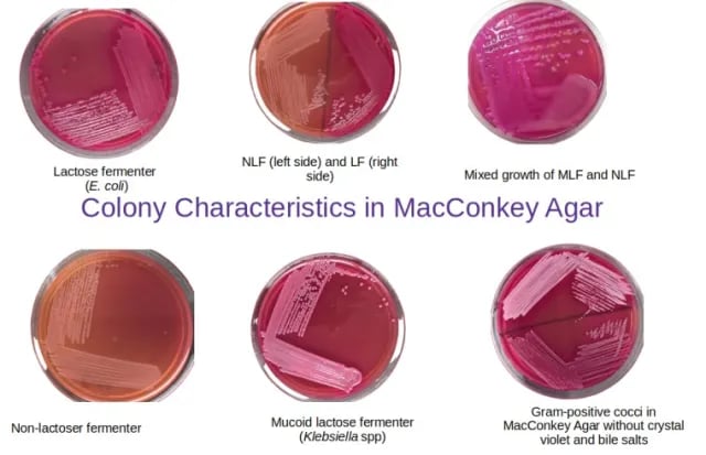 Figure: Colony characteristics in MacConkey Agar
Composition of MacConkey Agar
Key components of the MacConkey medium include crystal violet dye, bile salts, lactose, and neutral red (pH indicator).
Ingredient | MacConkey Agar (g/L) |
Peptone (pancreatic digest of gelatin) | 17 g |
Proteose peptone (meat and casein | 3 g |
Lactose | 10 g |
Bile salts | 1.5 g |
Sodium chloride | 5 g |
Agar | 13.5 g |
Neutral red | 0.03 g |
Crystal violet | 0.001 g |
Distilled water | 1 L |
Final pH | 7.1 |
Crystal violet dye and bile salts prevent the growth of Gram-positive bacteria and fastidious Gram-negative bacteria (such as Neisseria and Pasteurella) making it favorable for the growth of gram-negative bacteria. Since Gram-negative enteric bacteria possess a bile-resistant outer membrane, they remain unaffected by bile salts.
Lactose present in the medium is fermented by bacteria to form lactic acid that decreases the pH of the agar, and turns the indicator (neutral red) pink, thus differentiating lactose fermenters from non-lactose fermenters.
Other ingredients such as enzymatic digest of gelatin, casein, and animal tissue provide nitrogen, vitamins, minerals, and amino acids essential for growth. Sodium chloride provides osmotic balance and supplies essential electrolytes for transport. Agar is incorporated as the solidifying agent.
Principle of MacConkey Agar
MacConkey agar contains four key ingredients (lactose, bile salts, crystal violet, and neutral red) that make it a selective and differential media. Bile salts and crystal violet act as selective agents that inhibit the growth of Gram-positive organisms, and aid in the selective growth of non-fastidious gram-negative bacteria. Lactose acts as a source of carbohydrates. Lactose-fermenting bacteria produce pink-red colonies, after fermenting the lactose to acids and dropping the pH of the indicator (neutral red) present in the medium. Since, non-fermenters can’t utilize lactose, colonies appear colorless or transparent.
Encapsulated bacteria such as Klebsiella and Enterobacter produce capsules using lactose. This gives sticky, wet-appearing colonies on MacConkey medium.
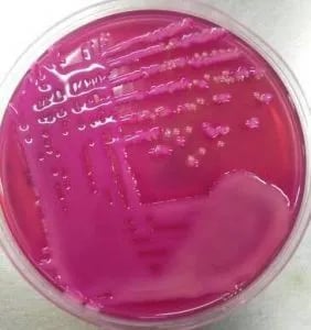 Figure: Mixed growth of mucoid lactose fermenting colonies and NLF colonies in MacConkey agar
Lactose Fermenters
Gram-negative enteric bacteria that grow on MacConkey medium are differentiated by their ability to ferment lactose. If the lactose is fermented the production of the acid drops the pH of the media. The drop in pH is indicated by the change of the neutral red indicator to pink (neutral red appears pink at pH below 6.8).
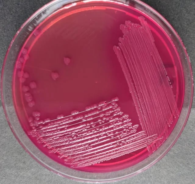 Figure: Dry pink colonies of E. coli in MacConkey Agar
Strongly lactose fermenting bacteria produce sufficient acid which causes precipitation of the bile salts around the growth. It appears as a pink halo surrounding colonies or areas of confluent growth. A pink halo is not seen around the colonies of weaker lactose fermenting bacteria.
Lactose non-fermenters
Gram-negative bacteria that grow on MacConkey agar but do not ferment lactose appear colorless on the medium and the agar surrounding the bacteria remains relatively transparent.
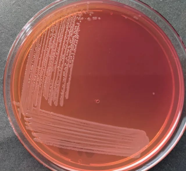 Figure: Pale, non-lactose fermenting colonies of Salmonella in MacConkey Agar
Uses of MacConkey Agar Medium
1. Urine culture — the most common clinical application
MacConkey agar is routinely used for urine culture. Urine specimens contain a mix of urinary pathogens and contaminating organisms; MacConkey selects for gram-negative uropathogens (the most common cause of urinary tract infections) while suppressing gram-positive contamination. It is typically used alongside blood agar and CLED agar in a standard urine culture setup.
2. Stool culture for enteric pathogens
MacConkey agar is a primary plating medium for fecal specimens when enteric pathogens such as Salmonella, Shigella, Campylobacter, and E. coli O157:H7 are suspected. The ability to rapidly differentiate lactose fermenters (normal gut flora like E. coli) from non-lactose fermenters (potential pathogens like Salmonella and Shigella) at 24 hours guides the laboratory directly toward the likely pathogen.
3. Wound, pus, and respiratory specimens
MacConkey agar is included in the routine workup of wound swabs, pus, sputum, tracheal aspirates, and bronchial washings — particularly when gram-negative organisms such as Pseudomonas aeruginosa, Klebsiella pneumoniae, Acinetobacter baumannii, or members of Enterobacteriaceae are suspected clinically.
4. Blood culture subcultures
When blood culture bottles flag positive, the broth is subcultured to a battery of media including MacConkey agar. The colony characteristics at 24 hours — color, size, mucoid appearance, pigmentation — provide the first presumptive identification while awaiting further biochemical or MALDI-TOF results.
5. Differentiation of Enterobacteriaceae
MacConkey agar is foundational for separating members of the Enterobacteriaceae family based on lactose fermentation speed and colony appearance. Strong fermenters (E. coli, Klebsiella), late fermenters (Citrobacter, Serratia), and non-fermenters (Salmonella, Shigella, Proteus) are distinguished at 18–48 hours without any additional testing.
6. Water and environmental microbiology
MacConkey agar is used in water quality testing to detect and enumerate coliforms — gram-negative, lactose-fermenting rods that indicate fecal contamination of water. The presence of pink lactose-fermenting colonies on MacConkey agar is the basis for the presumptive coliform test in standard water analysis.
7. Food microbiology
MacConkey agar is approved by the Bacteriological Analytical Manual (FDA-BAM) for the detection of gram-negative pathogens in food products, particularly for Salmonella and coliforms in meats, dairy, and processed foods.
8. Detection of E. coli O157:H7 (Sorbitol MacConkey)
The sorbitol variant of MacConkey agar (SMAC) replaces lactose with sorbitol and is specifically used to detect E. coli O157:H7 — the cause of haemorrhagic colitis and haemolytic uraemic syndrome (HUS). Unlike most E. coli strains, O157:H7 does not ferment sorbitol and therefore appears as colorless colonies on SMAC, while other E. coli produce pink colonies.
Preparation of MacConkey Agar
- Weigh and suspend 50 grams of MacConkey agar powder in 1 Litre of purified water and mix thoroughly. Read and follow the instruction of the manufacturer if you have purchased dehydrated agar media from a commercial supplier.
- Heat with frequent agitation and boil for 1 minute to completely dissolve the powder.
- Autoclave at 121°C for 15 minutes.
- Cool to 45-50°C, mix well, pour about 20- 25ml into sterile Petri plates and allow to solidify.
- After solidification of the plates, label the media plates with the name and date of preparation. Labeling should always be done on the backside of the media plate, as lids could be interchanged.
- Store inverted (with lids down) at 2-8°C until use.
Ready-made MacConkey agar plates are also available from commercial suppliers.
Result and Interpretation
Pink-red colonies: Pink-red colonies on MAC medium indicate the presence of lactose fermenting bacteria. Examples include Escherichia coli, Klebsiella spp, Citrobacter, Enterobacter, etc.
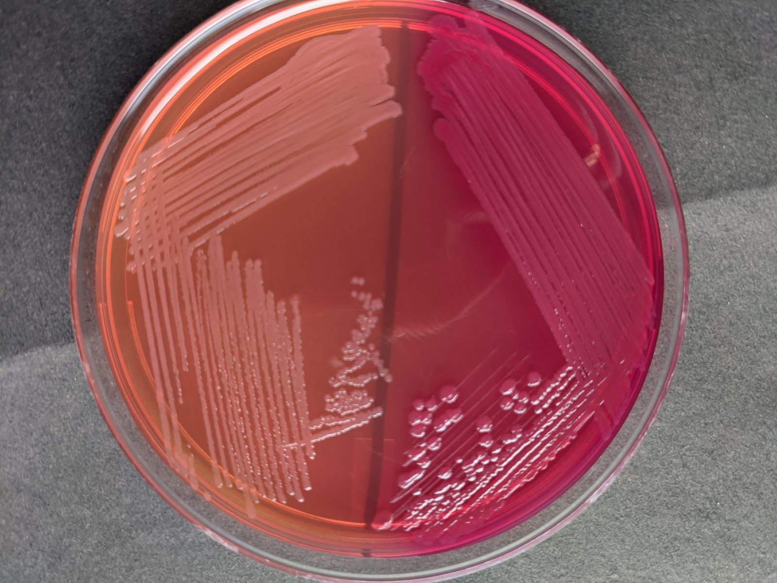 Figure: Pale (NLF) and pink (LF) colonies on MacConkey Agar
Colorless colonies/pale colonies (colonies similar to the color of the media): Colorless or pale colonies on MAC medium indicate that the test organism is a non-lactose fermenter. Examples include species of Salmonella, Shigella, Proteus, Providencia, Pseudomonas, Morganella, etc.
Colony Morphology on MacConkey Agar
Besides differentiating on the basis of color, colonies on MacConkey medium can further be presumptively identified based on their colonial appearances (shape, size, margin, time of growth, etc). Some of them are enlisted below:
Lactose fermenters (pink/red colonies)
| Organism | Fermentation | Colony characteristics | Additional notes |
|---|---|---|---|
| Escherichia coli | Strong LF | Flat, dry, pink to dark pink, non-mucoid, 2–3 mm; surrounded by darker pink halo of precipitated bile salts; classic "donut" appearance | Most common gram-negative on MacConkey; bile salt precipitation = strong acid production |
| Klebsiella pneumoniae | Strong LF | Large (4–6 mm), mucoid, pink to dark pink; pigment diffuses into surrounding agar; colonies appear wet and dome-shaped | Mucoid appearance due to polysaccharide capsule; may string when touched with loop |
| Klebsiella oxytoca | Strong LF | Similar to K. pneumoniae — large, mucoid, pink; may be slightly darker | Indole positive; distinguished from K. pneumoniae biochemically |
| Enterobacter aerogenes (Klebsiella aerogenes) | Strong LF | Pink, mucoid colonies, 3–4 mm; smaller and less mucoid than Klebsiella | May show mild pigment diffusion into agar |
| Enterobacter cloacae | Strong LF | Pink, mucoid colonies; similar to E. aerogenes | Clinically important nosocomial pathogen; AmpC beta-lactamase producer |
| Serratia marcescens | Late LF | Initially colorless at 24 hours; pink at 48 hours; may show distinctive red/pink pigment (prodigiosin) especially at 25°C | If plate left at room temperature, red pigmentation becomes very prominent |
| Citrobacter freundii | Late LF | Colorless at 24 hours (mimics NLF); light pink at 48 hours; 2–3 mm flat colonies | Late fermentation commonly causes misidentification if only 24-hour reading taken |
| Citrobacter koseri | Late LF | Similar to C. freundii; colorless to faint pink at 24 hours | Clinically significant in neonatal meningitis |
| Hafnia alvei | Late/weak LF | Colorless to faint pink; 1–2 mm | Weak lactose fermentation; may appear NLF at 24 hours |
Non-lactose fermenters (colorless/pale colonies)
| Organism | Colony characteristics | Distinguishing features |
|---|---|---|
| Salmonella Typhi | Colorless, translucent, convex, 1–2 mm, smooth margin | Smaller than non-typhoidal Salmonella; non-swarming |
| Salmonella spp. (non-typhoidal) | Colorless, convex, 2–3 mm, smooth to slightly serrated margin | Cannot distinguish from Shigella by color alone; further biochemical testing required |
| Shigella spp. | Colorless, flat, 1–2 mm, jagged/irregular edges; more translucent than Salmonella | S. sonnei is a late LF — may show faint pink at 48 hours; most other Shigella remain colorless |
| Proteus mirabilis | Colorless to pale; characteristic swarming on less selective areas; strong putrid fishy odor | Swarming may be reduced on MacConkey due to bile salts; odor is diagnostic |
| Proteus vulgaris | Similar to P. mirabilis; pale, swarming, odorous | Indole positive; distinguished from P. mirabilis biochemically |
| Pseudomonas aeruginosa | Colorless to pale yellow-brown; flat, smooth, 2–3 mm; characteristic sweet grape-like odor; may show blue-green pigmentation (pyocyanin) | Pyocyanin production is distinctive; oxidase positive |
| Pseudomonas aeruginosa (mucoid) | Large, mucoid, colorless colonies — commonly seen in cystic fibrosis isolates | Mucoid due to alginate overproduction; may be mistaken for Klebsiella without pigment check |
| Acinetobacter baumannii | Pale to colorless, opaque, convex, 1.5–2.5 mm, smooth; may appear slightly lavender/pink due to light refraction | Non-motile; oxidase negative; important MDR nosocomial pathogen in ICU patients |
| Acinetobacter spp. | Similar to A. baumannii; pale, opaque, convex | Species-level identification requires MALDI-TOF or molecular methods |
| Stenotrophomonas maltophilia | Pale to light lavender, small (1–2 mm), smooth colonies; may show pale yellow pigment | Oxidase negative; intrinsically resistant to carbapenems; important in immunocompromised patients |
| Burkholderia cepacia complex | Pale to faint yellow, wrinkled or rough texture, 1–2 mm; slow growing (may need 48–72 hours) | Oxidase positive; important pathogen in cystic fibrosis; intrinsically resistant to many antibiotics |
| Morganella morganii | Colorless, flat, 2–3 mm, smooth; no swarming | Urease positive; important nosocomial pathogen in elderly and ICU patients |
| Providencia spp. | Colorless, flat, 2–3 mm; does not swarm | Distinguished from Proteus by absence of swarming and different biochemical profile |
| Yersinia enterocolitica | Small (0.5–1 mm), colorless to faint peach/pink; requires 48–72 hours at 25–28°C | Better recovered at room temperature than 37°C on MacConkey |
| Haemophilus influenzae | No growth | Requires X and V factors not present in MacConkey agar |
| Neisseria gonorrhoeae | No growth | Inhibited by crystal violet and bile salts; requires Thayer-Martin or chocolate agar |
| Neisseria meningitidis | No growth | Same as N. gonorrhoeae |
| Gram-positive bacteria (MSSA, MRSA, Streptococcus, Enterococcus) | No growth on standard MacConkey | Inhibited by crystal violet and bile salts; grow on MacConkey without crystal violet (see Modifications section) |
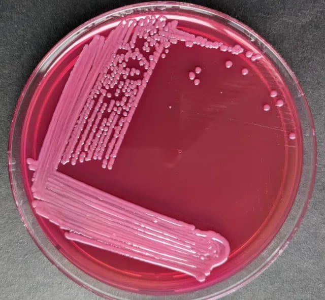 Figure: Pink-mucoid colonies of Klebsiella pneumoniae in MacConkey Agar
In MacConkey agar without crystal violet and bile salts
Staphylococcus spp | Lactose fermenter | Small pink colonies, 1-2mm in diameter, opaque |
Enterococcus spp | Lactose fermenter | Dark pink to red, very minute, translucent colonies |
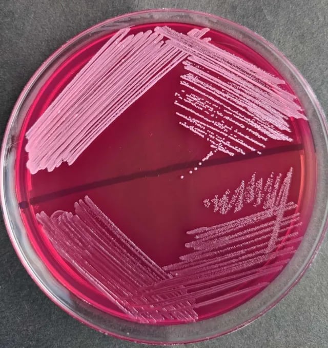 Figure: Light pink, small colonies of S. aureus(above) and dark pink-red, minute colonies of Enterococcus(below) on MacConkey agar without bile salts and crystal violet.
Note: Gram-positive organisms are inhibited in MacConkey agar with bile salts and crystal violet, however, in a different formulation where bile salt and crystal violet are not incorporated, Gram-positive organisms also appear as lactose fermenters but are smaller in size than gram-negative ones.
Quality Control of MacConkey agar
- Sterility testing: Incubate uninoculated plates of MacConkey for 48 hours at 35-37°C and observe for any growth. After 48 hours, the sterility test plate should remain clear. Discard the whole lot if any colonies are seen.
- Performance testing: Inoculate known standard strains on MacConkey agar plates, incubate for 18-24 hours at 35-37°C, and observe for growth and colony characteristics.
Organism | Used as | Colony morphology |
Escherichia coli ATCC 25922 | Positive control for lactose fermentation | Lactose fermenting pink colonies |
Pseudomonas aeruginosa ATCC 27853 | Positive control for non-lactose fermentation | Non-lactose fermenting colonies with or without pigmentation. |
Streptococcus pneumoniae ATCC 49619 | Negative control | No growth |
Modifications of MacConkey Agar
- MacConkey Agar without Crystal Violet It is a differential medium but is less selective than MacConkey agar. The lack of crystal violet permits the growth of Staphylococcus and Enterococcus. Staphylococci produce pale pink to red colonies and enterococci produce compact tiny red colonies either on or beneath the surface of the medium. The medium is also used to separate Mycobacterium fortuitum and M. chelonae from other rapidly growing mycobacteria.
- MacConkey Agar, CS (“Controlled Swarming”): MacConkey agar without crystal violet or salt is used to prevent the swarming of Proteus spp.
- Sorbitol MacConkey Agar: Sorbitol MacConkey agar is a variant of MacConkey agar, it contains sorbitol instead of lactose as fermentable sugar. The contents of Sorbitol MacConkey agar are sorbitol, peptone, bile salts, sodium chloride, neutral red, crystal violet, and agar. E. coli O157:H7 (a verotoxigenic strain) is non-sorbitol-fermenting and produces colorless colonies. Most other E. coli strains and other enterobacteria ferment sorbitol. Sorbitol-fermenting organisms produce pink colonies
**Quality control of Sorbitol MacConkey agar**
Escherichia coli ATCC® 25922: Good growth, pink colonies are sorbitol positive Escherichia coli ATCC® 35150: Good growth, colorless colonies are sorbitol negative.
How to Remember
- Pink means acid, pale means none. On MacConkey, color is a pH report. Lactose fermenters make acid, the neutral red turns them pink; non-fermenters make no acid and stay the color of the medium. If you remember only one thing, remember that the plate is reading pH, not counting sugar.
- The halo means "strong." A pink halo of precipitated bile salts around a colony means so much acid was produced that the bile salts fell out of solution. Only vigorous fermenters like E. coli do this, so the halo is a quick "strong fermenter" flag in a mixed plate.
- Two selective agents, one job: keep Gram-positives out. Bile salts and crystal violet are the doormen. Take them away (MacConkey without crystal violet) and Gram-positives like Staphylococcus and Enterococcus walk in. This is why the "without crystal violet" formulation exists.
- Late fermenters are the trap. Citrobacter, Serratia, and Shigella sonnei can look colorless at 24 hours and pink by 48. Reading a plate too early is the classic way a fermenter gets misfiled as a non-fermenter.
- Mucoid and stringy = capsule. Klebsiella looks wet and mucoid and strings when you touch it with a loop, because it pours lactose into capsule production. Mucoid pink is a Klebsiella signature.
Key exam facts in one table
| Concept | Key exam fact and why it holds |
|---|---|
| Type of medium | Selective AND differential. Selective for Gram-negatives (bile salts + crystal violet inhibit Gram-positives); differential by lactose fermentation. |
| Selective agents | Bile salts and crystal violet. Remove them and Gram-positives grow. This single pair explains both the "no Gram-positive growth" rows and the "without crystal violet" modification. |
| Differential agent + indicator | Lactose is the fermentable sugar; neutral red is the pH indicator, turning pink below pH ~6.8. |
| Lactose fermenter result | Pink to red colonies (E. coli, Klebsiella, Enterobacter, Citrobacter). Strong fermenters add a pink halo of precipitated bile salts. |
| Non-lactose fermenter result | Colorless or pale colonies (Salmonella, Shigella, Proteus, Pseudomonas, Providencia, Morganella). |
| The pink halo | A marker of strong acid production (E. coli). Acid drops local pH enough to precipitate bile salts. Useful rapid flag in mixed cultures. |
| Klebsiella clue | Large, mucoid, stringy pink colonies from capsule production. Mucoid pink = think Klebsiella. |
| The 24 vs 48 hour trap | Late fermenters (Citrobacter, Serratia, Shigella sonnei) can read colorless at 24 h and pink at 48 h. Read too early and a fermenter is misclassified. |
| No-growth organisms | Gram-positives, Neisseria, Haemophilus, and other fastidious organisms do not grow (inhibited, or need factors MacConkey lacks). |
| Sorbitol MacConkey (SMAC) | Sorbitol replaces lactose. E. coli O157:H7 is sorbitol-negative → colorless; most other E. coli ferment sorbitol → pink. This inversion is the whole point of SMAC. |
| Developed by | Alfred Theodore MacConkey, 1900, originally to separate typhoid bacilli from coliforms in water. |
References and further readings
- Tille, P. M. (2022). Bailey & Scott's Diagnostic Microbiology (15th ed.). Elsevier.
- Procop, G. W., Church, D. L., Hall, G. S., Janda, W. M., Koneman, E. W., Schreckenberger, P. C., & Woods, G. L. (2017). Koneman's Color Atlas and Textbook of Diagnostic Microbiology (7th ed.). Wolters Kluwer.
- Jung, B., & Hoilat, G. J. (2023). MacConkey Medium. In StatPearls. StatPearls Publishing. https://www.ncbi.nlm.nih.gov/books/NBK557394/
- Public Health England. (2015). Identification of Enterobacteriaceae (UK Standards for Microbiology Investigations, ID 16). https://www.gov.uk/government/publications/smi-id-16-identification-of-enterobacterales
- Atlas, R. M., & Snyder, J. W. (2014). Handbook of Media for Clinical and Public Health Microbiology. CRC Press.
Frequently Asked Questions
What is the difference between lactose fermenters and non-lactose fermenters on MacConkey agar?
Why do Klebsiella colonies appear mucoid on MacConkey agar?
How do you differentiate Salmonella from Shigella on MacConkey agar?
Why does Proteus swarm on some media but not on MacConkey agar?
Can fungi or yeast grow on MacConkey agar?
Why is MacConkey agar incubated at 35-37°C and not at room temperature?
What does a pink halo around E. coli colonies on MacConkey agar indicate?
Why is Pseudomonas aeruginosa described as a non-lactose fermenter on MacConkey agar?

Tankeshwar Acharya, MSc (Medical Microbiology)
Tankeshwar Acharya is an Assistant Professor in the Department of Microbiology at Patan Academy of Health Sciences (PAHS), Nepal, where he has been teaching and practicing clinical microbiology for over 14 years. He is the founder of Microbe Online, one of the leading free microbiology education resources on the web, covering bacteriology, mycology, parasitology, immunology, and clinical laboratory diagnostics written from direct experience in both the classroom and the diagnostic laboratory.