
Laboratory Diagnosis of Syphilis
Laboratory Diagnosis of Syphilis
Syphilis is a sexually transmitted disease caused by the bacterium *Treponema pallidum.*Syphilis has diverse clinical manifestations and shares many clinical features with other treponemal and nontreponemal diseases (because of this, syphilis has been called “The Great Pretender”). The etiological agent, Treponema pallidum, cannot be cultured, and there is no single optimal alternative test.
Commonly used laboratory diagnostic testing available for syphilis are:
Direct Diagnosis
Direct laboratory methods for diagnosing syphilis include the detection of Treponema pallidum by microscopic examination of fluid or smears from lesions, histological examination of tissues, or nucleic acid amplification methods such as polymerase chain reaction (PCR). Tests used for the direct detection of Treponema pallidum are;
Dark-field microscopy
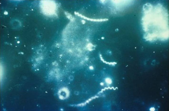 Figure: Darkfield micrograph of Treponema pallidum (source: CDC)
Dark-field microscopy is used to demonstrate the presence of motile Treponema pallidum in lesions or aspirates in early-stage (primary or secondary) syphilis before healing lesions.
Direct fluorescent antibody test for *Treponema pallidum*
It is a diagnostic method used to detect the presence of T. pallidum in clinical specimens. This test relies on fluorescent-labeled antibodies that specifically bind to T. pallidum antigens. Here’s an overview of how the DFA test for Treponema pallidum is typically performed:
DFA Test Procedure for *T. pallidum*
1. Specimen & Slide Preparation
Specimen Collection: Clinical specimens commonly collected for the DFA test include samples from syphilitic lesions, such as genital sores or mucous patches, as well as cerebrospinal fluid in cases of neurosyphilis.
Slide Preparation: A thin smear or impression of the specimen is prepared on a glass slide. The specimen is usually fixed to the slide to preserve the integrity of the T. pallidum cells.
2. Staining Process
Blocking Solution: The slide is treated with a blocking solution to block non-specific binding of antibodies to the specimen.
Fluorescent-Labeled Antibodies: Fluorescent-labeled antibodies specific to T. pallidum antigens are applied to the slide. These antibodies are conjugated with a fluorescent dye, allowing for the visualization of the bacteria under a fluorescence microscope.
Incubation: The slide is incubated to allow the fluorescent antibodies to bind specifically to T. pallidum antigens in the specimen.
Washing: Unbound or non-specifically bound antibodies are washed away to reduce background fluorescence.
3. Examination & Result
Microscopic Examination: A fluorescence microscope is used to examine the slide. When exposed to the appropriate wavelength of light, the bound fluorescent antibodies emit light, allowing for the visualization and identification of T. pallidum.
Interpretation: The presence of characteristic fluorescence in a spirochete-like pattern confirms the presence of T. pallidum in the specimen.
Molecular Methods for *Treponema pallidum* Detection
Nucleic acid amplification (PCR-based) and hybridization techniques offer excellent sensitivity and specificity for identifying T. pallidum, especially in early or primary stages where serology might lag.
1. Polymerase Chain Reaction (PCR)
Principle: A widely used amplification technique targeting specific regions of T. pallidum genomic DNA.
Procedure:
Collection: A clinical specimen (such as a lesion swab or blood) is collected.
Extraction: T. pallidum DNA is isolated from the sample.
Amplification: Primers specific to T. pallidum DNA copy the target region.
Detection: The amplified DNA is visualized using gel electrophoresis, real-time PCR, or hybridization assays.
2. Transcription-Mediated Amplification (TMA)
Principle: An isothermal (constant temperature) nucleic acid amplification technique designed to target and multiply RNA transcripts.
Procedure:
Reverse Transcription: T. pallidum RNA is copied into complementary DNA (cDNA).
Amplification: The cDNA is amplified isothermally via TMA.
Detection: The resulting RNA or cDNA is measured using specific detection probes.
3. Loop-Mediated Isothermal Amplification (LAMP)
Principle: An isothermal amplification technique that rapidly copies DNA at a single, constant temperature using a specialized set of four to six distinct primers.
Procedure:
Amplification: T. pallidum DNA is amplified directly in a heating block without thermal cycling.
Detection: Results are read visually via turbidity (cloudiness), color change indicators, or fluorescence.
4. Reverse Transcription-PCR (RT-PCR)
Principle: Combines reverse transcription of RNA into cDNA with standard PCR amplification to detect active gene transcription.
Application: Highly valuable for detecting T. pallidum RNA, particularly during the window of early syphilis.
5. In Situ Hybridization (ISH)
Principle: Utilizes a labeled nucleic acid probe that binds directly to complementary T. pallidum DNA or RNA strands directly within intact tissue sections.
Application: Excellent for localizing and visualizing the spatial distribution of spirochetes directly within biopsy or clinical tissue specimens.
Indirect diagnosis/serological methods
It is based on serological tests for the detection of antibodies. Serological testing is the mainstay in the laboratory diagnosis and follow-up of syphilis. Serological tests fall into two categories: nontreponemal tests for screening, and treponemal tests for confirmation.
Nontreponemal tests
They measure both immunoglobulin (IgG and IgM) antiphospholipid antibodies formed by the host in response to lipoidal material released by damaged host cells early in infection and lipid from the cell surfaces of the treponeme itself. Commonly used nontreponemal tests are
Rapid plasma reagin (RPR) test
Toluidine red unheated serum test (TRUST)
Venereal Disease Research Laboratory (VDRL) test
Treponemal Tests
All treponemal tests use Treponema pallidum or its components as the antigen.
Treponemal tests are used as confirmatory tests to verify reactivity in non-treponemal tests. Once positive, treponemal tests remain positive throughout life with or without treatment, so these tests can not be used to know the response to treatment.
Commonly used treponemal tests are:
Fluorescent treponemal antibody absorption test (FTA-ABS)test
Treponema pallidum particle agglutination (TP-PA) test
Treponema pallidum Hemagglutination Assay (TPHA)
Flow-chart for laboratory diagnosis of syphilis
If lesion exudate or tissue is available, a direct examination is performed, followed by a nontreponemal serology test. A treponemal test then confirms a reactive nontreponemal test.
Figure: Testing algorithm for Primary Syphilis
A confirmed serological test result indicates the presence of treponemal antibodies. Still, it does not indicate the stage of disease and, depending on the test, may not differentiate between past and current infections.
References
- Madigan Michael T, Bender, Kelly S, Buckley, Daniel H, Sattley, W. Matthew, & Stahl, David A. (2018). Brock Biology of Microorganisms (15th Edition). Pearson.
- Meyer, J. Ch. (1996). Laboratory diagnosis of syphilis. Sexually Transmitted Diseases: Advances in Diagnosis and Treatment, 1–11. https://doi.org/10.1159/000424876
- Larsen, S. A., Steiner, B. M., & Rudolph, A. H. (1995). Laboratory diagnosis and interpretation of tests for syphilis. Clinical Microbiology Reviews, 8(1), 1–21. https://doi.org/10.1128/cmr.8.1.1

Tankeshwar Acharya, MSc (Medical Microbiology)
Tankeshwar Acharya is an Assistant Professor in the Department of Microbiology at Patan Academy of Health Sciences (PAHS), Nepal, where he has been teaching and practicing clinical microbiology for over 14 years. He is the founder of Microbe Online, one of the leading free microbiology education resources on the web, covering bacteriology, mycology, parasitology, immunology, and clinical laboratory diagnostics written from direct experience in both the classroom and the diagnostic laboratory.