
Wuchereria bancrofti: Life Cycle, Lymphatic Filariasis, and Laboratory Diagnosis
How does a mosquito-borne worm cause elephantiasis decades after infection? Complete Wuchereria bancrofti life cycle, the lymphatic damage mechanism, clinical staging, and full laboratory diagnosis — from blood smear to antigen testing.
A man in coastal India has had recurring episodes of fever, chills, and painful swelling of his leg for years; each episode resolving after about a week, only to recur months later. By his forties, the leg has become permanently swollen, the skin thickened and fissured. He has elephantiasis, a condition whose name and appearance have not changed in centuries, caused by a thread-like worm he was almost certainly infected with as a child, decades before any visible disease appeared.
This delay between a mosquito bite carrying microscopic larvae and a grossly disfiguring, life-altering disability appearing years to decades later is the central feature that makes lymphatic filariasis pedagogically and clinically distinct. Understanding the organism's full life cycle, and specifically why the damage is delayed and progressive rather than immediate, is what allows both prevention programmes and individual clinicians to intervene before the irreversible chronic stage sets in.
What Is Wuchereria bancrofti?
Wuchereria bancrofti is a filarial nematode (roundworm) and the causative agent of more than 90% of cases of lymphatic filariasis worldwide; the disease popularly known as elephantiasis. A related organism, Brugia malayi (and less commonly B. timori), causes a similar disease (Brugian filariasis) in parts of South and Southeast Asia, but this article focuses on W. bancrofti, the dominant global cause.
Morphology: Adult worms are long, thread-like, and white, living coiled together in the lymphatic vessels and lymph nodes. Females measure roughly 80–100 mm in length, males considerably shorter at around 40 mm. The organism produces microfilariae — sheathed, motile larval forms — which circulate in the peripheral blood and are the form most relevant to laboratory diagnosis. Microfilariae are sheathed and measure 240–300 μm on stained blood smears.
Vector: Transmitted by various mosquito genera depending on geographic region, most commonly Culex species in urban areas and Anopheles or Aedes species in some rural/island settings.
Life Cycle
- A female mosquito takes a blood meal from an infected human, ingesting circulating microfilariae.
- Inside the mosquito, microfilariae develop through two larval stages (L1 → L2 → L3) over approximately 10–14 days, ultimately becoming infective third-stage (L3) larvae that migrate to the mosquito's proboscis.
- During a subsequent blood meal, the mosquito deposits L3 larvae onto the human's skin, and the larvae enter through the bite wound.
- Larvae migrate to the lymphatic vessels and lymph nodes, where they develop over several months into adult worms.
- Adult worms mate and live coiled together in the lymphatics, with females releasing microfilariae into the lymphatic and then peripheral blood circulation.
- Microfilariae exhibit nocturnal periodicity — for the classic W. bancrofti pattern, they are most abundant in peripheral blood between approximately 10 pm and 4 am, retreating to the pulmonary capillaries during the day. (Subperiodic strains, found in some Pacific Island populations, do not show this strict nocturnal pattern.)
- The cycle continues when another mosquito takes a blood meal during the period of peak microfilarial circulation.
Adult worm lifespan is typically 5–8 years, during which a single mated pair can produce microfilariae continuously — this prolonged adult lifespan, combined with the slow, cumulative nature of lymphatic damage described below, is central to understanding why disease often appears only after years of ongoing or repeated infection.
Why the Damage Is Delayed and Progressive: Pathogenesis
This is the conceptual core of lymphatic filariasis, and it resolves an apparent puzzle: how does a worm living quietly in a lymphatic vessel eventually produce something as dramatic as elephantiasis?
The damage is not primarily caused by the worm's physical presence alone — it is driven by the host's immune and inflammatory response, compounded over years. Living adult worms are relatively well tolerated immunologically and can persist with minimal symptoms for long periods. Pathology develops through a combination of mechanisms:
- Direct lymphatic vessel dysfunction: the presence of worms, and the host inflammatory response to them (and to their associated symbiotic Wolbachia bacteria, which provoke a strong immune reaction when worms die), progressively impairs lymphatic vessel function and valve competence
- Recurrent acute inflammatory episodes: as detailed in the clinical staging below, repeated bouts of acute inflammation cause cumulative scarring of lymphatic vessels
- Secondary bacterial infection: once lymphatic drainage is impaired, the affected limb becomes more susceptible to recurrent bacterial skin infections, which further damage lymphatics and accelerate the progression to chronic lymphoedema and elephantiasis — a self-reinforcing cycle
This explains why lymphatic filariasis is often described as an iceberg disease: a large proportion of infected individuals are microfilaraemic but asymptomatic for years, while lymphatic damage may already be developing subclinically (as demonstrated by lymphoscintigraphy studies showing abnormalities even in asymptomatic infected children) — and importantly, this early subclinical damage can be reversible with treatment, which is the rationale behind mass drug administration programmes treating entire at-risk populations regardless of symptom status.
Clinical Disease
Acute Manifestations
Acute adenolymphangitis (ADLA) / "filarial fever": episodes beginning with malaise, fever, and chills, with tender, enlarged lymph nodes draining the affected area (most often a lower limb), which becomes warm and swollen. Episodes typically resolve spontaneously after about a week but recur multiple times per year, and are often followed by superficial skin peeling as the swelling subsides.
Chronic Manifestations
Lymphoedema and elephantiasis: repeated ADLA episodes, over years, lead to cumulative lymphatic obstruction and chronic swelling, most commonly of the limbs. In advanced disease (elephantiasis), the skin becomes grossly thickened, hardened, and pitted, with frequent secondary bacterial infection.
Hydrocele: lymphatic obstruction affecting the scrotal lymphatics causes fluid accumulation in the scrotum — a common manifestation in infected males, while equivalent genital manifestations (such as labial oedema) are comparatively uncommon in females.
Chyluria: lymphatic damage can occasionally cause lymph to leak into the urinary tract, producing milky-appearing urine.
Tropical Pulmonary Eosinophilia (TPE)
A distinct and less common manifestation, occurring in under 1% of patients with lymphatic filariasis, caused by a hypersensitivity reaction to microfilariae trapped in the pulmonary microcirculation rather than by adult worms directly. TPE particularly affects young adult males (male-to-female ratio of roughly 4:1) and presents with nocturnal cough, wheeze, and marked blood eosinophilia, with a notably strong IgE response, as already noted in the diagnosis section of this article. As this article's existing X-ray section states, chest imaging shows interstitial thickening and diffuse nodular mottling. TPE responds to specific anti-filarial treatment (see Treatment below), though relapse occurs in up to 25% of patients, sometimes requiring repeat courses.
Laboratory Diagnosis
Lymphatic filariasis can be diagnosed clinically and through laboratory techniques.
Demonstration of Microfilariae in Peripheral Blood
Collection of blood specimen: The microfilariae that cause lymphatic filariasis circulate in the blood at night (nocturnal periodicity). The optimal time for drawing blood to detect nocturnal periodic W. bancrofti infections is between 10 pm and 4 am. Blood used to detect subperiodic W. bancrofti may be drawn at any time.
Sites of blood collection: finger prick, earlobe, or venous blood (using EDTA anticoagulant).
Thick blood smear: The standard method for diagnosing active infection is identification of microfilariae in a blood smear (multiple thin and thick smears) by microscopic examination. Blood collection should be timed to coincide with the appearance of microfilariae, and a thick smear should be made and stained with Giemsa or haematoxylin and eosin. Giemsa stain does not stain the microfilarial sheath adequately, and sheathed microfilariae often lose their sheath when drying on thick films. At least two thick smears and two thin smears must be prepared. Concentration techniques can be used for increased sensitivity.
Thin blood smear: Examination should include a low-power review of the entire film, not just the feathered edge.
Membrane filtration method: A concentration technique used to trap microfilariae on a polycarbonate filter after red blood cells are lysed. 1–2 mL of intravenous blood is filtered through a 3 μm pore size membrane filter; the filter may be examined directly on a microscope slide, as filters are transparent when wet.
DEC (diethylcarbamazine) provocative test (2 mg/kg): Since obtaining blood late at night is often impractical, this test brings microfilariae into peripheral circulation during the daytime — after consuming DEC, microfilariae appear in peripheral blood within 30–45 minutes.
Quantitative Buffy Coat (QBC): Filarial infection can be diagnosed rapidly using a microhaematocrit tube coated with acridine orange, which identifies microfilariae and aids morphological study. Though quick, it is less sensitive than blood smear examination, and the major disadvantage is the requirement for a fluorescence microscope.
→ For full QBC procedure and principle, see: Quantitative Buffy Coat (QBC) Test
Immunochromatographic Test (ICT)
Antigen detection can be performed by card test or ELISA. Circulating filarial antigen detection is regarded as the "gold standard" for diagnosing W. bancrofti infection: specificity is near-complete, sensitivity is greater than all other parasite detection assays, and it will detect antigen in amicrofilaraemic individuals as well as those with clinical manifestations such as lymphoedema and elephantiasis.
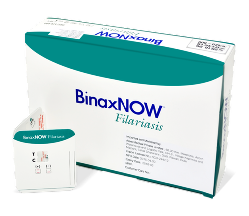 Figure: This test kit detects Wuchereria bancrofti antigen in whole blood, serum, or plasma.
Serological Tests
A wide variety of serological tests are available for diagnostic and epidemiological purposes, providing an alternative to microscopic detection of microfilariae. Patients with active filarial infection typically have elevated antifilarial IgG4, detectable using routine assays; detecting IgG4 specifically reduces cross-reactivity with non-filarial antibodies. IgG2 levels appear elevated specifically in patients with elephantiasis.
Limitation: except in patients not native to the area of endemicity, immunodiagnostic tests are of limited value, since lifelong exposure in endemic populations complicates interpretation.
PCR Amplification Methods
PCR assays are useful diagnostic tools as they can discriminate between past and present infection, monitor therapy response, and detect/differentiate multiple filarial infections. These procedures are currently often limited to research facilities.
Ultrasonography
Using 7.5 MHz or 10 MHz probes, ultrasonography can locate and visualise the movements of living adult W. bancrofti worms in the scrotal lymphatics of asymptomatic microfilaraemic males — the characteristic constant thrashing movements are described as the "filaria dance sign."
X-ray Diagnosis
Helpful in diagnosing tropical pulmonary eosinophilia, where chest imaging shows interstitial thickening and diffuse nodular mottling.
Haematology
Increased eosinophil count is typical; patients with tropical pulmonary eosinophilia show a marked IgE response in addition to elevated eosinophilia.
Treatment
Individual treatment of confirmed infection and mass drug administration (MDA) for population-level control use overlapping but distinct drug strategies:
- Diethylcarbamazine (DEC) combined with albendazole, or ivermectin combined with albendazole, are the standard MDA combinations used by the WHO's Global Programme to Eliminate Lymphatic Filariasis (GPELF), administered to entire at-risk populations regardless of individual infection status, since the goal is interrupting transmission by reducing the microfilarial reservoir available to mosquitoes
- Doxycycline has macrofilaricidal activity (it kills adult worms, unlike DEC and albendazole, which primarily target microfilariae) by depleting the worm's symbiotic Wolbachia bacteria, which the worm depends on — this is an important and increasingly used adjunct, particularly relevant given that adult worm survival is what perpetuates the cycle of lymphatic damage described in the pathogenesis section above
- Tropical pulmonary eosinophilia responds to DEC (2 mg/kg orally, three times daily for 14–21 days), though relapse occurs in up to 25% of patients, sometimes requiring repeat courses
- Surgical management has a role in advanced disease: nodal-venous shunt procedures can offer long-term benefit in extreme elephantiasis cases, and surgical management of massive hydrocele is sometimes performed, though recurrence is common
Morbidity management, separate from antifilarial drug treatment, is a major component of care for patients with established lymphoedema or elephantiasis — including hygiene measures to prevent the secondary bacterial infections that accelerate disease progression (as discussed in the pathogenesis section), since this self-reinforcing cycle of infection and lymphatic damage can be substantially slowed even when the underlying worm infection has already been treated.
How to Remember
Nocturnal periodicity: "Microfilariae work the night shift" — peak circulation between 10 pm and 4 am for classic W. bancrofti; this is why blood collection timing is the single most important practical detail in diagnosis, and why the DEC provocative test exists as a workaround.
Why damage is delayed: "It's the scar tissue, not the worm" — adult worms themselves are relatively well tolerated; it is the cumulative effect of repeated inflammatory episodes (ADLA) and subsequent lymphatic scarring, compounded over years, that produces irreversible elephantiasis. This is why early treatment, even in asymptomatic microfilaraemic individuals, can reverse early lymphatic damage.
ICT vs microscopy: "Antigen doesn't sleep" — unlike microfilariae, circulating filarial antigen does not show nocturnal periodicity, which is part of why the ICT antigen test can be performed at any time of day and remains the gold standard, including in amicrofilaraemic patients with clinical disease.
Where Students Actually Get Confused
1. "Elephantiasis appears soon after infection." The opposite is true, and this is the central teaching point of this article. Elephantiasis is a late, chronic-stage manifestation resulting from years of cumulative lymphatic damage from repeated acute inflammatory episodes — not a direct or immediate effect of the worm's presence. A person can be microfilaraemic and entirely asymptomatic for years before any visible chronic disease develops.
2. "A negative blood smear taken at midday rules out infection." For classic nocturnal periodic W. bancrofti, blood drawn outside the 10 pm–4 am window will substantially underestimate or miss circulating microfilariae. Either the timing must be corrected, the DEC provocative test used to bring microfilariae into daytime circulation, or an antigen-based test (ICT) used instead, since antigen detection does not depend on this nocturnal timing.
3. "QBC is the best test since it's the fastest." As this article's diagnosis section already states, QBC, despite being quick, is less sensitive than standard blood smear examination, and it requires a fluorescence microscope — a significant practical limitation in many field settings. Speed is not the same as diagnostic performance.
4. "Tropical pulmonary eosinophilia means the worms have invaded the lungs." TPE is a hypersensitivity reaction to microfilariae trapped in the pulmonary microcirculation, not a direct lung infection by adult worms (which remain in the lymphatics). This is an immunological phenomenon, which is also why it responds to anti-filarial treatment (DEC) rather than requiring a fundamentally different approach.
5. "DEC and albendazole kill the adult worms." DEC and albendazole, the standard MDA combination, primarily target microfilariae, reducing the reservoir available for mosquito transmission — they are not reliably macrofilaricidal. Doxycycline, by contrast, has genuine macrofilaricidal activity via its effect on the worm's symbiotic Wolbachia bacteria. This distinction matters because killing only microfilariae controls transmission at the population level without necessarily resolving an individual's ongoing lymphatic damage from surviving adult worms.
Key Exam Facts in One Table
| Fact | Detail | Memory hook |
|---|---|---|
| Causative organism | Wuchereria bancrofti (>90% of cases); also Brugia malayi, B. timori | "Bancroftian" filariasis |
| Vector | Culex (urban), Anopheles/Aedes (some rural/island settings) | Varies by region |
| Adult worm lifespan | 5–8 years | Long-lived — sustains chronic disease |
| Microfilariae periodicity | Nocturnal — peak 10 pm–4 am (classic); subperiodic strains exist | "Night shift" microfilariae |
| Microfilariae size | 240–300 μm (stained blood smear) | Sheathed |
| Optimal blood collection time | 10 pm–4 am | DEC provocative test if impractical |
| DEC provocative test dose/timing | 2 mg/kg; microfilariae appear in 30–45 min | Daytime workaround |
| QBC limitation | Less sensitive than blood smear; needs fluorescence microscope | Fast ≠ most sensitive |
| Gold standard diagnostic test | ICT (circulating filarial antigen) | Detects even amicrofilaraemic disease |
| IgG4 | Elevated in active infection | Reduces cross-reactivity with non-filarial antibodies |
| IgG2 | Elevated specifically in elephantiasis | |
| "Filaria dance sign" | Thrashing adult worm movement on scrotal ultrasound | 7.5–10 MHz probes |
| Acute manifestation | ADLA ("filarial fever") — recurs, resolves in ~1 week each episode | Repeated episodes → chronic damage |
| Chronic manifestations | Lymphoedema, elephantiasis, hydrocele, chyluria | Years of cumulative damage |
| TPE frequency | <1% of LF patients; male:female ~4:1 | Hypersensitivity, not direct lung infection |
| TPE treatment | DEC 2mg/kg TID for 14–21 days | Relapse in up to 25% |
| MDA drug combinations | DEC + albendazole, OR ivermectin + albendazole | WHO GPELF programme |
| Macrofilaricidal drug | Doxycycline (via Wolbachia depletion) | DEC/albendazole mainly target microfilariae, not adults |
| Why early treatment matters | Subclinical lymphatic damage can be REVERSIBLE | Even in asymptomatic microfilaraemic children |
Self-Check Questions
- A 30-year-old man from an endemic area has chronic leg lymphoedema but his blood smear, drawn at noon, is negative for microfilariae. Does this rule out W. bancrofti infection, and what test would be more appropriate at this stage of disease?
- Explain why elephantiasis develops only after years of infection rather than appearing soon after the initial mosquito bite.
- A patient with suspected tropical pulmonary eosinophilia asks whether the worms have invaded his lungs. How would you explain what is actually happening?
- Why is doxycycline increasingly used in filariasis treatment regimens alongside or instead of DEC and albendazole?
- A national filariasis elimination programme treats an entire village with DEC and albendazole, including many residents who have no symptoms and have never been tested. What is the rationale for treating asymptomatic, untested individuals?
- What is the "filaria dance sign," and in what diagnostic context is it observed?
Answers
- No — this does not rule out infection. A blood smear drawn at noon, outside the nocturnal periodicity window (10 pm–4 am) for classic W. bancrofti, will substantially underestimate or miss circulating microfilariae regardless of true infection status. Additionally, in chronic-stage disease with established lymphoedema, the patient may be amicrofilaraemic even with correct timing, since chronic disease can persist after microfilarial circulation has declined. The ICT antigen test, which does not depend on nocturnal timing and detects infection even in amicrofilaraemic patients with clinical manifestations, would be more appropriate at this stage.
- Elephantiasis results from cumulative lymphatic vessel damage caused by repeated acute inflammatory episodes (ADLA) and the host's immune response to the worms (and their Wolbachia symbionts) over years, rather than from any immediate or direct destructive effect of the worm's presence alone. Each acute episode contributes incremental scarring; it is the accumulation of this damage, compounded by recurrent secondary bacterial infections in lymphatically compromised tissue, that eventually produces the chronic, often irreversible swelling and skin changes characteristic of elephantiasis.
- Tropical pulmonary eosinophilia is a hypersensitivity (allergic) reaction to microfilariae trapped in the pulmonary microcirculation, not a direct infection or invasion of lung tissue by adult worms (which remain in the lymphatic system). The lung findings result from the immune system's response to the microfilariae, which is also why the condition responds to anti-filarial drug treatment (DEC) rather than requiring an entirely separate approach to lung infection.
- Doxycycline has macrofilaricidal activity — it kills adult worms by depleting their symbiotic Wolbachia bacteria, which the worms depend on for survival. This is a meaningful advantage over DEC and albendazole, which primarily target microfilariae and do not reliably kill adult worms; since adult worm survival perpetuates ongoing lymphatic damage, doxycycline addresses a problem that the standard MDA combination does not.
- The rationale is interrupting community-level transmission by reducing the microfilarial reservoir available for mosquitoes to pick up and transmit to others, regardless of whether any individual currently has symptoms. Since a large proportion of infected individuals are asymptomatic for years despite potentially having ongoing subclinical lymphatic damage (which can be reversible with treatment), mass treatment also offers a morbidity-prevention benefit beyond its primary transmission-control goal.
- The "filaria dance sign" refers to the constant thrashing movements of living adult Wuchereria bancrofti worms, visualised on scrotal ultrasonography (using 7.5 MHz or 10 MHz probes) in asymptomatic microfilaraemic males. It is observed specifically when imaging the scrotal lymphatics, where adult worm pairs commonly reside, and serves as a direct, non-invasive way to confirm the presence of living adult worms.
References
- Nuchprayoon, S. (2009). DNA-based diagnosis of lymphatic filariasis. The Southeast Asian Journal of Tropical Medicine and Public Health, 40(5), 904–913.
- Kapil, A. (1989). Laboratory diagnosis of lymphatic filariasis. Indian Journal of Pediatrics, 56(3), 314–317. https://doi.org/10.1007/BF02722290
- Mendoza, N., Li, A., Gill, A., & Tyring, S. (2009). Filariasis: diagnosis and treatment. Dermatologic Therapy, 22(6), 475–490. https://doi.org/10.1111/j.1529-8019.2009.01271.x
- World Health Organization. (2023). Lymphatic filariasis. WHO Fact Sheet. https://www.who.int/news-room/fact-sheets/detail/lymphatic-filariasis
- Babu, S., & Nutman, T. B. (2014). Immunology of lymphatic filariasis. Parasite Immunology, 36(8), 338–346. https://doi.org/10.1111/pim.12081
- Taylor, M. J., Hoerauf, A., Townson, S., Slatko, B. E., & Ward, S. A. (2014). Anti-Wolbachia drug discovery and development: safe macrofilaricides for onchocerciasis and lymphatic filariasis. Parasitology, 141(1), 119–127. https://doi.org/10.1017/S0031182013001108
- Vijayan, V. K. (2007). Tropical pulmonary eosinophilia: pathogenesis, diagnosis and management. Current Opinion in Pulmonary Medicine, 13(5), 428–433. https://doi.org/10.1097/MCP.0b013e328climate
- Babu, S., & Nutman, T. B. (2014). Filariasis: Lymphatic filariasis and Onchocerciasis. Annals of the New York Academy of Sciences — see also CDC DPDx: Lymphatic Filariasis. https://www.cdc.gov/dpdx/lymphaticfilariasis/index.html
Frequently Asked Questions
Why does elephantiasis develop years after Wuchereria bancrofti infection rather than immediately?
Why must blood for microfilaria detection be collected between 10pm and 4am?
What is the gold standard test for diagnosing Wuchereria bancrofti infection?
What is tropical pulmonary eosinophilia and how is it related to lymphatic filariasis?
Why is doxycycline used in lymphatic filariasis treatment alongside DEC and albendazole?

Tankeshwar Acharya, MSc (Medical Microbiology)
Tankeshwar Acharya is an Assistant Professor in the Department of Microbiology at Patan Academy of Health Sciences (PAHS), Nepal, where he has been teaching and practicing clinical microbiology for over 14 years. He is the founder of Microbe Online, one of the leading free microbiology education resources on the web, covering bacteriology, mycology, parasitology, immunology, and clinical laboratory diagnostics written from direct experience in both the classroom and the diagnostic laboratory.