Hydrogen Sulfide (H₂S) Production Test: Why the Same Organism Reads Positive on One Medium and Negative on Another
H₂S turns a medium black when the gas meets an iron or lead salt. But the same organism can read H₂S-positive on lead acetate paper and negative on TSI, because the methods differ enormously in sensitivity. Here are the two ways bacteria make H₂S, why lead acetate beats SIM beats TSI, and why acid in the TSI butt suppresses the reaction.
A stool culture grows a non-lactose fermenter that could be Salmonella. On the TSI tube, the butt is barely darkened — maybe a faint line at the stab, easy to read as negative. The technologist reports "H₂S negative," and the Salmonella line of reasoning stalls. But the same colony, tested with a lead acetate paper strip, blackens it unmistakably. Nothing about the organism changed. What changed is how sensitively each method looks for the gas.
That is the single most important idea in this test: a negative H₂S result means "negative on this medium," not "this organism makes no H₂S." Positivity is trustworthy — black is black. Negativity is only as good as the method's sensitivity. Once you understand how bacteria make H₂S and why the media differ in sensitivity, apparently contradictory results collapse into one sensible picture.
This is why H₂S is worth understanding as a concept, not just memorizing per medium. Once you know how bacteria make H₂S and why the detection methods differ in sensitivity, the apparently contradictory results resolve into a single, sensible picture. This article covers the two biochemical routes to H₂S, the indicator chemistry that makes the gas visible, and the sensitivity hierarchy that explains why the same organism reads differently on different media. For reading H₂S in a specific medium, it links to the TSI, KIA, and SIM articles, which own those readings.
Certain bacterial species liberate sulfur from sulfur-containing amino acids or other compounds in the form of H₂S. This ability of these bacteria can be used as an important characteristic of their identification.
Principle: two different ways bacteria make H₂S
Bacteria produce H₂S by two biochemically distinct routes, and the distinction matters for detection:
1. Cysteine desulfhydration (from an amino acid). The enzyme cysteine desulfhydrase removes the sulfhydryl (–SH) and amino groups from the amino acid cysteine, yielding H₂S, ammonia, and pyruvate. This is a putrefaction reaction, the source of the rotten-egg smell of decaying protein, and it does not require anaerobic conditions.
2. Thiosulfate reduction (anaerobic respiration). The enzyme thiosulfate reductase reduces thiosulfate (S₂O₃²⁻), using it as a terminal electron acceptor in anaerobic respiration, releasing H₂S. This requires reducing (anaerobic) conditions.
Both routes yield the same product, colorless H₂S gas. Because the gas is invisible, the medium contains a metal-salt indicator; usually an iron salt (ferrous ammonium sulfate or ferric ammonium citrate), or else lead acetate or a bismuth salt. The H₂S reacts with the metal ion to form an insoluble black precipitate (ferrous sulfide, lead sulfide, or bismuth sulfide), which blackens the medium. Black = H₂S produced.
Which sulfur source a medium supplies (cysteine, thiosulfate, or both) and which indicator it uses determine how sensitively it detects H₂S, which is the key to the sensitivity hierarchy below.
The sensitivity hierarchy: why methods disagree
Different H₂S detection methods have very different sensitivities. From most to least sensitive:
| Method | Relative sensitivity | Why |
|---|---|---|
| Lead acetate paper strip | Most sensitive (~10× the media) | Lead sulfide forms even from trace H₂S; the strip sits in the gas phase above the medium, catching gas that never reacts in the agar |
| SIM medium | More sensitive than TSI/KIA | Semisolid (traps gas near the growth), no interfering carbohydrates, peptonized iron indicator |
| TSI / KIA agar | Least sensitive | Carbohydrate fermentation acidifies the butt, and H₂S detection needs near-neutral pH; the acid suppresses the reaction. Ferrous sulfate indicator is less sensitive than peptonized iron |
Two consequences worth holding onto:
- A weak producer can read positive on lead acetate but negative on TSI. S. Typhi, a characteristically weak H₂S producer, is the classic case. This is not a contradiction; it is the sensitivity difference. Always interpret a "negative" H₂S in light of how sensitive the method was.
- On TSI/KIA, acid suppresses H₂S. Because H₂S needs a near-neutral pH to react with the iron and blacken, an organism that vigorously ferments the sugar and acidifies the butt can mask its own H₂S. This is why H₂S on TSI is read at the junction (where the pH is less acidic) and why a fully acidic butt can hide the reaction. (See the TSI and KIA articles for reading H₂S in those media.)
Media for the detection of Hydrogen Sulfide (H₂S)
Commonly used media for the detection of hydrogen sulfide production and the sources for sulfur and the sulfide indicators are as follows:
| Media | Sulfur source | H₂S indicator |
|---|---|---|
| Bismuth sulfite agar | Peptones plus sulfite | Bismuth sulfite / ferric citrate |
| Citrate sulfide agar | Sodium thiosulfate | Ferric ammonium citrate |
| Deoxycholate citrate agar (DCA) | Peptones | Ferric citrate |
| Lysine iron agar (LIA) | Sodium thiosulfate | Ferric ammonium citrate |
| Kligler iron agar (KIA) | Sodium thiosulfate | Ferrous sulfate |
| Triple sugar iron (TSI) agar | Sodium thiosulfate | Ferrous sulfate |
| Lead acetate agar | Sodium thiosulfate | Lead acetate |
| Salmonella-Shigella (SS) agar | Sodium thiosulfate | Ferric citrate |
| Sulfide-indole-motility (SIM) Medium | Sodium thiosulfate | Peptonized iron |
| Xylose-lysine-deoxycholate (XLD) agar medium | Sodium thiosulfate | Ferric ammonium citrate |
| Hektoen enteric agar | Sodium thiosulfate | Ferric ammonium citrate |
As various types of media are available for the detection of H₂S production with varying degrees of sensitivity, microbiologists can choose a specific detection system based on their needs and characteristics of the test isolate. For example, lead acetate, the most sensitive indicator, should be used whenever bacteria that produce only trace amounts of H₂S are tested.
Note: When incorporated in culture media, lead acetate may inhibit the growth of many fastidious bacteria so while testing, instead of incorporating it into the media, a lead acetate impregnated filter paper should be draped under the cap of a culture tube.
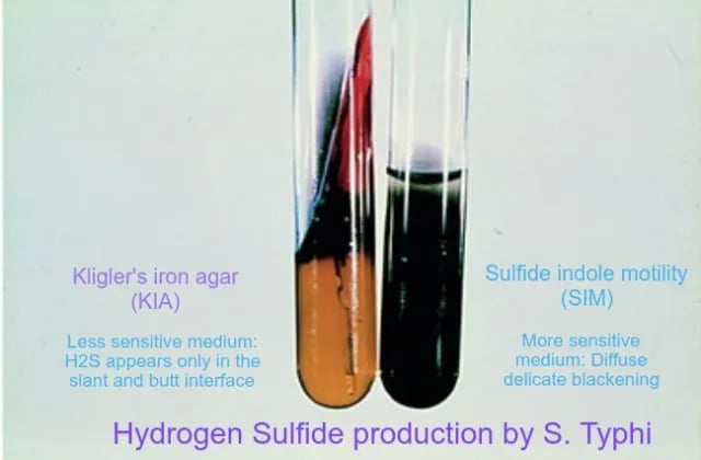 Figure: SIM is more sensitive in the detection of H₂S than either TSI or KIA, because of its semisolid nature, its lack of interfering carbohydrates, and the use of peptonized iron as an indicator.
As H₂S detected in one medium may not be detected in another, it is necessary to know the test system used when interpreting identification charts. In diagnostic microbiology, SIM, KIA, or TSI tubes are commonly used for the detection of H₂S production.
SIM is more sensitive than TSI and KIA. Its lack of fermentable carbohydrate means no competing acid, and its peptonized-iron indicator detects H₂S more readily, making SIM the better tube method when a weak producer is suspected.
With all H₂S detection systems, the endpoint is an insoluble, heavy metal sulfide, which produces a black precipitate in the medium or on the filter paper strip. Because hydrogen ions must be available for H₂S formation, the blackening is first seen in test media in which acid formation is maximal, that is, along the inoculating line, within the deeps of slanted agar media, or in the centers of colonies growing on agar surfaces.
Reading H₂S in different media
H₂S is read in several media, each covered in full in its own article. This article owns the concept; those own the medium-specific reading:
- Triple Sugar Iron (TSI) agar — H₂S read as blackening of the butt/junction, alongside sugar and gas reactions. See the TSI article.
- Kligler's Iron Agar (KIA) — similar to TSI without sucrose. See the KIA article.
- SIM medium — H₂S read alongside indole and motility; more sensitive than TSI/KIA. See the SIM article.
- Lead acetate paper — a strip suspended above the medium; the most sensitive method, used to catch weak producers.
- Other media — Cystine Trypticase Agar (CTA), bismuth sulfite agar, and Salmonella-Shigella (SS) agar also detect H₂S for specific purposes.
Procedure
- Tube Media
- Warm medium to room temperature and examine for cracks. Do not use if cracks appear.
- Using a sterile inoculating needle, touch the center of a well-isolated colony.
- Stab to within 3 to 5 mm from the bottom of the tube. Withdraw the needle.
- For KIA or TSI, streak the entire surface of the agar slant. Optional for fastidious organisms: add a strip of lead acetate paper to top of tube and hold in place with the cap of the tube so that it extends 1 in. into the tube.
- Place cap loosely on tube. Do not tighten the cap to allow for release of gas in the tube. Incubate aerobically at 35 to 37°C for 18 to 24 h.
- Observe for black precipitate indicating hydrogen sulfide production.
- If desired, extend incubation only to detect H₂S production. Campylobacters may take 3 days for production of H₂S.
- Plate Media
Streak plate so as to obtain isolated colonies. Incubate aerobically at 35 to 37°C Observe for blackened colonies
Result and Interpretation
- Positive reactions: H₂S production in tube media: black color throughout the medium, a black ring at the junction of the butt and slant, or any black precipitate in the butt. Blackening usually begins at the line of inoculation. H₂S production in plate media: black colonies surrounded by a brownish-black zone or metallic sheen Lead acetate paper: brownish-black coloration of the paper strip
- Negative reactions: H₂S production in tube media: no blackening in tube H₂S production in plate media: no blackening and no metallic-sheen colonies Lead acetate paper: no change in color of the strip
H₂S-producing and non-producing bacteria
Generally H₂S-positive:
- Salmonella (most serovars; S. Typhi weakly, S. Paratyphi A negative)
- Proteus species
- Citrobacter (species-dependent: C. freundii positive, C. koseri negative)
- Edwardsiella
- Some Klebsiella, Enterobacter (variable)
Generally H₂S-negative:
- Escherichia coli
- Shigella
- Klebsiella pneumoniae (typically)
- Yersinia, Serratia (typically)
The classic enteric use: H₂S separates the H₂S-positive Salmonella and Proteus from the H₂S-negative Shigella and E. coli. This is one of the most useful single discriminations in a stool workup. Note that "positive" and "negative" here are general tendencies; the actual result depends on the medium's sensitivity (see the hierarchy above) and the specific serovar.
Limitations
- H₂S can be masked on TSI/KIA when vigorous sugar fermentation acidifies the butt, because the iron-sulfide reaction needs a near-neutral pH. This suppresses the visible blackening, not the organism's H₂S production itself.
- Lead acetate is toxic to bacteria and may inhibit the growth of some bacteria.
How to remember
Two routes, one gas: chop or breathe. Bacteria make H₂S either by chopping the amino acid cysteine (cysteine desulfhydrase — the putrefaction, rotten-egg route) or by breathing thiosulfate as a terminal electron acceptor in anaerobic respiration (thiosulfate reductase). Two different enzymes, same colorless gas. The metal indicator is what makes it visible: H₂S + iron (or lead) → black metal sulfide. The black is the precipitate, never the gas.
Sensitivity ladder: lead acetate > SIM > TSI/KIA. Lead acetate paper sits in the gas phase above the medium and is roughly ten times more sensitive than the agars; SIM beats TSI and KIA because it is semisolid, carries no interfering sugars, and uses peptonized iron. So a weak producer — think S. Typhi — can be clearly positive on lead acetate, faint on SIM, and negative on TSI. A negative is only as trustworthy as the method that produced it.
Acid hides H₂S. The FeS reaction needs a near-neutral pH. An organism that vigorously ferments sugar acidifies the butt and can mask its own H₂S. That is why H₂S is read at the less-acidic junction, and why a fully acid butt can hide the reaction.
The H₂S-positive enterics — "Please Send Cats, Extra Kittens": Proteus, Salmonella, Citrobacter, Edwardsiella, some Klebsiella. The first four are the ones you separate from the reliably negative Shigella and E. coli in a stool workup; the "some Klebsiella" tail is the reminder that the list has soft edges.
Key exam facts in one table
| Question | Answer | The reason behind it |
|---|---|---|
| What does the test detect? | H₂S gas production | Seen as blackening from a metal-sulfide precipitate |
| Two mechanisms | Cysteine desulfhydration; thiosulfate reduction | Amino-acid route vs anaerobic-respiration route |
| Cysteine route enzyme | Cysteine desulfhydrase | Yields H₂S + ammonia + pyruvate; needs no anaerobic conditions |
| Thiosulfate route enzyme | Thiosulfate reductase | Thiosulfate as terminal electron acceptor; needs reducing conditions |
| Why the medium turns black | H₂S + iron/lead → insoluble metal sulfide | The gas itself is colorless |
| Common indicators | Ferrous/ferric iron salts, lead acetate, bismuth | Trap H₂S as a black precipitate |
| Most sensitive method | Lead acetate paper (about tenfold) | Gas-phase detection catches trace H₂S |
| More sensitive than TSI/KIA | SIM | Semisolid, no interfering sugars, peptonized iron |
| Least sensitive | TSI / KIA | Fermentation acid lowers pH and suppresses the FeS reaction |
| Why acid suppresses H₂S | The FeS reaction needs near-neutral pH | An acidic butt masks the reaction; read at the junction |
| Classic weak producer | S. Typhi | Positive on sensitive methods, weak/negative on TSI (faint line at the stab) |
| H₂S-negative typhoidal serovar | S. Paratyphi A | Important exception; gas weakly positive |
| High-H₂S typhoidal serovar | S. Paratyphi B | Abundant H₂S — the counterpoint to Paratyphi A |
| Species-split genus | Citrobacter | C. freundii positive, C. koseri negative |
| Enteric discrimination | Salmonella/Proteus (+) vs Shigella/E. coli (−) | High-yield stool-workup split |
| Where to read per-medium | TSI, KIA, SIM articles | This article owns the concept; they own the reading |
| Key principle | Negativity is method-dependent | A negative on a weak medium does not rule out a weak producer |
Where students get confused
Thinking H₂S is a fixed property of the organism. It is not fixed across methods. The same organism can read positive on a sensitive method and negative on a less sensitive one. H₂S positivity is always relative to the detection method, and a weak producer like S. Typhi is the classic trap.
Trusting a negative on a low-sensitivity medium. A negative H₂S on TSI does not rule out a weak producer, because TSI is among the least sensitive methods. When it matters, confirm with SIM or lead acetate paper. Positivity is reliable; negativity is method-dependent.
Forgetting that acid suppresses H₂S on TSI/KIA. The FeS reaction needs a near-neutral pH. An organism that vigorously ferments the sugar acidifies the butt and can mask its own H₂S. This is why H₂S is read at the less-acidic junction, and why a fully acid butt can hide the reaction. (The TSI and KIA articles cover this reading in detail.)
Assuming all typhoidal Salmonella behave alike on H₂S. They do not. S. Typhi is a weak producer (faint line), S. Paratyphi A is H₂S-negative, and S. Paratyphi B produces H₂S abundantly. Reading H₂S as if the whole group were uniform is a common exam and bench error.
Confusing the black of H₂S with other dark reactions. The black here is a metal sulfide from H₂S. It is not the same as the black of bile esculin (esculetin + iron) or the darkening in some other tests. Context and medium tell you which reaction you are reading.
Reading the two mechanisms as interchangeable. Cysteine desulfhydration (amino-acid route) and thiosulfate reduction (anaerobic-respiration route) are biochemically distinct, and a medium's sulfur source determines which it detects. Cysteine is attacked more readily than thiosulfate, which is part of why cysteine-containing sensitive methods detect more organisms.
Over-reading H₂S as an identification. H₂S production is one characteristic among many. It contributes to enteric identification (notably the Salmonella/Shigella split) but is read alongside the rest of the biochemical panel, not as a standalone ID.
References and further reading
- Skarnes RC, Watson DW. Hydrogen sulphide production by bacteria. J Gen Microbiol. 1953;8(3):397–407. doi:10.1099/00221287-8-3-397
- Procop GW, Church DL, Hall GS, Janda WM, Koneman EW, Schreckenberger PC, Woods GL. Koneman's Color Atlas and Textbook of Diagnostic Microbiology. 7th ed. Philadelphia: Wolters Kluwer; 2017.
- Tille PM. Bailey and Scott's Diagnostic Microbiology. 15th ed. St. Louis: Elsevier; 2022.
- Leber AL, editor. Clinical Microbiology Procedures Handbook. 4th ed. Washington, DC: ASM Press; 2016. doi:10.1128/9781683670438
Frequently Asked Questions
Why is Salmonella Typhi H₂S positive on SIM but negative on TSI?
What are the two ways bacteria produce H₂S?
Why does the medium turn black in a positive H₂S test?
Which is the most sensitive method for detecting H₂S?
Are all typhoidal Salmonella serovars the same on the H₂S test?
Why is H₂S read at the junction of a TSI tube rather than in the butt?

Tankeshwar Acharya, MSc (Medical Microbiology)
Tankeshwar Acharya is an Assistant Professor in the Department of Microbiology at Patan Academy of Health Sciences (PAHS), Nepal, where he has been teaching and practicing clinical microbiology for over 14 years. He is the founder of Microbe Online, one of the leading free microbiology education resources on the web, covering bacteriology, mycology, parasitology, immunology, and clinical laboratory diagnostics written from direct experience in both the classroom and the diagnostic laboratory.