
Gelatin Hydrolysis Test: Why You Have to Put It in the Fridge to Read It

On this page
The enzyme that eats connective tissue
When a wound infection spreads faster than it should, tracking along tissue planes and refusing to stay walled off, one of the tools the bacterium is using is an enzyme that dissolves collagen.
Collagen is the structural protein of connective tissue. It is the mesh that holds tissue together and, from the host's point of view, the fence that is supposed to keep an infection local. Gelatin is what you get when you boil collagen: the same protein, denatured into the substance that sets a dessert. An organism that can hydrolyze gelatin can hydrolyze collagen, and an organism that can hydrolyze collagen can take the host's fence apart.
This is why gelatinase is not merely an identification marker. It is a virulence factor. Pseudomonas aeruginosa, Serratia, Proteus, and pathogenic Staphylococcus aureus all produce it. Their non-invasive relatives often do not, which is exactly why the test helps separate S. aureus from S. epidermidis.
The gelatin hydrolysis test puts that enzyme in a tube. You give the organism a tube of set gelatin and ask whether it can turn the solid back into liquid. But there is a catch that trips up nearly everyone who runs it for the first time, and it has to do with temperature. This article is about reading the test correctly despite that catch, and about the enzyme that makes it matter in the first place.
Gelatin is a protein derived from the animal protein collagen-a component of vertebrate connective tissue. It has been used as a solidifying agent in food for a long time.
Organisms producing proteolytic enzymes called gelatinases hydrolyze gelatin into polypeptides and then into individual amino acids, which the cell absorbs and uses. In the process, gelatin loses its structure and becomes liquid. That liquefaction is what the test detects, and because the same enzyme degrades the collagen of host connective tissue, gelatinase doubles as a virulence factor in several invasive pathogens.
Principle of Gelatin hydrolysis test
A gelatin hydrolysis test is used to detect the ability of an organism to produce gelatinases that liquefy gelatin. This process takes place in two sequential reactions. In the first reaction, gelatinases degrade gelatin to polypeptides. Then, the polypeptides are further converted into amino acids. The bacterial cells can then take up these amino acids and use them in their metabolic processes.
Why gelatinase matters: nutrition and virulence in one enzyme
Gelatin is a large protein. It is far too big to cross the bacterial cell membrane, so an organism cannot simply import and eat it. To use gelatin as a nutrient, the organism must break it down outside the cell first, then take up the small fragments. That is what an exoenzyme does: it is secreted into the surrounding medium and works on a substrate the cell cannot bring inside.
Gelatinase is that exoenzyme, and it works in two steps: gelatin to polypeptides, then polypeptides to absorbable amino acids. The bacterium secretes the enzyme, waits for the extracellular digestion, and imports the pieces.
The same activity has a second face. The collagen that gelatinase evolved to digest is also the collagen that structures host tissue. In an invasive infection, a gelatinase-producing organism is degrading connective tissue, dismantling the barriers that would otherwise contain it, and easing its own spread. This is why gelatinase production correlates with pathogenicity, and why the test's ability to separate Staphylococcus aureus (gelatinase positive, invasive) from Staphylococcus epidermidis (gelatinase negative, usually commensal) reflects a real difference in what the two organisms can do to a host, not just a convenient lab marker.
So the tube result carries two meanings at once: this organism can feed on protein it cannot import, and this organism can take apart the tissue that is supposed to hold you together.
Quality Control
- Inspect gelatin agar tubes for contamination, dehydration, and lack of liquefaction at refrigeration temperatures before storage and before use.
- Perform QC on each new lot of media prior to using them.
- Use an uninoculated control with each use.
- Organisms
- Pseudomonas aeruginosa ATCC 10145: liquefaction (positive)
- Escherichia coli ATCC 25922: no liquefaction (negative)
Procedure of Gelatin hydrolysis test
There are several methods for determining gelatinase production, all of which make use of gelatin as the substrate. The standard and most commonly employed method is the nutrient gelatin stab method.
- Inoculate a heavy inoculum of test bacteria (18- to 24-hour-old) by stabbing 4-5 times (half an inch) on the tube containing nutrient gelatin medium.
- Incubate the inoculated tube along with an uninoculated medium at 35°C, or at the test bacterium’s optimal growth temperature, for 48 hours.
- Gently remove the tubes daily from the incubator and place them in an ice bath or refrigerator (4°C) for 30 minutes or until the control tube solidifies.(Gelatin normally liquefies at 28°C and above, so to confirm that liquefaction was due to gelatinase activity, the tubes are immersed in an ice bath or kept in a refrigerator at 4°C).
- After 30 minutes of refrigeration, tilt the tubes gently to observe liquefaction by the test organism.
- Reincubate a negative test for up to 2 weeks if indicated by the nature of the organism, and examine at regular intervals.
This is a slow test.
Strong gelatinase producers may liquefy the medium within 48 hours, but weak producers can take up to two weeks, with daily chilling and reading along the way. A negative should not be called until the recommended incubation has fully elapsed. In a modern lab this slowness is why gelatin hydrolysis has largely been replaced by faster tests and by automated identification panels.
Why you have to chill the tube to read it
Here is the catch that makes this test different from almost every other biochemical tube. Gelatin is solid only when cold. It liquefies on its own at about 28°C and above, with no organism and no enzyme involved. A nutrient gelatin tube incubated at 35°C is therefore liquid no matter what, whether the organism produced gelatinase or not.
If you read the tube straight out of the incubator, every tube looks positive. The incubator temperature alone melted the gelatin.
So the test is read the other way around. After incubation, you chill the tube in an ice bath or refrigerator at 4°C for 30 minutes, alongside an uninoculated control tube.
- The control always re-solidifies in the cold. That is your proof the gelatin can still gel.
- A tube that re-solidifies with it was never hydrolyzed. The gelatin is intact; the cold set it firm again. Negative.
- A tube that stays liquid in the cold cannot re-gel because the gelatin protein has been chemically taken apart by gelatinase. Nothing left to set. Positive.
The rule: read the test cold, never hot, and always chill an uninoculated control beside it. The control is not optional. Gelatin varies batch to batch in how firmly it gels, and the control is what tells you the medium was capable of setting at all. Without it, a tube that stays liquid could mean gelatinase, or could just mean weak gelatin.
One more consequence: do not shake or agitate a warm tube before chilling. Gelatinase acts mainly at the surface, and swirling the warm, already-liquid tube can disperse partially hydrolyzed gelatin in a way that reads as a false negative once chilled. Move the tubes gently.
Expected results
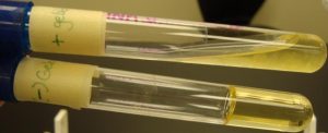 Figure: Gelatin Hydrolysis Test: Above tube: Positive; Below Tube: Negative
| Species | Growth | Liquefaction |
|---|---|---|
| Bacillus subtilis | + | + |
| Pseudomonas aeruginosa | + | + |
| Clostridium perfringens | + | + |
| Escherichia coli | + | – |
| Proteus vulgaris | + | + |
| Serratia liquefaciens | + | + |
| Staphylococcus aureus | + | + |
Positive: Partial or total liquefaction of the inoculated tube (uninoculated control medium must be completely solidified) even after exposure to the cold temperature of ice bath or refrigerator (4°C).
Negative: Complete solidification of the inoculated tube even after exposure to the cold temperature of an ice bath or refrigerator (4°C)
Common bacteria and their reactions to the gelatin hydrolysis test performed on nutrient gelatin.
Uses of Gelatin Hydrolysis Test
Gelatinase production is useful as a taxonomic aid in the classification and identification of both Gram-negative rods and Gram-positive rods.
- Gelatin liquefaction test can also be used to differentiate genera of gelatinase-producing bacteria such Serratia and Proteus from other members of the family Enterobacteriaceae. Proteus spp. and Serratia spp. are usually gelatinase positive.
- Gelatin hydrolysis test is helpful in identifying and differentiating species of Bacillus, Clostridium, and, Pseudomonas. Gram-positive, spore-forming, rod-shaped, aerobic, or anaerobic bacteria such as Bacillus anthracis, Bacillus cereus, Bacillus subtilis, Clostridium perfringens, and Clostridium tetani, are also positive for gelatin hydrolysis. P. fluorescens is gelatinase positive, but P. putida is gelatinase negative.
- It distinguishes the gelatinase-positive, pathogenic Staphylococcus aureus from the gelatinase-negative, non-pathogenic S. epidermidis.
Limitations
- Gelatinase usually acts at the surface of the tube medium. Shaking the tube while it is warm may result in a false-negative interpretation.
- Gelatin may vary in its gelling ability; therefore, incubate an uninoculated control with the test. The control must be refrigerated along with the test, prior to reading.
How to remember
Hot lies, cold tells the truth.
A warm gelatin tube is always liquid, so a hot reading tells you nothing. Only the cold reading is honest: if it stays liquid in the fridge, the enzyme did it; if it sets, it did not. Whenever you pick up a gelatin tube, ask yourself: am I reading this cold? If not, you are about to call every tube positive.
The control is the truth-teller.
The uninoculated tube chilled beside the test is the whole test's honesty check. It must re-solidify. If it does, then any test tube that stayed liquid did so because of the organism, not because of weak gelatin. No control, no valid result.
Gelatinase is a demolition enzyme.
Collagen holds tissue together; gelatin is boiled collagen; gelatinase dissolves both. Picture the positive organisms, Pseudomonas, Serratia, Proteus, invasive S. aureus, as the ones equipped to knock down the host's walls. The commensals that stay put, like S. epidermidis, are the ones without the wrecking ball. That image is also the reason the test separates them.
Key exam facts
| Question | Answer | The reason behind it |
|---|---|---|
| What does the test detect? | Production of gelatinase, an exoenzyme that hydrolyzes gelatin | Gelatin is too large to import; it must be digested outside the cell |
| What is gelatin? | Denatured collagen, the connective-tissue protein | Boiling collagen yields gelatin |
| Two-step reaction | Gelatin → polypeptides → amino acids | The cell absorbs the small amino acids |
| Why is gelatinase a virulence factor? | It degrades host collagen, dismantling connective-tissue barriers | Same enzyme, nutrition and invasion |
| Medium | Nutrient gelatin (peptone, beef extract, gelatin) | Gelatin is both substrate and solidifying agent |
| Method | Stab inoculation, incubate, then chill to read | Gelatinase acts at the surface; cold reveals hydrolysis |
| Why read cold? | Gelatin liquefies at 28°C and above on its own | A warm tube is liquid regardless of the organism |
| Positive result | Stays liquid after chilling to 4°C | Gelatin protein destroyed; cannot re-gel |
| Negative result | Re-solidifies on chilling | Gelatin intact; cold sets it |
| Why an uninoculated control? | Gelatin gel strength varies by batch | The control proves the medium can still set |
| Why not shake the warm tube? | Can disperse partial hydrolysis and read false negative | Gelatinase acts mainly at the surface |
| How long? | 48 hours to 2 weeks | Weak producers are slow; do not call negative early |
| Key separation | S. aureus (positive) vs S. epidermidis (negative) | Reflects a real difference in invasiveness |
| Other positives | Proteus, Serratia, Bacillus, Clostridium perfringens, C. tetani | Broad among proteolytic organisms |
Where students get confused
Reading the tube straight out of the incubator. The defining mistake. At 35°C the gelatin is liquid whether or not the organism produced anything. Every tube looks positive. The result only exists after you chill the tube back down. Read cold, never hot.
Skipping the uninoculated control. Gelatin gels with different firmness from batch to batch. If you do not chill a control tube alongside the test, a tube that stays liquid is ambiguous: it could be gelatinase, or it could be a batch of gelatin that simply gels poorly. The control, which must re-solidify, is what removes that doubt. It is not optional.
Getting the liquid/solid logic backwards. A negative tube re-solidifies; a positive tube stays liquid. Students sometimes reason that "solid gelatin" sounds like a positive reaction because something is visibly there. It is the reverse. Liquid that will not re-set is the positive, because the protein has been destroyed.
Calling a negative too early. This is a slow test. Some organisms take up to two weeks. A tube that is still solid at 48 hours is not necessarily negative; it may just be a weak producer. Follow the full incubation before recording a negative.
Shaking or tilting the warm tube roughly. Gelatinase works mostly at the surface of the medium. Agitating the warm, liquefied tube before chilling can disperse partially hydrolyzed gelatin and produce a false negative. Handle gently, chill, then tilt once to read.
Treating gelatinase as only an ID marker. The reason the test separates pathogenic from commensal species is that gelatinase is a genuine virulence factor. It is the same enzyme activity that lets an organism degrade host connective tissue. That is the point worth remembering, not just the color of the tube.
References and further readings
- Tille PM. Bailey and Scott's Diagnostic Microbiology. 15th ed. St. Louis: Elsevier; 2022.
- Procop GW, Church DL, Hall GS, Janda WM, Koneman EW, Schreckenberger PC, Woods GL. Koneman's Color Atlas and Textbook of Diagnostic Microbiology. 7th ed. Philadelphia: Wolters Kluwer; 2017.
- Leber AL, editor. Clinical Microbiology Procedures Handbook. 4th ed. Washington, DC: ASM Press; 2016. doi:10.1128/9781555818814
- MacFaddin JF. Biochemical Tests for Identification of Medical Bacteria. 3rd ed. Philadelphia: Lippincott Williams & Wilkins; 2000.
Frequently Asked Questions
Why do I have to refrigerate the gelatin tube to read the result?
Is a solid tube positive or negative?
Why do I need an uninoculated control tube?
How long does the gelatin hydrolysis test take?
Why is gelatinase considered a virulence factor?
Because the collagen that gelatinase digests is the structural protein of host connective tissue. Gelatin is simply denatured collagen. An organism that hydrolyzes gelatin can hydrolyze collagen, degrading the connective-tissue barriers that would otherwise contain an infection and easing its own spread. This is why gelatinase production correlates with invasiveness, and why the test helps separate pathogenic Staphylococcus aureus, which is positive, from commensal Staphylococcus epidermidis, which is negative.
Why shouldn't I shake the warm gelatin tube?
Which organisms are gelatinase positive?
Common positives include Pseudomonas aeruginosa, Proteus species, Serratia species, Bacillus species including B. anthracis, B. cereus and B. subtilis, and Clostridium perfringens and C. tetani. Among staphylococci, pathogenic Staphylococcus aureus is positive while commensal Staphylococcus epidermidis is negative. Within the fluorescent pseudomonads, P. fluorescens is positive but P. putida is negative. Escherichia coli is negative.

Tankeshwar Acharya, MSc (Medical Microbiology)
Tankeshwar Acharya is an Assistant Professor in the Department of Microbiology at Patan Academy of Health Sciences (PAHS), Nepal, where he has been teaching and practicing clinical microbiology for over 14 years. He is the founder of Microbe Online, one of the leading free microbiology education resources on the web, covering bacteriology, mycology, parasitology, immunology, and clinical laboratory diagnostics written from direct experience in both the classroom and the diagnostic laboratory.
Comments
No comments yet. Be the first to share your thoughts.
Leave a comment
All comments are reviewed before they appear.