
Entamoeba histolytica: Life Cycle, Pathogenesis, Liver Abscess, and Laboratory Diagnosis
How does a swallowed cyst cause both bloody dysentery and a liver abscess months later? Complete Entamoeba histolytica life cycle, flask-shaped ulcer mechanism, and lab diagnosis including how to tell it apart from E. dispar and E. coli.

On this page
A 34-year-old man returns from working in an area with poor sanitation, presenting six weeks later with right upper quadrant pain, fever, and a tender, enlarged liver. He has no history of diarrhea at all. An ultrasound reveals a large abscess in the right lobe of the liver. Aspiration yields thick, brownish-yellow pus with the consistency of anchovy paste.
This presentation is deceptive precisely because, on the surface, it has nothing to do with the intestine, yet it began there. Entamoeba histolytica, ingested as a microscopic cyst in contaminated food or water, can travel silently from the gut to the liver via the portal circulation, causing a destructive abscess in a patient who never had the bloody diarrhea. In fact, in roughly half of liver abscess cases, there is no preceding history of dysentery at all.
Understanding the complete journey of this organism, from cyst to trophozoite to tissue invasion to distant organ spread, is what allows a clinician to recognize amoebiasis in its many disguises.
Entamoeba histolytica is an enteric protozoan parasite with worldwide distribution. It is responsible for amoebic dysentery (bloody diarrhea) and invasive extraintestinal amebiasis (such as liver abscess, peritonitis, and pleuropulmonary abscess). Other species of Entamoeba; Entamoeba hartmanni, Entamoeba coli, and Entamoeba dispar are generally considered non-pathogenic but their trophozoite is difficult to distinguish from those of E. histolytica by light microscopy.
Mode of transmission
Feco-oral route, via the ingestion of contaminated food or water containing mature quadrinucleate cyst of Entamoeba histolytica. Trophozoites if ingested would not survive exposure to the gastric environment.
Mnemonic: EntAmoeba HistoLytica
- Ent: enterocytes (a cell of the intestinal lining);
- Amoeba: protozoa;
- Histo: tissue;
- Lytica: lysis.
Why the Mnemonic Is the Whole Disease in Four Syllables
The mnemonic above is not just a memory device, it is literally the pathophysiology of every clinical manifestation. Histo-lytica (tissue lysis) is the mechanism behind:
- Amoebic dysentery: lysis of colonic mucosa produces the flask-shaped ulcer and the resulting blood and mucus in stool
- Liver abscess: the same cytolytic action, carried by trophozoites to the liver, destroys hepatocytes and produces the anchovy-paste pus
- Ameboma: even the granulomatous mass that can mimic colon cancer is a consequence of chronic tissue destruction and the host's reparative response
Every destructive lesion this organism causes, wherever in the body it occurs, traces back to the same enzymatic tissue-lysing capability. The organism does not have a separate mechanism for the liver and for the gut. It is the same lytic process happening in a different location.
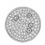 Figure: Quadrinucleate cyst of Entamoeba histolytica
Infective form: Mature quadrinucleate cyst; it is spherical in shape with a refractile wall
Note: Giardia lamblia cyst also has four nuclei, but the cyst is oval in shape.
Geographical distribution: Worldwide, more common in the tropics and subtropics, especially in areas with poor sanitation (developing and under-developed countries).
Habitat: Trophozoites of E. histolytica live in the mucosal and submucosal layers of the large intestine of man. The life cycle of Entamoeba histolytica has two stages: the motile trophozoite and the non-motile cyst. Trophozoites are found in intestinal lesions, extra-intestinal lesions, and diarrheal stools whereas cyst predominates in non-diarrheal stools.
Morphology of Entamoeba histolytica
Entamoeba histolytica exists in two forms: the trophozoite, the active, motile, feeding stage that invades tissue and causes disease, and the cyst, the dormant, infective stage that transmits the infection. The trophozoite causes the disease; the cyst spreads it.
The trophozoite has a single nucleus with a small central karyosome and fine, evenly distributed chromatin along the nuclear membrane. This even chromatin and central karyosome distinguish it from the nuclei of the non-pathogenic amoebae. It moves by extending finger-like pseudopodia and, when actively invading tissue, characteristically contains ingested red blood cells, a feature that signals active disease.
The mature cyst is spherical with a refractile wall and contains four nuclei, which is why it is called the quadrinucleate cyst. This four-nucleus mature cyst is the infective form and the diagnostic hallmark of the species. Immature cysts may contain one or two nuclei and may show chromatoid bodies with rounded ends.
| Feature | Trophozoite | Cyst |
|---|---|---|
| Role | Active, invasive stage; causes disease | Dormant, infective stage; transmits disease |
| Motility | Motile, with pseudopodia | Non-motile |
| Nuclei | One | Four in the mature cyst (quadrinucleate) |
| Shape | Irregular, changing | Spherical, with a refractile wall |
| Nucleus detail | Small central karyosome; fine, even peripheral chromatin | Same nuclear pattern, in each of the four nuclei |
| Ingested red blood cells | Present during active tissue invasion | Absent |
| Found in | Diarrheal stools, intestinal and extraintestinal lesions | Formed, non-diarrheal stools |
| Survival outside the body | Rapidly destroyed | Survives days to weeks |
The single most reliable identifying features are the spherical quadrinucleate cyst and the trophozoite containing ingested red blood cells, the latter being the finding that confirms active tissue invasion.
Life cycle of Entamoeba histolytica
Infection by Entamoeba histolytica occurs by the ingestion of mature quadrinucleate cysts in fecally contaminated food, water, or hands. The quadrinucleate cyst is resistant to the gastric environment and passes unaltered through the stomach
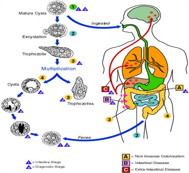 Figure: Life Cycle of Entamoeba histolytica (Source: CDC)
- When the cyst of E. histolytica reaches caecum or lower part of ileum excystation occurs and an amoeba with four nuclei emerges and that divides by binary fission to form eight trophozoites.
- Trophozoites migrate to the large intestine and lodge into the submucosal tissue.
- Trophozoites grow and multiply by binary fission in the large intestine (Trophozoite phase of the life cycle is responsible for producing characteristics lesion of amoebiasis).
- Certain numbers of trophozoites are discharged into the lumen of the bowel and are transformed into cystic forms.
- The cysts thus formed are unable to develop in the same host and therefore necessitate a transference to another susceptible host. The cysts are passed in the feces.
Note: Because of the protection conferred by their walls, the cysts can survive days to weeks in the external environment. Cysts are not highly resistant and are readily killed by boiling. But they are resistant to chlorination or can be removed by filtration. Trophozoites can also be passed in diarrheal stools, but are rapidly destroyed once outside the body.
Trophozoites are responsible for disease conditions:
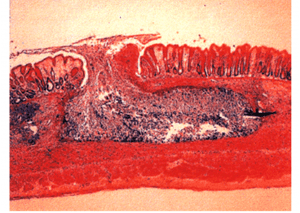 Figure: Flask Shaped Ulcer (Histopathology, UFPA, Araujo R.)
- The trophozoites invade the colonic epithelium and secrete enzymes that cause localized necrosis. Little inflammation occurs at the site.
- As the lesion reaches the muscularis layer, a typical “flask-shaped” ulcer forms, which can undermine and destroy large areas of the intestinal epithelium.
- Progression into submucosa leads to invasion of the portal circulation by the trophozoites.
Diseases
- Non-invasive infection: In many cases, the trophozoites remain confined to the intestinal lumen of individuals who are thus asymptomatic carriers and cysts passers.
- Intestinal disease: In some patients, the trophozoites invade the intestinal mucosa, producing amoebic colitis and dysentery.
- Extra-intestinal disease: through the bloodstream, trophozoites invade extraintestinal sites such as the liver, brain, and lungs, with resultant pathologic manifestations.
Amoebic liver abscess
- About 2-10% of individuals infected with E. histolytica suffer from hepatic complications.
- In 50% of cases, history of amoebic dysentery may not be seen.
- The trophozoites of E. histolytica are carried as emboli by the radicles of the portal vein from the base of amoebic ulcer in the large intestine.
- Capillary system of the liver acts as an efficient filter and holds the trophozoites, which multiply and carry out the same cytolytic destruction of hepatocytes.
- This leads to obstruction to the circulation and produces thrombosis of the portal venules (sinusoids) resulting in anemic necrosis of the liver cells
- Progressive destruction of concentric layers of liver cells occurs. A large-sized abscess is formed by the coalescence of miliary abscesses.
- Amebic abscess of the liver is characterized by right upper quadrant pain, weight loss, fever, and a tender enlarged liver.
- Right-lobe abscesses can penetrate the diaphragm and cause lung disease (pulmonary amoebiasis)
- Other metastatic lesions: Cerebral amoebiasis, Amoebic pericarditis, Cutaneous amoebiasis, Splenic abscess etc.
- Aspiration of the liver abscess yields brownish-yellow pus with the consistency of anchovy-paste.
Clinical Findings
- Acute intestinal amebiasis dysentery (i.e. bloody, mucus containing diarrhea), lower abdominal discomfort, flatulence.
- Chronic amebiasis: low-grade symptoms such as occasional diarrhea, weight loss, and fatigue also occurs.
- Roughly 90% of infected individuals have an asymptomatic infection but they may be carriers.
- Ameboma, a granulomatous lesion may form in the cecal or rectosigmoid areas of the colon in some patients. These lesions resemble an adenocarcinoma of the colon and must be distinguished from them.
Laboratory diagnosis
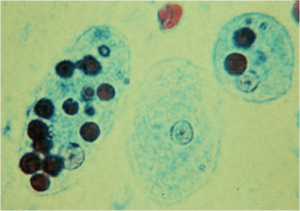 Figure: E. histolytica with ingested RBCs
Diagnosis of intestinal amebiasis rests on finding either trophozoites in diarrheal stools or cysts in formed stools. Diarrheal stools should be examined within one hour of collection to see the ameboid motility of the trophozoite. The trophozoite characteristically contains ingested red blood cells.
Characteristics of Stool
- Macroscopic appearance of stool: Offensive dark brown semisolid stool, acid in reaction, admixed with blood, mucus, and much fecal matter.
- General microscopic examination: Presence of Charcot-Leyden crystals. Scanty cellular exudates, and consists of only the nuclear masses (pyknotic bodies). E. histolytica infection is distinguished from bacillary dysentery by the lack of high fever and the absence of PMN leukocytosis.
A. Microscopy
E. histolytica can be distinguished from other amoebas by two major criteria
- The nature of the nucleus. As described in the morphology section, the Entamoeba histolytica nucleus has a small central karyosome and fine, even peripheral chromatin, a pattern that differs from the nuclei of the other amoebae.
Note: The trophozoites of Entamoeba dispar, a nonpathogenic species of Entamoeba, are morphologically indistinguishable from those of E. histolytica - Cyst size and the number of its nuclei. Mature cysts of E. histolytica are smaller and contain four nuclei, whereas E. coli cysts are larger and contain eight nuclei (occasionally up to 16). Any Entamoeba cyst with more than four nuclei is E. coli, not E. histolytica.
B. Antigen Detection
Stool antigen detection tests (typically ELISA-based) detect E. histolytica-specific antigens, most notably the Gal/GalNAc lectin. This is a critical advance over light microscopy because antigen detection can distinguish E. histolytica from the non-pathogenic E. dispar. This makes antigen detection the preferred confirmatory method when microscopy reveals amoebic cysts or trophozoites of uncertain pathogenicity.
C. Serologic Testing
Serology (typically ELISA or indirect hemagglutination, detecting anti-amoebic antibodies) is most useful for invasive disease, particularly amoebic liver abscess, where stool examination is frequently negative (since the trophozoites have left the intestine) and where antigen detection sensitivity is lower than in intestinal disease.
Serology is positive in the large majority of liver abscess cases, making it a key diagnostic tool when imaging suggests a hepatic abscess but the cause is not yet confirmed.
Limitation: Antibodies can persist for years after infection clears, so a positive serology in an endemic-area patient does not necessarily indicate current active disease.
D. Molecular Methods (PCR)
PCR-based assays detect E. histolytica nucleic acid directly from stool, tissue, or aspirate samples, offering high sensitivity and specificity, and like antigen detection, the ability to distinguish E. histolytica from E. dispar and E. moshkovskii definitively.
PCR is increasingly used in reference laboratories and is particularly valuable in research and outbreak settings, though it remains less widely available than microscopy or antigen testing in resource-limited settings.
Differentiating Entamoeba histolytica from Other Intestinal Amoebae
Several non-pathogenic amoebae live in the human intestine and are seen in stool, and they must be told apart from Entamoeba histolytica so that a harmless commensal is not mistaken for the pathogen. The most important is Entamoeba coli, a common commensal whose cyst is frequently seen on stool microscopy.
The mature cyst is the most useful stage for this distinction. The mature cyst of Entamoeba histolytica is smaller and contains four nuclei, whereas the mature cyst of Entamoeba coli is larger and contains eight nuclei, occasionally up to sixteen. A simple and reliable rule follows: any Entamoeba cyst with more than four nuclei is Entamoeba coli, not Entamoeba histolytica. The nuclear detail also differs: Entamoeba histolytica has a small central karyosome with fine, even peripheral chromatin, while Entamoeba coli has a larger, eccentric (off-center) karyosome with coarse, unevenly clumped peripheral chromatin.
| Feature | Entamoeba histolytica | Entamoeba coli |
|---|---|---|
| Pathogenicity | Pathogenic | Non-pathogenic commensal |
| Mature cyst size | Smaller | Larger |
| Nuclei in mature cyst | Four | Eight (occasionally up to sixteen) |
| Karyosome position | Small, central | Larger, eccentric (off-center) |
| Peripheral chromatin | Fine and evenly distributed | Coarse and unevenly clumped |
| Chromatoid bodies (cyst) | Rounded, blunt ends | Splinter-like, pointed ends |
| Ingested red blood cells (trophozoite) | Present during invasion | Absent |
Two further species complete the picture. Entamoeba dispar is genetically distinct and non-pathogenic, but its trophozoites and cysts are morphologically identical to those of Entamoeba histolytica, so they cannot be separated by light microscopy and require antigen detection or PCR to tell apart. Entamoeba hartmanni resembles Entamoeba histolytica but is smaller, and careful measurement of size is used to distinguish it.
Treatment
Treatment depends on whether the infection is luminal (asymptomatic cyst passage) or invasive (intestinal or extra-intestinal disease). This distinction matters because the two drug classes used have different targets:
| Disease state | Treatment | Why |
|---|---|---|
| Asymptomatic cyst passer (luminal infection only) | Luminal agent e.g., diloxanide furoate or paromomycin | Clears intestinal colonization; tissue-acting drugs are not needed since there is no invasion |
| Invasive intestinal disease (amoebic dysentery) | Tissue-acting agent (metronidazole or tinidazole) followed by a luminal agent | Metronidazole treats active tissue invasion; the luminal agent afterward eliminates any remaining intraluminal cysts/trophozoites to prevent relapse and continued transmission |
| Amoebic liver abscess | Tissue-acting agent (metronidazole or tinidazole) followed by a luminal agent | Same two-drug logic, metronidazole resolves the abscess; the luminal agent clears the intestinal reservoir that seeded it |
Why the two-drug sequence matters clinically: Metronidazole alone treats invasive tissue disease effectively but has poor activity against organisms remaining in the bowel lumen. Treating only with metronidazole and stopping once symptoms resolve can leave intraluminal cysts behind, leading to relapse and, just as importantly from a public health perspective, continued transmission to others through cyst shedding. This is the most commonly tested treatment principle for this organism.
Most amoebic liver abscesses respond to medical therapy alone; percutaneous or surgical drainage is reserved for large abscesses, abscesses at risk of rupture, or those not responding to medical treatment.
How to Remember
Four nuclei, spherical, that is the infective cyst. The mature cyst of Entamoeba histolytica is the quadrinucleate (four-nucleus), spherical, refractile-walled cyst, and that is what infects the next person. Hold "four and spherical" together, because Giardia is also four-nucleus but oval, and E. coli is spherical but eight-nucleus.
More than four means the harmless one. In the histolytica-versus-coli comparison, more nuclei points to the harmless species: four is pathogenic histolytica, eight (up to sixteen) is commensal E. coli. This reverses the intuition that "more looks more dangerous."
Where Students Get Confused
1. "If the trophozoite looks like E. histolytica under the microscope, it is E. histolytica." Not necessarily. Entamoeba dispar, a genetically distinct, non-pathogenic species, has trophozoites and cysts that are morphologically indistinguishable from E. histolytica by light microscopy. E. dispar is in fact more common than E. histolytica in many populations. This is precisely why antigen detection or PCR is required to confirm pathogenic infection when treatment decisions depend on it.
2. "More nuclei in a cyst always means it's a different, more dangerous species." The opposite logic applies here. E. histolytica mature cysts have four nuclei and are smaller; Entamoeba coli (a non-pathogenic commensal) have eight nuclei (up to 16) and are larger, so any Entamoeba cyst with more than four nuclei is E. coli. More nuclei, in this specific comparison, points to the harmless species, not a more dangerous one.
3. "A four-nucleated cyst in stool is always Entamoeba histolytica." Giardia lamblia cysts also contain four nuclei but Giardia cysts are oval, while E. histolytica cysts are spherical. Nuclei count alone is not sufficient; shape must be considered together with nuclei count, especially when differentiating from Giardia.
4. "A patient with a liver abscess must have had bloody diarrhea first." Roughly half of amoebic liver abscess cases have no preceding history of dysentery. The absence of a diarrheal history should not lower clinical suspicion for amoebic liver abscess in the right epidemiological context (travel history, endemic area, compatible presentation).
5. "Ameboma is a tumor." An ameboma is a granulomatous inflammatory mass, not a neoplasm. It can closely resemble colonic adenocarcinoma both clinically and on imaging, particularly in the caecal or rectosigmoid regions. Distinguishing the two is clinically important precisely because the appropriate management is entirely different (antiamoebic therapy vs oncological workup and treatment), and misdiagnosis in either direction carries real consequences.
6. "Amoebic dysentery and bacillary dysentery present identically." Amoebic dysentery characteristically lacks the high fever and peripheral PMN leukocytosis seen in bacillary (e.g., Shigella) dysentery. This is a frequently tested bedside distinction, since both can present with bloody, mucoid stool.
7. "Treating with metronidazole alone is sufficient for invasive amoebiasis." Metronidazole is highly effective against invasive tissue disease but has limited activity against organisms remaining in the intestinal lumen. Without a follow-up luminal agent, residual intraluminal infection can cause relapse and ongoing transmission via cyst shedding. This is a commonly tested treatment principle (see Treatment section above).
Key Exam Facts
| Fact | Detail | Memory hook |
|---|---|---|
| Mnemonic | Ent (enterocytes) + Amoeba + Histo (tissue) + Lytica (lysis) | The name IS the pathophysiology |
| Infective form | Mature quadrinucleate cyst | Spherical, refractile wall |
| Cyst vs Giardia cyst | Both 4 nuclei, but E. histolytica is spherical, Giardia is oval | Shape distinguishes them |
| E. histolytica vs E. coli cyst | Histolytica: 4 nuclei, smaller; E. coli: 8 nuclei (up to 16), larger | >4 nuclei = E. coli (harmless) |
| E. histolytica vs E. dispar | Morphologically IDENTICAL trophozoites/cysts | Antigen detection or PCR required to differentiate |
| Trophozoite habitat | Mucosal/submucosal layers of large intestine | Cysts in formed stool; trophozoites in diarrheal stool |
| Diagnostic hallmark on smear | Trophozoite with ingested RBCs | Confirms active tissue invasion |
| Classic lesion | Flask-shaped ulcer | Narrow neck, wide base in submucosa |
| % asymptomatic carriers | ~90% | Most infections are silent |
| % with hepatic complications | 2–10% | Liver abscess is the major extraintestinal disease |
| % liver abscess with no prior dysentery history | ~50% | Absence of diarrhea history doesn't exclude it |
| Classic liver abscess pus | Brownish-yellow, "anchovy paste" consistency | Distinctive aspirate description |
| Amoebic vs bacillary dysentery | Amoebic: no high fever, no PMN leukocytosis | Key bedside distinguishing feature |
| Stool microscopy finding | Charcot-Leyden crystals; scanty exudate (pyknotic bodies) | Different pattern from bacterial dysentery |
| Luminal-only treatment | Diloxanide furoate or paromomycin | For asymptomatic cyst passers |
| Invasive disease treatment | Metronidazole/tinidazole + luminal agent afterward | Two-drug sequence prevents relapse/transmission |
| Ameboma mimics | Colonic adenocarcinoma | Granulomatous mass, not a tumor |
Frequently Asked Questions
How does Entamoeba histolytica cause both intestinal disease and liver abscess?
How does Entamoeba histolytica cause both intestinal disease and liver abscess?
How is Entamoeba histolytica distinguished from Entamoeba dispar?
How is Entamoeba histolytica distinguished from Entamoeba dispar?
Trophozoites and cysts of E. histolytica and E. dispar are morphologically identical under light microscopy. E. dispar is a genetically distinct, non-pathogenic species. They can only be reliably distinguished using antigen detection tests (such as ELISA for the Gal/GalNAc lectin) or PCR-based molecular methods, both of which are specific to E. histolytica and do not cross-react with E. dispar.
Why is the treatment of invasive amoebiasis a two-drug sequence rather than metronidazole alone?
Can a patient have an amoebic liver abscess without ever having had diarrhoea?
What is the difference between Entamoeba histolytica and Entamoeba coli?
What is the difference between Entamoeba histolytica and Entamoeba coli?
Entamoeba histolytica is the pathogen that causes amoebic dysentery and liver abscess, while Entamoeba coli is a harmless commensal. The most reliable way to tell them apart is the mature cyst. The cyst of Entamoeba histolytica is smaller and has four nuclei, whereas the cyst of Entamoeba coli is larger and has eight nuclei, occasionally up to sixteen. A simple rule is that any Entamoeba cyst with more than four nuclei is Entamoeba coli. Their nuclear detail also differs: Entamoeba histolytica has a small central karyosome with fine even chromatin, while Entamoeba coli has a larger, off-center karyosome with coarse, clumped chromatin.
What is the infective form of Entamoeba histolytica?
What is the infective form of Entamoeba histolytica?
The infective form is the mature quadrinucleate cyst, meaning the cyst that contains four nuclei. It is spherical with a refractile wall and is resistant to the acidic stomach environment, so it passes through the stomach unharmed to reach the intestine. The trophozoite, by contrast, is not infective, because it is destroyed by stomach acid if swallowed and is rapidly killed outside the body.
Can a liver abscess occur without amoebic dysentery first?
Can a liver abscess occur without amoebic dysentery first?
Yes. In roughly half of amoebic liver abscess cases, there is no preceding history of dysentery. The trophozoites can travel from the intestine to the liver through the portal circulation and cause a destructive abscess in a patient who never had bloody diarrhea. For this reason, the absence of a diarrhea history should not lower suspicion for an amoebic liver abscess in someone with right upper quadrant pain, fever, and a tender enlarged liver who has been in an endemic area.
References
- Garcia, L. S. (2016). Diagnostic Medical Parasitology (6th ed.). ASM Press.
- Procop, G. W., Church, D. L., Hall, G. S., Janda, W. M., Koneman, E. W., Schreckenberger, P. C., & Woods, G. L. (2017). Koneman's Color Atlas and Textbook of Diagnostic Microbiology (7th ed.). Wolters Kluwer.
- Haque, R., Huston, C. D., Hughes, M., Houpt, E., & Petri, W. A. Jr. (2003). Amebiasis. New England Journal of Medicine, 348(16), 1565–1573. https://doi.org/10.1056/NEJMra022710
- Stanley, S. L. Jr. (2003). Amoebiasis. The Lancet, 361(9362), 1025–1034. https://doi.org/10.1016/S0140-6736(03)12830-9
- Shirley, D. T., Farr, L., Watanabe, K., & Moonah, S. (2018). A review of the global burden, new diagnostics, and current therapeutics for amebiasis. Open Forum Infectious Diseases, 5(7), ofy161. https://doi.org/10.1093/ofid/ofy161
- World Health Organization. (1997). Amoebiasis. Weekly Epidemiological Record, 72(14), 97–99.

Tankeshwar Acharya, MSc (Medical Microbiology)
Tankeshwar Acharya is an Assistant Professor in the Department of Microbiology at Patan Academy of Health Sciences (PAHS), Nepal, where he has been teaching and practicing clinical microbiology for over 14 years. He is the founder of Microbe Online, one of the leading free microbiology education resources on the web, covering bacteriology, mycology, parasitology, immunology, and clinical laboratory diagnostics written from direct experience in both the classroom and the diagnostic laboratory.
Comments
No comments yet. Be the first to share your thoughts.
Leave a comment
All comments are reviewed before they appear.