
Cytopathic Effect (CPE) of Viruses: How Cell Damage Reveals the Culprit
Why different viruses leave different fingerprints on infected cells, and how to read those patterns to identify an unknown virus.
A diagnostic lab in a district hospital without same-day PCR access receives a cell culture sample from a patient with suspected viral encephalitis. Days later, the technologist examines the inoculated cell monolayer under the microscope and sees cells fusing together into large, multinucleated masses, syncytia. That single visual clue, with no sequencing, no antigen test, no expensive reagent, already narrows the likely culprit to a specific group of viruses, because not every virus damages cells the same way.
Why would a virus bother killing or visibly altering the cell it depends on for survival in the first place? It seems almost self-defeating; a virus that destroys its host cell also destroys its own replication factory. The answer is that CPE isn't really the virus's "goal," it's a side effect of the virus hijacking cellular machinery so completely, and producing so much viral protein and genome, that the cell simply can't function normally anymore. What the cell looks like while dying, rounding up and detaching, fusing into a syncytium, or accumulating dense inclusion bodies, depends on exactly how a given virus subverts the cell, and that "how" differs enough between virus families to be diagnostically recognizable, in much the same way bacterial colonies on agar have characteristic shapes that hint at species identity before any further test is run.
PCR has largely replaced CPE as the primary diagnostic method, and for good reason: it's faster and more specific. But in laboratories without reliable, affordable access to molecular testing, a setting still common across much of the world, recognizing CPE patterns under a standard light microscope remains a genuinely useful, low-cost first step, which is exactly why this skill is still worth learning properly rather than treating it as a historical curiosity.
Visible morphological changes in cell cultures caused by viral infections are called cytopathic effects (CPE). The degree of visible changes to cells caused by viral infection varies with the type of virus, type of host cells, the multiplicity of infection (MOI), and other factors.
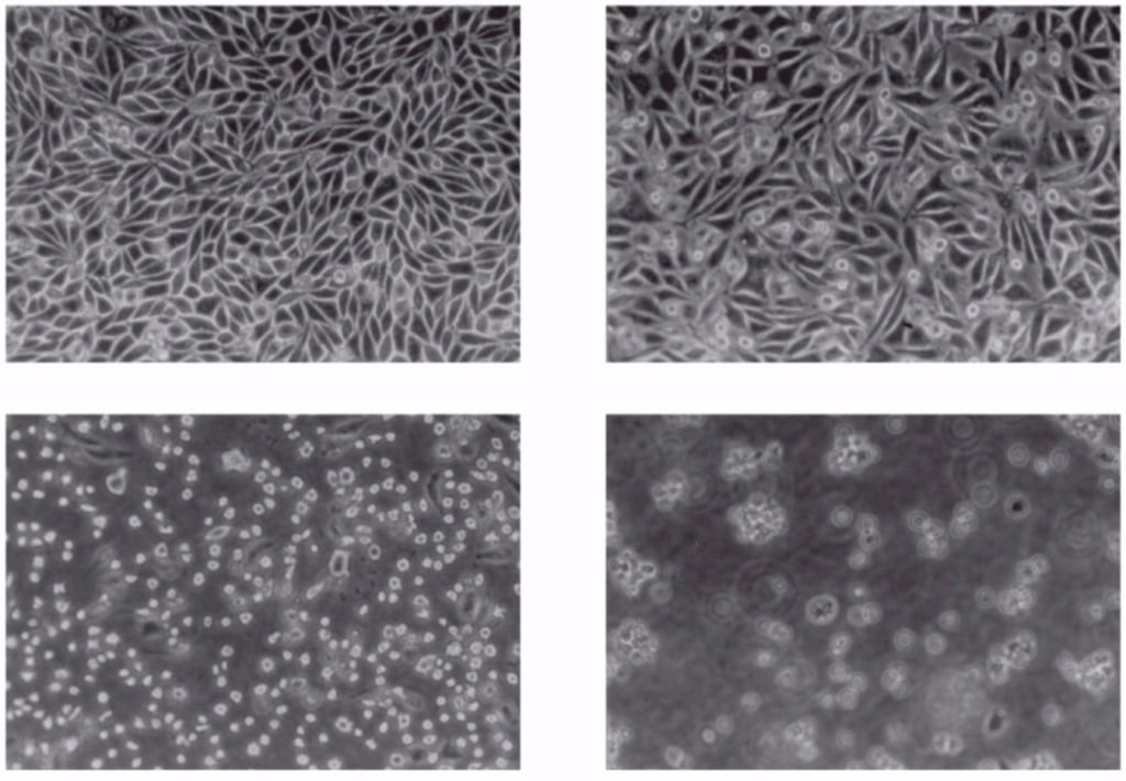 Figure: HeLa cell lines infected with poliovirus (From top left, normal cells, 4 hours, 8 hours and 12 hours after infections)
Some viruses cause very little or no CPE in the cells of their natural host, while others cause complete and rapid destruction of the cell monolayer after infection. Cell line supporting virus replication, the time required to produce CPE, and the microscopic appearance of the CPEs may be sufficiently characteristic to allow the provisional identification of an unknown virus.
Viruses have distinct CPEs, just as colonies of bacteria on agar plates have unique morphologies.
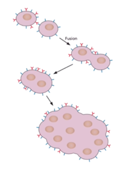 Figure: Syncytia cytopathic effects
Some CPE can be readily observed in unfixed, unstained cells under the low power of the light microscope. Still, several types of CPE are distinguishable in living cultures, thus requiring fixation and staining of the cells. Cell cultures are stained with hematoxylin, a basic dye, and eosin, an acidic dye.
Recognizing CPE and using it as a diagnostic tool requires much experience examining both stained and unstained cultures of many cell types. Uninfected cells should always be run as a control to distinguish age-related changes in infected cells from the cytopathic effects.
Types of Cytopathic effects
Summary of Cytopathic effect(s) of common, clinically encountered viruses
| Cytopathic effect(s) | Virus(es) |
|---|---|
| Morphological alterations | |
| Nuclear shrinking (pyknosis), proliferation of membrane | Picornaviruses |
| The proliferation of nuclear membrane | Alphaviruses, herpesviruses |
| Vacuoles in cytoplasm | Polyomaviruses, papillomaviruses |
| Syncytium formation (cell fusion) | Paramyxoviruses, coronaviruses |
| Margination and breaking of chromosomes | Herpesviruses |
| Rounding up and detachment of cultured cells | Herpesviruses, rhabdoviruses, adenoviruses, picornaviruses |
| Inclusion bodies | |
| Virions in nucleus | Adenoviruses |
| Virions in the cytoplasm (Negri bodies) | Rabies virus |
| “Factories” in the cytoplasm (Guarnieri bodies) | Poxviruses |
| “Owl’s eye” inclusion (large, basophilic, intranuclear, with a clear surrounding halo) | Cytomegalovirus (CMV) |
| Clumps of chromatin in the nucleus | Herpesviruses |
| Clumps of ribosomes in virions | Arenaviruses |
How to Remember the High-Yield Pairings
Inclusion bodies are visible viral factories, not random debris. Whether intranuclear or intracytoplasmic, an inclusion body usually marks the physical site where a virus is mass-producing genome copies or structural proteins, dense enough to be visible under the microscope as an area of altered staining. Once you see inclusion bodies this way, as a literal photograph of "this is where the virus factory is running," the high-yield name-virus pairs become much easier to retain:
- Negri bodies (cytoplasmic) → Rabies virus
- Guarnieri bodies (cytoplasmic) → Poxviruses ("Guarnieri's factories run in the cytoplasm, like a workshop in the yard rather than the main house.")
- Owl's eye inclusion (large, basophilic, intranuclear, with a surrounding clear halo) → Cytomegalovirus (CMV). This pairing is extremely high-yield and is missing from the current article, see section 7 below for the recommended addition.
Syncytium formation is the virus's way of spreading without ever leaving the cell. Fusing multiple cells into one giant, multinucleated mass lets a virus move directly from cell to cell through the fused cytoplasm, bypassing the extracellular space entirely, where neutralizing antibodies would otherwise be waiting. This is why paramyxoviruses (measles, RSV) and herpesviruses, both of which need to keep spreading despite a host actively making antibodies against them, favor this strategy. If you remember "syncytia = stealth spread, avoiding antibodies," the virus list attached to it stops being an arbitrary memorization task.
Destruction-type CPE belongs to viruses built for fast, lytic replication: enteroviruses are the classic example. Picture an enterovirus as a fast-replicating "smash and grab" virus, fast in, fast out, with no investment in keeping the host cell alive longer than necessary. That replication speed is exactly what produces complete monolayer destruction within 72 hours rather than a slower, more contained pattern.
Destruction
It is the most severe form of CPE. All cells in the monolayer rapidly shrink, become dense (pyknosis), and detach from the glass within 72 hours. This CPE is typical of most enteroviruses.
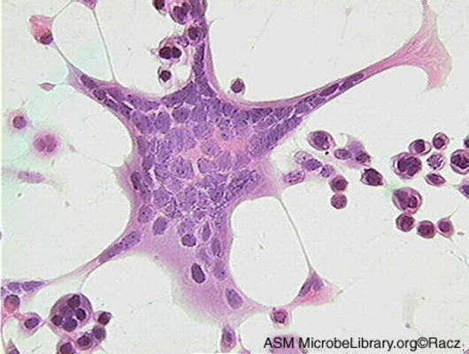 Figure: Cytopathic effect HSV
Subtotal destruction
It consists of detachment (death) of some but not all of the cells in the monolayer, which can be observed using the 20X objective. Some togaviruses (alphaviruses), some picornaviruses, and some paramyxoviruses may cause this type of CPE.
Focal degeneration
Instead of causing generalized cell monolayer destruction, some viruses produce localized areas (foci) of infection. Cells become enlarged, rounded, and refractile, eventually detaching from the growth surface, leaving cleared areas surrounded by rounded-up cells as the infection spreads concentrically. Focal degeneration is characteristic of the herpesviruses and poxviruses.
Swelling and clumping
Infected cells greatly enlarge and clump together in “grape-like” clusters. For example, adenoviruses.
Foamy degeneration (vacuolization)
Several virus families produce large and/or numerous cytoplasmic vacuoles. Vacuolation is visible only after staining. Retroviruses, paramyxoviruses, and flaviviruses may cause vacuolization.
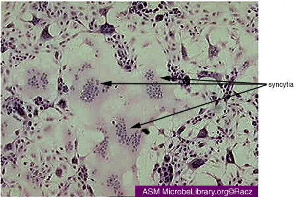 Figure: Syncytia formation
Cell fusion (syncytium or polykaryon formation)
It involves the fusion of the plasma membranes of four or more cells to produce an enlarged cell with four or more nuclei. Some paramyxoviruses; and herpesviruses may produce syncytia. Syncytia are much easier to observe after staining.
Inclusion bodies
These are areas of altered staining in cells that cannot be seen in live cell cultures. Depending on the causative virus, these inclusions may be single or multiple, large or small, round or irregularly shaped, intranuclear or intracytoplasmic, eosinophilic (pink staining), or basophilic (blue-purple staining). One of the most diagnostically recognizable examples is the "owl's eye" inclusion of cytomegalovirus (CMV): a large, basophilic, intranuclear inclusion surrounded by a clear halo, giving infected cells an owl-eye appearance under the microscope.
A related but distinct cytologic finding worth knowing is the koilocyte, seen on Pap smears in human papillomavirus (HPV) infection: a squamous epithelial cell with a shrunken, irregular nucleus and a clear perinuclear halo. Koilocytes aren't classic cell-culture CPE, HPV doesn't readily grow in standard cell culture, but the underlying principle is the same: a virus leaving a recognizable visual signature on infected cells, in this case forming the basis of cervical cytology screening.
Before, virologists used to rely on cytopathic effects to identify viruses. The reliance has decreased substantially due to a rise in nucleic acid amplification tests such as polymerase chain reaction (PCR),which is faster and more specific.
Where Students Get Confused
The summary table and the named CPE categories below it don't map one-to-one, and that's confusing if you don't expect it. The table at the top groups effects by morphological description (nuclear shrinking, vacuoles, syncytium formation, inclusion bodies) across virus families, while the prose sections that follow (Destruction, Subtotal destruction, Focal degeneration, and so on) describe overlapping phenomena using different category names. Read the table as a quick-reference summary of effects by virus family, and the prose sections as the same set of effects described in more diagnostic depth, not as two separate, competing classification systems you need to reconcile perfectly.
"No visible CPE" does not mean "no virus present." Some viruses replicate efficiently in cell culture without producing any visible morphological change, a property called being non-cytopathogenic in that particular cell line. Absence of CPE is a reason to use a more sensitive confirmation method (such as hemadsorption or immunofluorescence), not a reason to conclude the sample is negative.
Nuclear shrinking (pyknosis) and nuclear membrane proliferation sound contradictory, but they describe different virus families' endgames. Picornaviruses cause the nucleus to shrink and condense as the cell dies quickly; alphaviruses and herpesviruses, by contrast, can cause nuclear membrane proliferation as part of a different, often slower replication strategy. Don't assume all CPE moves cellular structures in the same direction, shrinking versus expanding nuclear material reflects genuinely different underlying viral strategies.
Key Exam Facts Table
| CPE type | Mechanistic idea | Classic virus example(s) |
|---|---|---|
| Total destruction | Fast, lytic replication kills the cell quickly | Enteroviruses |
| Subtotal destruction | Some cells in monolayer survive, slower/incomplete spread | Some togaviruses, picornaviruses, paramyxoviruses |
| Focal degeneration | Localized spread outward from an infection focus | Herpesviruses, poxviruses |
| Swelling/clumping | Cells enlarge into "grape-like" clusters | Adenoviruses |
| Foamy degeneration (vacuolization) | Cytoplasmic vacuoles, visible after staining | Retroviruses, paramyxoviruses, flaviviruses |
| Syncytium formation | Cell-to-cell fusion enables antibody-evading spread | Paramyxoviruses, herpesviruses, RSV, coronaviruses |
| Negri bodies (cytoplasmic inclusion) | Site of viral nucleocapsid accumulation | Rabies virus |
| Guarnieri bodies (cytoplasmic inclusion) | Site of viral protein/genome synthesis | Poxviruses |
| Owl's eye inclusion (intranuclear) | Large basophilic inclusion with clear halo | Cytomegalovirus (CMV) |
| Koilocytes (not classic CPE, but a related cytologic clue) | Nuclear atypia with perinuclear clearing, seen on Pap smear | Human papillomavirus (HPV) |
References and further readings
- Suchman, E., & Blair, C. (2007). Cytopathic effects of viruses protocols. American Society for Microbiology. (Verify current URL resolves in CMS before inserting.)
- Tille, P. M. (2017). Bailey & Scott's Diagnostic Microbiology (14th ed.). Elsevier Mosby.
- Mattes, F. M., McLaughlin, J. E., Emery, V. C., Clark, D. A., & Griffiths, P. D. (2000). Histopathological detection of owl's eye inclusions is still specific for cytomegalovirus in the era of human herpesviruses 6 and 7. Journal of Clinical Pathology, 53(8), 612–614. https://doi.org/10.1136/jcp.53.8.612
Frequently Asked Questions
If a virus depends on a host cell to survive, why does it damage or kill that cell?
Is recognizing CPE under a microscope still useful if PCR is available?
What is an "owl's eye" inclusion and which virus causes it?
Are koilocytes the same thing as cytopathic effect?

Tankeshwar Acharya, MSc (Medical Microbiology)
Tankeshwar Acharya is an Assistant Professor in the Department of Microbiology at Patan Academy of Health Sciences (PAHS), Nepal, where he has been teaching and practicing clinical microbiology for over 14 years. He is the founder of Microbe Online, one of the leading free microbiology education resources on the web, covering bacteriology, mycology, parasitology, immunology, and clinical laboratory diagnostics written from direct experience in both the classroom and the diagnostic laboratory.