
Cell Wall–Deficient Bacteria
Cell wall deficient bacteria — Mycoplasma, L-forms, protoplasts, and spheroplasts. Why they are completely resistant to beta-lactam antibiotics, how L-forms form during antibiotic treatment, and their role in chronic and recurrent infections. With clinical stories and exam tips.
Although most prokaryotes cannot survive in nature without their cell walls, some do so naturally. These include the mycoplasmas, a group of pathogenic bacteria that causes several infectious diseases of humans and other animals, and the Thermoplasma group, species of Archaea that naturally lack cell walls.
These bacteria are able to survive without cell walls because they either contain unusually tough cytoplasmic membranes or because they live in osmotically protected habitats such as the animal body.
Cell Wall-Deficient Bacteria
- Mollicutes (often known as Mycoplasma species)
- L-forms
- Spheroplasts
- Protoplasts
Why cell wall deficient bacteria matter clinically
This topic falls squarely into the category of "interesting but why should I care?" The honest answer is: cell wall deficient bacteria are one of the key explanations for why some bacterial infections never fully resolve, why some antibiotic treatments fail despite in vitro susceptibility, and why Mycoplasma infections are so commonly misdiagnosed.
Three clinical scenarios that make this topic immediately relevant:
1. The atypical pneumonia that won't respond to amoxicillin
Mycoplasma pneumoniae causes "walking pneumonia," and prescribing amoxicillin for it is one of the most common, entirely avoidable errors in primary care. Mycoplasma has no cell wall, so amoxicillin has zero mechanism of action against it. See the full clinical case in our dedicated Mycoplasma pneumoniae article.
2. The recurrent UTI that keeps coming back
Some "recurrent" UTIs aren't reinfections at all: cell wall-targeting antibiotics can push E. coli into wall-less L-forms that hide inside uroepithelial cells, invisible to both antibiotics and standard culture, then revert and relapse once treatment stops. (Full case below, under "How to Learn and Remember.")
3. The Mycoplasma that looks like a virus Early in the history of microbiology, Mycoplasma was classified as a virus because it passed through bacteriological filters and could not be seen by light microscopy. This misclassification had real consequences — infections were treated with antiviral measures rather than antibiotics. Understanding what makes Mycoplasma genuinely unique (no cell wall, membrane stabilised by cholesterol, smallest free-living organism) explains not just its biology but why its diagnosis and treatment differ fundamentally from all other bacteria.
Mycoplasmas
The Mollicutes, often called mycoplasmas, are naturally occurring stable bacteria that lack cell walls (mollis is latin for “soft”). These pleomorphic bacteria are not stained by Gram-stain but are phylogenetically related to Firmicutes (a phylum of bacteria, most of which have Gram-positive cell wall structure).
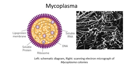 Figure: Mycoplasma (cell wall-deficient bacteria)
Mycoplasma (cell wall-deficient bacteria) resemble protoplasts (bacteria treated to remove their cell walls) but are more resistant to osmotic lysis than protoplasts. Species of medical importance include Mycoplasma pneumoniae and Ureaplasma urealyticum.
Characteristics feature
- They are among the smallest living microorganisms capable of independent existence, ranging in size from 0.1-0.2 μm, approximately the size of the largest viruses (the poxviruses).
- Cytoplasmic membranes of mycoplasmas are more stable than that of other bacteria due to the presence of sterols. Sterols add strength and rigidity to the cytoplasmic membranes.
- The genomes of mycoplasmas are between 500 and 1100 kilobase pairs of DNA in most cases. This is smaller than those of most bacteria, comparable to the genome size of the obligately parasitic chlamydia and rickettsia.
- Mycoplasmas give a characteristic “fried-egg” appearance in solid culture media consisting of a dense central core that penetrates downward into the agar, surrounded by a circular spreading area that is lighter in color.
- Growth of mycoplasmas is not inhibited by antibiotics such as penicillin that inhibit cell wall synthesis. However, mycoplasmas are as sensitive as most Bacteria to antibiotics whose targets are other than the cell wall.
- Certain mycoplasma contains compounds called lipglycans, a lipopolysaccharide-like biomolecule that lacks lipid A backbone. Lipoglycans are long-chain heteropolysaccharides covalently linked to membrane lipids and embedded in the cytoplasmic membrane of many mycoplasmas. Lipoglycans stabilize the cytoplasmic membrane and also facilitates attachment of mycoplasms to cell surface receptors of animal cells.
- Media for the culture of most mycoplasmas are typically quite complex requiring unsaturated fatty acids, sterols, vitamins, amino acids, purines, and pyrimidines as growth factors.
- Oxygen requirements of mycoplasmas vary widely, some are strictly respiratory while others are facultative or even obligate anaerobes.
| Requires sterols | Do not require sterols |
|---|---|
| Mycoplasma | Acholeplasma |
| Anaeroplasma | Asteroleplasma |
| Spiroplasma | Mesoplasma |
| Ureaplasma | |
| Entomoplasma |
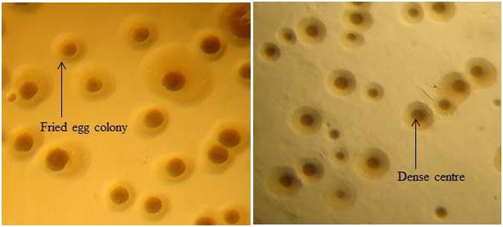 Figure: Fried egg colonies of Mycoplasma (source)
Some mycoplasmas require sterols in their growth media. Based on sterol requirements, mycoplasmas can be differentiated into two groups.
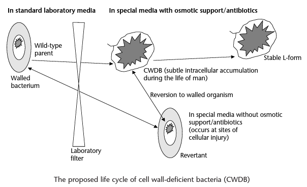 Figure: (Imagesource)
Why Mycoplasma is clinically unique — a summary
| Feature | Mycoplasma | All other bacteria |
|---|---|---|
| Cell wall | Absent | Present (peptidoglycan) |
| Gram stain | Not visible (no cell wall to retain dye) | Gram-positive or negative |
| Size | 0.1–0.2 μm — smallest free-living organism | Most 0.5–5 μm |
| Membrane sterols | Yes (cholesterol from host) | No (except some archaea) |
| Bacteriological filter passage | Yes — passes 0.22 μm filters | No |
| Beta-lactam antibiotics | Completely resistant — no target | Susceptible (varying degrees) |
| Glycopeptides (vancomycin) | Completely resistant — no target | Gram-positive susceptible |
| Treatment options | Macrolides, tetracyclines, fluoroquinolones | Dependent on species |
| Colony appearance | Fried-egg (central dense core + lighter periphery) | Variable |
| Fastidiousness | Requires rich media with cholesterol | Variable |
The critical clinical table from this: Beta-lactams have zero activity against Mycoplasma — not reduced activity, zero activity. No dose of amoxicillin, ceftriaxone, or meropenem has any effect. This is intrinsic, complete, and permanent resistance.
L-forms
L-forms bacteria, also known as L-phase bacteria are mutant bacteria without a cell wall, usually produced in the laboratory but sometimes formed in the body of patients being treated with penicillin. They can reproduce on ordinary culture media.
L-forms are completely resistant to most antibiotics working specifically on cell wall synthesis, such as penicillins and cephalosporins. L‐forms are able to grow as spheroplasts or protoplasts.
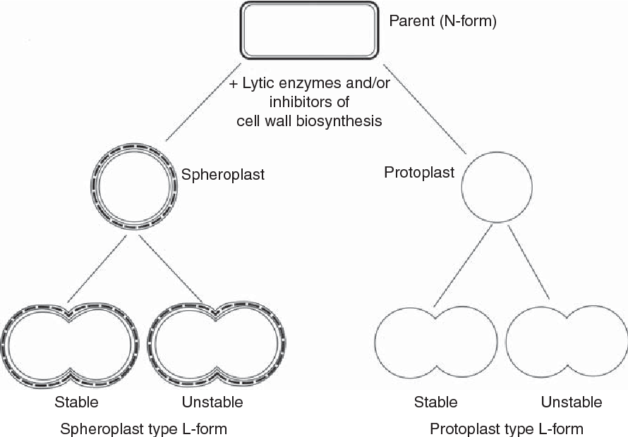 Figure: L form of bacteria
L-forms — the clinical significance students need to understand
L-forms are bacterial variants that have lost their cell wall — either partially (spheroplasts from gram-negative bacteria) or completely (protoplasts from gram-positive bacteria, or true L-forms that can stably replicate without a wall). They are named after the Lister Institute in London where they were first described.
How L-forms form in patients: When bacteria are exposed to cell wall-targeting antibiotics (penicillins, cephalosporins, vancomycin) or lysozyme (in tissues or body fluids), the peptidoglycan cell wall is damaged or prevented from being built. In standard conditions this kills the bacterium. But in certain environments — particularly within host cells (intracellular L-forms) or in osmotically protected niches (certain body cavities, urinary epithelium) — the bacterium can survive without its cell wall by relying on its plasma membrane alone.
These L-forms:
- Are completely resistant to all cell wall-active antibiotics (the target no longer exists)
- Are difficult or impossible to culture on standard media (they lyse in hypotonic conditions)
- Are not detected by standard clinical microbiology culture
- Can revert to normal walled bacteria when antibiotic pressure is removed
- May persist intracellularly in macrophages, uroepithelial cells, and renal tubular cells
The clinical consequence: A patient treated with a beta-lactam antibiotic for a UTI or endocarditis may appear to have responded — symptoms improve, cultures become negative. But L-forms that have retreated intracellularly are invisible to culture and immune to the antibiotic. When treatment stops, L-forms revert and the infection relapses — sometimes with the same organism, sometimes with partial antibiotic susceptibility changes. This mechanism is increasingly recognised as contributing to:
- Recurrent UTIs
- Relapsing endocarditis
- Treatment-refractory chronic infections
What this means practically: This is not a reason to panic or avoid beta-lactams — they remain essential antibiotics. But it does explain why:
- Antibiotic courses must be long enough to suppress any residual L-forms
- Recurrent infections with the same organism should prompt investigation for intracellular persistence
- Some chronic infections require combination therapy with agents that penetrate intracellularly (fluoroquinolones, macrolides, tetracyclines)
Protoplasts
Protoplasts are unstable cells with all the rigid wall layers lost artificially. The cell wall is lost due to the action of lysozyme enzymes which destroy peptidoglycan. Protoplasts are metabolically active but unable to reproduce and are easily lysed.
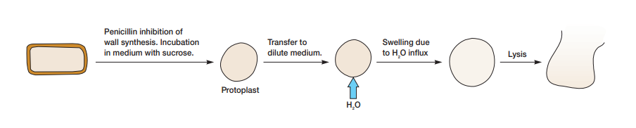 If bacteria are incubated with penicillin in an isotonic solution, gram-positive bacteria are converted to protoplasts and continue to grow normally when isotonicity is maintained.
Spheroplasts
A spherical, osmotically sensitive cell derived from a bacterium by loss of some but not all of the rigid wall layer.
Spheroplasts are spherical, osmotically sensitive cells derived from Gram-negative bacteria by loss of some but not all of the rigid wall layer. The damage in the wall is caused by a toxic chemical or antibiotic such as penicillin (gram-negative bacteria retain their outer membrane after penicillin treatment). They are able to change back to their normal form when the toxic agent is removed.
Archaea lacking cell walls
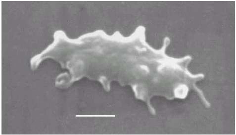 Figure: Thermoplasma Photo by Dr. William Hixon
Cells of some Archaea, such as Thermoplasma and Ferroplasma, lack cell walls. Thermoplasma is a chemoorganotroph that grows optimally at 55°C and pH 2 in complex media. The cytoplasmic membrane of Thermoplasma contains a lipopolysaccharide-like material called lipoglycan. This substance consists of a tetraether lipid monolayer membrane with mannose and glucose. The membrane also contains glycoproteins but not sterols. These molecules render the Thermoplasma membrane stable to hot, acidic conditions.
Ferroplasma is a chemolithotrophic relative of Thermoplasma and is a strong acidophile.
How to Learn and Remember Cell Wall Deficient Bacteria
The calibration: theory — address the "when does this matter?" question
The difficulty here is not complexity — the concepts are simple. The difficulty is relevance. The three clinical scenarios in the opening section address this directly. Below is the consolidation.
One sentence that captures the entire clinical relevance
"Cell wall deficient bacteria are the bacteria that beta-lactam antibiotics cannot touch — either because they were born without a wall (Mycoplasma) or because they shed their wall during treatment (L-forms)."
Key exam facts in one table
| Question | Answer |
|---|---|
| Which organisms naturally lack a cell wall? | Mycoplasma and Ureaplasma (genus Mollicutes) |
| What replaces the cell wall structurally in Mycoplasma? | Cholesterol-containing plasma membrane |
| Why is Mycoplasma resistant to all beta-lactams? | No peptidoglycan — no transpeptidase target |
| What antibiotics treat Mycoplasma infections? | Macrolides (azithromycin), tetracyclines (doxycycline), fluoroquinolones |
| What are L-forms? | Bacteria that have lost their cell wall during antibiotic treatment or lab passage |
| Can L-forms revert to walled bacteria? | Yes — when antibiotic pressure is removed |
| What is a protoplast? | Gram-positive bacterium with cell wall completely removed (by lysozyme); osmotically fragile |
| What is a spheroplast? | Gram-negative bacterium with partial cell wall removal; retains outer membrane |
| What characteristic colony does Mycoplasma produce? | Fried-egg — dense central core + lighter spreading periphery |
| Why did Mycoplasma pass through bacteriological filters? | 0.1–0.2 μm diameter — smaller than 0.22 μm filter pore size |
A clinical story that makes this unforgettable
The infection that survived inside the bladder wall
A microbiologist studying recurrent UTI takes bladder biopsies from women who have had more than 3 UTIs per year with the same E. coli strain. Using fluorescence microscopy and specialised culture techniques, she finds E. coli L-forms within uroepithelial cells — invisible to standard urine culture, invulnerable to the amoxicillin courses the women have been receiving. The cells show them as faint, wall-less shadows deep within the epithelium, waiting for antibiotic pressure to subside. This is not a hypothetical — L-form persistence in the bladder epithelium has been demonstrated in mouse models and increasingly in human studies. It changes our understanding of why some women have truly recurrent rather than reinfection-based UTIs.
References and further readings
- Madigan, M. T., Bender, K. S., Buckley, D. H., Sattley, W. M., & Stahl, D. A. (2018). Brock Biology of Microorganisms (15th ed.). Pearson.
- Tille, P. M. (2017). Bailey & Scott's Diagnostic Microbiology (14th ed.). Mosby Elsevier.
- Errington, J., Mickiewicz, K., Kawai, Y., & Wu, L. J. (2016). L-form bacteria, chronic diseases and the origins of life. Philosophical Transactions of the Royal Society B, 371(1707). https://doi.org/10.1098/rstb.2015.0494
- Hayward, R. J., & Schneewind, O. (2019). Atypical bacterial development: Mycoplasma. Nature Reviews Microbiology, 17(6), 381–391.
Frequently Asked Questions
Why are Mycoplasma species completely resistant to all beta-lactam antibiotics?
What is the significance of Mycoplasma's fried-egg colony appearance?
What is the difference between L-forms, protoplasts, and spheroplasts?
Can cell wall deficient bacteria be detected by standard culture?
What is the clinical significance of Ureaplasma urealyticum?
What is the role of L-forms in recurrent infections?

Tankeshwar Acharya, MSc (Medical Microbiology)
Tankeshwar Acharya is an Assistant Professor in the Department of Microbiology at Patan Academy of Health Sciences (PAHS), Nepal, where he has been teaching and practicing clinical microbiology for over 14 years. He is the founder of Microbe Online, one of the leading free microbiology education resources on the web, covering bacteriology, mycology, parasitology, immunology, and clinical laboratory diagnostics written from direct experience in both the classroom and the diagnostic laboratory.