
Bacterial Spores: Structure and Spore-Forming Bacteria
Bacterial endospores — structure, sporulation stages, germination, spore positions with mnemonics, resistance mechanisms, clinically important spore-forming bacteria (Bacillus, Clostridium), and why endospores matter in infection control.
Bacterial spores are highly resistant, dormant structures (i.e. no metabolic activity) formed in response to adverse environmental conditions. As bacterial spores are formed within the parent cell, these are called endospores.
When vegetative cells of certain bacteria such as Bacillus spp and Clostridium spp are subjected to environmental stresses such as nutrient deprivation, they produce metabolically inactive or dormant form-endospore. The formation of endospores circumvents the problems associated with environmental stress and ensures the survival of the organisms.
Note:Spores of fungi have a reproductive role.
Why endospores matter
Before we explore structure and formation, understand why endospore biology is one of the most practically important topics in infection control and clinical microbiology:
1. Clostridioides difficile (the healthcare epidemic) C. difficile produces endospores that survive on hospital surfaces, medical equipment, and hands for months. Standard alcohol-based hand sanitisers — effective against vegetative bacteria and viruses — do not kill C. difficile spores. This is why C. difficile outbreaks spread so readily in hospitals and why soap-and-water handwashing (mechanical removal of spores) is essential for C. difficile control. Globally, C. difficile infection causes hundreds of thousands of hospital-acquired infections annually.
2. Anthrax & bioterrorism Bacillus anthracis spores can survive in soil for over 50 years. In 2001, anthrax spores were deliberately sent through the US postal system, killing five people and infecting 22 others from inhalation of airborne spores. Inhalation anthrax has a mortality rate above 80% if untreated — and spores can be inhaled without the victim knowing exposure occurred. The extreme persistence and hardiness of spores make B. anthracis a Tier 1 bioterrorism agent.
3. Tetanus and its association with soil and wounds Clostridium tetani spores are ubiquitous in soil, dust, and animal faeces worldwide. They persist for years. A wound contaminated with soil — even a minor puncture — can introduce C. tetani spores. In the anaerobic environment of a deep wound, spores germinate to vegetative cells that produce tetanospasmin — one of the most potent biological toxins known. Without vaccination, tetanus is fatal in up to 70% of cases in resource-limited settings.
4. Autoclave validation The most heat-resistant organism used to validate autoclave sterilization is Geobacillus stearothermophilus — a thermophilic, spore-forming bacterium. If autoclave conditions (121°C, 15 min) kill these spores, the cycle is validated as effective. Every hospital and laboratory that uses an autoclave relies on spore biology for its quality control.
The core clinical message: endospores are not just a fascinating biological curiosity. They are the reason hospital infection control is so difficult, the reason some wounds cause tetanus and gas gangrene, the reason autoclaves must reach 121°C, and the reason standard disinfectants are ineffective against certain pathogens.
Endospores size, shape, and location are particularly useful for identifying Clostridium, Bacillus, and related species. Bacterial spores (e.g., spores of Bacillus stearothermophilus) are used as an indicator for proper sterilization of autoclave.
Structure of the Bacterial Spore
An endospore is structurally and chemically more complex than a vegetative cell. It contains more layers than vegetative cells. Resistance of Bacterial spores may be mediated by dipicolinic acid, a calcium ion chelator found only in spores. Following are the constituents of bacterial spores;
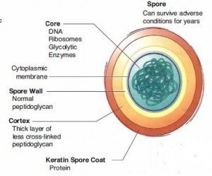 Figure: Structure of Bacterial Spore
Exosporium
Exosporium, a thin protein covering, is the outermost layer of a bacterial endospore.
Spore coat
It lies below the exosporium. The spore coat is composed of layers of spore-specific proteins.
Cortex
The cortex lies below the spore coat and consists of loosely cross-linked peptidoglycan.
Core
The core is the innermost region of a bacterial endospore surrounded by a core wall. Cytoplasmic membrane, cytoplasm, nucleoid, ribosomes, and other cellular essentials are found inside the core wall. The core of the mature endospore differs greatly from the vegetative cell from which it was formed.
- Dipicolinic acid, absent in vegetative cells, accumulates in the bacterial endospore’s core.
- Endospores are also enriched in calcium, most of which are complexed with dipicolinic acid. The calcium-dipicolinic acid complex represents about 10% of the dry weight of the endospore, thus helping to dehydrate it. This complex intercalates (inserts between bases) in DNA and stabilizes the DNA against heat denaturation.
- The core of a mature endospore has only 10-25% of the water content of the vegetative cell, and thus the consistency of the core cytoplasm is that of a gel.
- In addition to the endospore’s low water content, the core’s pH is about one unit lower than the vegetative cell cytoplasm.
Sporulation
Bacterial cell undergoes spore formation in nutritionally deprived conditions, and this process is called sporulation. Spore develops from a portion of protoplasm (forespore) near one end of the cell. The remaining part of the cell is called sporangium. Spore-forming bacilli form endospores during unfavorable conditions (especially when carbon and nitrogen become depleted or unavailable).
The analogy that makes sporulation memorable
Think of sporulation as the bacterial equivalent of building a nuclear bunker.
When conditions become hostile (no food, no water, extreme heat, radiation), the cell does not simply die. Instead, it:
- Duplicates its DNA and seals one copy in the innermost vault (the core)
- Builds layer after layer of protective walls around it (cortex, spore coat, exosporium)
- Fills the vault with a special chemical preservative (calcium-dipicolinic acid) that prevents desiccation and chemical damage
- Dehydrates the contents to near-zero water activity, halting all metabolism
- Shuts the vault door permanently — and the vegetative cell dies around it
The completed spore can then survive for decades with zero metabolic activity, impervious to heat, radiation, chemicals, and desiccation — waiting for conditions to improve.
When conditions become favourable again (nutrients, water, correct temperature), the spore germinates:
- The vault door opens (activation)
- Water floods in, dissolving the dipicolinic acid complex
- Metabolic activity resumes
- The spore swells and a vegetative cell emerges — ready to start growing and potentially causing disease
Spore Germination
The process of conversion of a spore into a vegetative cell under suitable conditions is known as germination. When favorable conditions prevail (i.e., availability of water, appropriate nutrients), spores germination occurs, forming vegetative cells of pathogenic bacteria.
This process involves three steps: activation, germination, and outgrowth. Activation occurs when endospores are heated for several minutes at an elevated but sub-lethal temperature. Activated endospores can germinate when placed in a suitable culture medium. Germination is a rapid process that involves loss of microscopic refractility of the endospore, increased ability to be stained by dyes, and loss of resistance to heat and chemicals. The final stage, outgrowth, involves visible swelling due to water uptake and synthesis of RNA, proteins, and DNA.
Resistance
Bacterial spores are highly resistant to ordinary boiling, disinfectants, and heating, but spores of all medically important bacteria are destroyed by autoclaving. Bacterial spores are highly resistant to;
- Heat
- Dehydration
- Radiation and
- Chemicals
The following factors/constituents play major roles in the resistance of bacterial spore:
- Calcium dipicolinate in core
- Keratin spore coat
- New enzymes (i.e., dipicolinic acid synthetase, heat-resistant catalase)
- Increases or decreases in other enzymes.
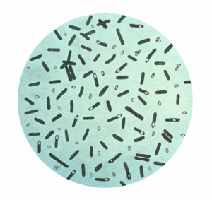 Figure: Spore of Clostridium botulinum
A mature endospore contains a complete set of genetic material (DNA) from the vegetative cell, ribosomes, and specialized enzymes.
Mature endospores are released from the vegetative cell to become free endospores. When the free endospores are placed in an environment that supports growth, the endospores will revert to vegetative cells in a process called germination. It should be noted that unlike the process of binary fission observed with vegetative cells, endospore formation is not a reproductive process but a process of differentiation that provides the bacteria with a mechanism for survival.
Medical Importance of Bacterial Spores
| Important features of Spores | Medical Implications |
|---|---|
| Spores are highly resistant to heating; spores are not killed by boiling (100°C) but are killed at 121°C. | Medical supplies must be heated to 121°C for at least 15 minutes to be sterilized. |
| Spores are highly resistant to many chemicals, including most disinfectants. | The only solution designated as sporicidal will kill spores. |
| Spores can survive for many years in soil and other inanimate objects. | Wounds contaminated with soils can be infected with spores and cause diseases such as tetanus and gas gangrene. |
| Spores do not exhibit measurable metabolic activity. | Antibiotics are ineffective against spores. |
| Spores are formed only when nutrients are insufficient. | Spores are not often found at the site of infection because nutrients are not limited. |
Clinically Important Spore-Forming Bacteria
Most endospore-forming bacteria are found in soil or aquatic environments. However, some species of Bacillus and Clostridium have medical significance. Clostridium perfringens, C. botulinum (a potential agent of bioterrorism), and C. tetani are the causative agents of gas gangrene, botulism, and tetanus, respectively. Bacillus anthracis and Bacillus cereus are the causative agents of anthrax and self-limiting food poisoning, respectively.
Aerobic spore-formers — genus Bacillus
| Organism | Spore position | Key disease | Why spores matter clinically |
|---|---|---|---|
| Bacillus anthracis | Central, oval, non-distending | Anthrax (cutaneous, inhalation, gastrointestinal) | Spores persist 50+ years in soil; bioterrorism agent; inhalation form >80% mortality |
| Bacillus cereus | Central/subterminal | Food poisoning (emetic and diarrheal types) | Spores survive cooking temperatures; cereulide toxin pre-formed in food; "reheated rice syndrome" |
| Bacillus subtilis | Central | Rare opportunistic infections; important research organism | B. stearothermophilus (now Geobacillus stearothermophilus) spores = autoclave biological indicator |
Anaerobic spore-formers — genus Clostridium
| Organism | Spore position | Key disease | Why spores matter clinically |
|---|---|---|---|
| Clostridium tetani | Terminal, round, distending ("drumstick") | Tetanus | Spores ubiquitous in soil; wound contamination triggers germination; tetanospasmin — one of the most toxic substances known (lethal dose ~1 ng/kg) |
| Clostridium perfringens | Subterminal, oval, non-distending | Gas gangrene, food poisoning, necrotising fasciitis | Most common clostridial wound infection; lecithinase (alpha-toxin) destroys cell membranes; double-zone haemolysis on blood agar |
| Clostridium botulinum | Subterminal/terminal | Botulism (food, infant, wound) | Botulinum toxin — the most toxic biological substance known (lethal dose ~1 ng/kg by injection); spores in home-canned foods at risk |
| Clostridioides difficile | Subterminal, oval | Antibiotic-associated diarrhoea, pseudomembranous colitis | Spores resist alcohol hand gel and most disinfectants; hospital surfaces remain contaminated for months; sodium hypochlorite (bleach) required |
| Clostridium novyi | Subterminal | Gas gangrene, "black disease" of liver | Type B produces alpha-toxin causing tissue necrosis; difficult anaerobic culture requirements |
Demonstration of Endospore
Endospores cannot be stained by ordinary methods, such as simple and Gram staining, because dyes do not penetrate the endospore wall. But endospores can be seen as an unstained refractile body within the cell, in gram-stained smears, or unstained preparations. Without a special stain, it’s hard to differentiate endospores from inclusions of stored material.
Though the modified Ziehl-Neelsen method can be used for endospore staining, the most commonly used endospore stain is the Schaeffer-Fulton.
Spore positions — What, Why, and How to Remember
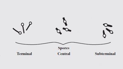 ### The mnemonic: "Big Cats Try"
| Letter | Organism | Spore position | Visual shape |
|---|---|---|---|
| Big | Bacillus anthracis | Central | Oval, does not distend sporangium — cell looks normal |
| Cats | Clostridium perfringens | Subterminal | Oval, does not distend — cell looks slightly swollen near one end |
| Try | Clostridium tetani | Terminal | Round, distends sporangium — "drumstick" or "tennis racket" shape |
The clinical story behind each position:
Central — B. anthracis "the invisible threat" Bacillus anthracis spores sit centrally within the cell and do not distend it — the vegetative cell looks completely normal on gram stain. You would never know from looking at a gram stain that this organism is already forming a spore that will survive for 50 years. This invisibility is part of what makes anthrax spores so insidious as a biological weapon.
Subterminal — C. perfringens "the gas gangrene clock" C. perfringens spores are subterminal and do not distend the cell. Interestingly, C. perfringens rarely sporulates in clinical tissue specimens — the nutrient-rich environment of infected tissue provides no trigger for sporulation. You will almost never see spores of C. perfringens in a wound smear. The clinical significance of its spores is in the environment — soil contamination of wounds — not in active infection.
Terminal — C. tetani "the drumstick" C. tetani produces a terminal, spherical spore that is wider than the cell body — forcing the cell to bulge into the characteristic drumstick shape. This is one of the most recognizable morphological appearances in clinical microbiology:
Gram-positive rod with a terminal, spherical spore producing a drumstick shape = Clostridium tetani
This single gram stain finding, in the context of a wound and progressive muscle spasms, is essentially diagnostic.
How to Learn and Remember Bacterial Spores
The three resistance mechanisms — "DCD"
The three main reasons endospores are so resistant can be remembered as DCD — the "Death-Cheating Defences":
D — Dipicolinic acid (calcium-dipicolinic acid complex)
- Present only in spores — not in vegetative cells
- Constitutes ~10% of spore dry weight
- Chelates DNA and stabilises it against heat denaturation
- Also removes water from the core (dehydration)
C — Cortex (thick peptidoglycan layer)
- Maintains the dehydrated state of the core
- Physically protects against osmotic damage
D — Dehydration of the core
- Core contains only 10–25% of the water of a vegetative cell
- Chemical reactions — including destruction by heat and disinfectants — require water
- Near-zero water activity means near-zero reactivity
Key exam facts in one table
| Question | Answer |
|---|---|
| Which two genera form clinically important endospores? | Bacillus (aerobic) and Clostridium (anaerobic) |
| Which has terminal, distending spore? | Clostridium tetani — drumstick |
| Which has central, non-distending spore? | Bacillus anthracis |
| What chemical is unique to spores? | Calcium-dipicolinic acid |
| Does boiling kill spores? | No — spores survive 100°C for hours |
| What kills spores reliably? | Autoclaving (121°C, 15 min) |
| Does alcohol hand gel kill C. difficile spores? | No — soap and water required (mechanical removal) |
| What is the biological indicator for autoclave validation? | Geobacillus stearothermophilus spores |
| Can antibiotics kill spores? | No — spores have no metabolic activity; antibiotics need active metabolism to work |
| What triggers sporulation? | Nutrient deprivation (especially carbon and nitrogen) |
| What triggers germination? | Return of nutrients and favourable conditions |
Three clinical stories that make spores unforgettable
Story 1 — The 50-year seed In 1979, animal carcasses were discovered in Sverdlovsk, USSR — the victims of a secret biological weapons accident. Bacillus anthracis spores had been accidentally released from a military facility. Decades later, similar spore contamination has been found in ancient burial sites. Some genuine anthrax spore samples from historical outbreaks are still viable after 100+ years of storage. The endospore does not age — it simply waits.
Story 2 — The hand gel illusion A nurse in a hospital with a C. difficile outbreak used alcohol hand gel conscientiously between every patient contact. The outbreak continued spreading. The problem: alcohol gel is highly effective against vegetative bacteria and viruses, but C. difficile spores have a 10–25% water core and an impermeable spore coat — alcohol cannot penetrate or denature them. Physical removal by soap-and-water handwashing and a 0.5% sodium hypochlorite surface disinfectant were required. Understanding spore biology would have changed this nurse's infection control practice.
Story 3 — The reheated rice A canteen prepares fried rice and leaves it at room temperature for several hours. Bacillus cereus spores, which survive cooking, germinate in the warm rice, producing emetic toxin (cereulide). When the rice is reheated, the vegetative cells are killed — but cereulide is heat-stable and remains in the food. Students eating the reheated rice develop nausea and vomiting within 1–5 hours. The lesson: killing the bacteria does not destroy the toxin pre-formed during sporulation and germination.
References and further readings
- Madigan, M. T., Bender, K. S., Buckley, D. H., Sattley, W. M., & Stahl, D. A. (2018). Brock Biology of Microorganisms (15th ed.). Pearson.
- Tille, P. M. (2017). Bailey & Scott's Diagnostic Microbiology (14th ed.). Mosby Elsevier.
- Shen, A. (2020). Clostridioides difficile spore formation and germination: New insights and opportunities for intervention. Annual Review of Microbiology, 74, 545–566. https://doi.org/10.1146/annurev-micro-011320-011321
- Driks, A., & Eichenberger, P. (2016). The spore coat. Microbiology Spectrum, 4(2). https://doi.org/10.1128/microbiolspec.TBS-0023-2016
Frequently Asked Questions
What is the difference between a bacterial endospore and a fungal spore?
Why do alcohol-based hand sanitisers not kill Clostridioides difficile spores?
What is the drumstick appearance of Clostridium tetani?
What is the role of calcium-dipicolinic acid in endospore resistance?
Can antibiotics kill bacterial endospores?
What triggers sporulation and what triggers germination?
Why does Clostridium perfringens rarely sporulate in clinical infections?
What makes the Bacillus cereus food poisoning story unusual?

Tankeshwar Acharya, MSc (Medical Microbiology)
Tankeshwar Acharya is an Assistant Professor in the Department of Microbiology at Patan Academy of Health Sciences (PAHS), Nepal, where he has been teaching and practicing clinical microbiology for over 14 years. He is the founder of Microbe Online, one of the leading free microbiology education resources on the web, covering bacteriology, mycology, parasitology, immunology, and clinical laboratory diagnostics written from direct experience in both the classroom and the diagnostic laboratory.