
Membrane Filtration Technique: Principle, Procedure, and Bacteriological Analysis of Water
Membrane filtration concentrates bacteria from large water volumes onto a 0.45 µm filter for direct colony counting. Learn the principle, step-by-step procedure, mEndo vs mFC agar colony interpretation, CFU/100 mL calculation, and how membrane filtration compares to MPN and plate count methods.
A community health worker in a flood-affected district collects water samples from five borehole sites for bacteriological testing. One site serves a school of 400 children. The question is straightforward but critical: does this water contain faecal coliforms? And if so, how many per 100 mL?
For a sample that is relatively clear, the membrane filtration technique can answer this question within 24 hours — faster than MPN, more precise than a statistical estimate, and capable of handling the large volumes (100 mL or more) needed to detect the very low bacterial counts that might still pose a health risk. Each bacterium trapped on the membrane grows into a countable colony. Each colony tells you something directly about the water's safety.
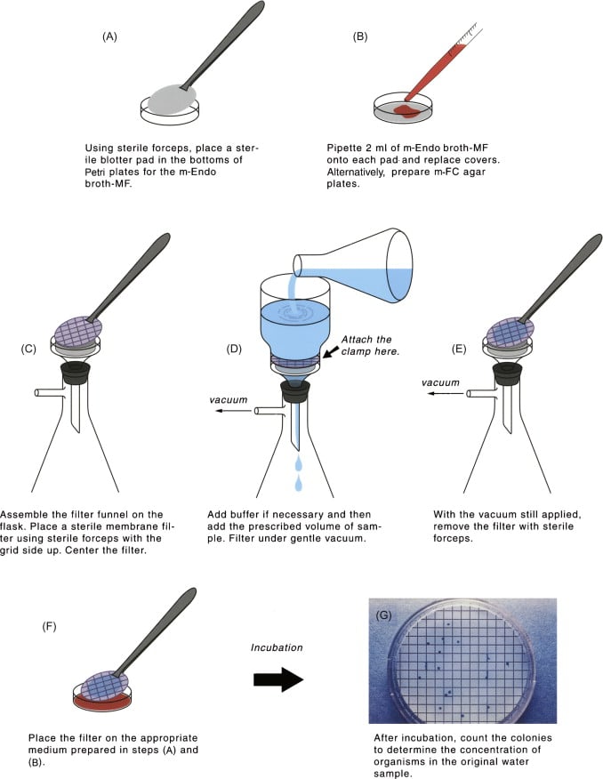 This article covers membrane filtration as both a general microbiological technique and as the standard method for bacteriological analysis of drinking water.
What Is the Membrane Filtration Technique?
Membrane filtration is a method of concentrating microorganisms from a liquid sample onto the surface of a membrane filter, then incubating the membrane on a nutrient medium to allow trapped organisms to grow into visible, countable colonies.
 Unlike the pour plate or spread plate methods — which limit inoculum to 0.1–1.0 mL — membrane filtration can process volumes of 100 mL, 500 mL, or even larger, making it far more sensitive for detecting low concentrations of organisms in water, pharmaceutical solutions, and environmental samples.
The technique serves two purposes simultaneously:
- Concentration — organisms from a large volume are deposited on a small surface
- Enumeration — each trapped organism grows into a discrete, countable colony
Principle: How the membrane filter works
A membrane filter is a thin disc of cellulose acetate, cellulose nitrate, or polycarbonate with a uniform, controlled pore size — typically 0.45 µm for bacteriological work. This size is small enough to retain all bacteria (which range from 0.5–10 µm in diameter) while allowing water, dissolved nutrients, and smaller particles to pass through freely.
Why 0.45 µm specifically:
- Most bacteria: 0.5–10 µm → retained on membrane
- Viruses: 0.01–0.3 µm → pass through (limitation of the technique)
- Mycoplasma: 0.1–0.3 µm → pass through (limitation)
- Water molecules and dissolved salts → pass through freely
How colonies grow on the membrane:
After filtration, the membrane (with trapped bacteria on its upper surface) is placed grid-side up on an absorbent pad saturated with nutrient medium, or directly onto a selective agar plate. During incubation, nutrients diffuse upward through the membrane from below, reaching the bacteria on the surface. Each viable bacterium absorbs these nutrients, divides by binary fission, and grows into a visible colony within 18–24 hours. The colony grows on the membrane surface, directly above where the original bacterium was trapped.
The four types of membrane filtration:
| Type | Pore size | What is retained | Primary application |
|---|---|---|---|
| Microfiltration | 0.1–10 µm | Bacteria, yeast, particles | Bacteriological water testing; pharmaceutical sterilization |
| Ultrafiltration | 0.001–0.1 µm | Macromolecules, viruses, colloids | Virus removal; protein concentration |
| Nanofiltration | 0.0001–0.001 µm | Multivalent ions, small organic molecules | Water softening; desalination |
| Reverse osmosis | <0.0001 µm | Essentially all solutes including monovalent ions | Desalination; ultra-pure water production |
For bacteriological work, microfiltration with 0.45 µm pore size is the standard. The other filter types are not used for bacteriological colony counting because their smaller pore sizes do not allow nutrient diffusion through the membrane efficiently.
Why E. coli and Coliforms? The Indicator Organism Rationale
In bacteriological water testing, it is impractical to test directly for every possible pathogen (Salmonella, Vibrio cholerae, Cryptosporidium, hepatitis A virus) that could contaminate water. Instead, indicator organisms are used — organisms whose presence signals the likelihood of faecal contamination.
An ideal indicator organism has two properties:
- It is not naturally present in the environment and appears in water only when faecal contamination has occurred
- Its density is proportional to the degree of contamination — more indicator = more contamination
E. coli and coliforms meet these requirements:
- E. coli is present in the intestine of all warm-blooded animals in large numbers and is shed continuously in faeces
- It survives in water for a similar duration to most intestinal pathogens
- It does not multiply in most natural water sources (unlike some environmental coliforms)
- It is easily detected by lactose fermentation and characteristic colony appearance
- Detection of E. coli therefore means recent faecal contamination and the probable presence of enteric pathogens
Total coliforms vs faecal (thermotolerant) coliforms:
| Group | Definition | Key genera | Indicator significance |
|---|---|---|---|
| Total coliforms | Gram-negative, non-spore-forming facultative anaerobes that ferment lactose with acid and gas at 35°C within 48 hrs | Escherichia, Enterobacter, Klebsiella, Citrobacter, Serratia | Broad indicator; includes environmental strains; less specific for faecal contamination |
| Faecal (thermotolerant) coliforms | Subset of total coliforms that ferment lactose with acid and gas at 44.5°C within 24 hrs | Escherichia, Enterobacter | More specific for faecal contamination; E. coli confirmed by positive indole test |
WHO drinking water guideline (2022): E. coli or thermotolerant coliforms must be absent in any 100 mL sample of drinking water. Total coliforms should also be absent in treated piped water. Any detection is a trigger for immediate investigation, regardless of count.
Materials Required
Equipment:
- Membrane filtration apparatus (filter funnel, support base, clamp)
- Vacuum pump or vacuum line; suction flask
- Incubator(s): 35°C (±0.5°C) for total coliforms and 44.5°C (±0.2°C) for faecal coliforms — water bath preferred for the 44.5°C incubation to maintain narrow temperature range
- Autoclave for sterilising media and equipment
- Flame source (Bunsen burner or spirit lamp) for forceps sterilization
- Colony counter with magnifying glass
- Refrigerator for media storage
Consumables:
- Membrane filters: 0.45 µm pore size, diameter appropriate for filtration apparatus (47 mm standard)
- Absorbent pads (if using pad-saturation method)
- Culture media: mEndo Agar LES (total coliforms) and mFC Agar (faecal coliforms)
- Phosphate-buffered dilution water (for diluting turbid samples)
- Sterile Petri dishes (glass, aluminium, or plastic)
- Blunt-edged sterile forceps
- Sterile pipettes (1 mL and 10 mL) or micropipettes
- Watertight plastic bags (for mFC plates in water bath)
- Wax pencil or waterproof marker
Controls (mandatory):
- Positive control: 100 mL sterile water + 5 mL of 1:100 dilution of overnight E. coli culture
- Negative control: 100 mL sterile water (sterility check of media and technique)
Sample Volumes for Different Water Types
Large volumes can be filtered when bacteria concentrations are expected to be low; small volumes (with dilution) are used when high contamination is expected. The goal is 20–80 countable colonies per membrane.
| Water source | Recommended volumes to filter |
|---|---|
| Treated tap water (expected low count) | 100 mL, 10 mL, 1 mL |
| Untreated borehole or well water | 10 mL, 1 mL, 0.1 mL |
| River or surface water | 1 mL, 0.1 mL, 0.01 mL |
| Heavily polluted water | 0.1 mL, 0.01 mL, 0.001 mL (serial dilutions) |
| Swimming pool / recreational water | 100 mL, 10 mL, 1 mL |
Practical rule: Always filter at least two volumes from each sample (e.g., 100 mL and 10 mL for tap water) to ensure at least one membrane falls in the countable range of 20–80 colonies. If a volume is less than 10 mL, add sterile diluent to bring it to at least 10 mL to ensure even distribution across the membrane surface.
Water Sample Collection Precautions
- Collect in a sterile, pre-labelled bottle
- Sample must be representative of the supply
- Avoid contamination during and after collection
- Test as promptly as possible after collection
- If delay is unavoidable, store at 0–10°C; test within 6 hours; absolute maximum 24 hours with cold storage
- For chlorinated water samples: add sodium thiosulfate to the collection bottle before autoclaving (neutralises residual chlorine that would inhibit or kill coliforms)
Step-by-Step Procedure of Membrane Filtration Technique
Aseptic technique throughout: Every surface the membrane contacts — forceps tips, funnel interior, Petri dish — must be sterile. A single contaminant introduced during handling can produce false-positive colonies, invalidate the entire sample, and lead to an incorrect water safety declaration.
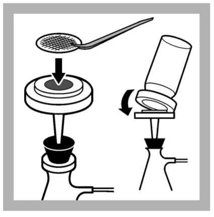
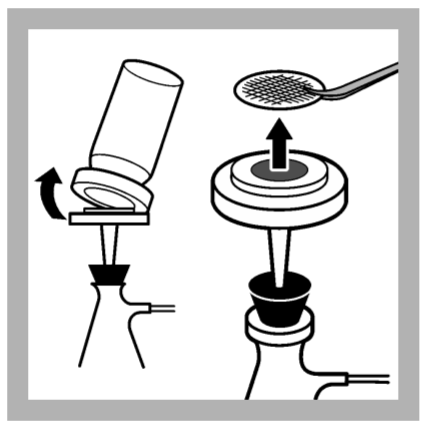 1. Sterilise the tips of the blunt-ended forceps in a flame and allow them to cool. 2. Carefully remove a sterile membrane filter from its package, holding it only by its edge. 3. Place the membrane filter in the filter apparatus, and clamp it in place. (If the apparatus has been disinfected by boiling, ensure that it has cooled down before inserting the membrane filter.)4. Mix the sample by inverting its container several times. Pour or pipette the desired volume of sample into the filter funnel. This volume should normally be chosen in the light of previous experience, but suggested volumes are given in Table 1. If the volume to be filtered is less than 10 ml, it should be made up to at least 10 ml with sterile diluent so that the sample will be distributed evenly across the filter during filtration. 5. Apply a vacuum to the suction flask and draw the sample through the filter; disconnect the vacuum. 6. Dismantle the filtration apparatus and remove the membrane filter using the sterile forceps, taking care to touch only the edge of the filter. 7. Remove the lid of a previously prepared mENDO agar LES plate and place the membrane, grid side uppermost, onto the agar. Lower the membrane, starting at one edge in order to avoid trapping air bubbles between membrane and agar.**Mark the petri dish with the sample number or other identification. The sample volume should also be recorded. Use a wax pencil or waterproof pen when writing on Petri dishes. 8. Repeat procedure (1 to 8) with the same volume, but place membranes on mFC plates.9. Incubate mEndo agar LES plates at 35 ± 0.5°C for 22 – 24 hours and mFC plates at 44.5 ± 0.2°C for 22 to 26 hours, all lid side down. In order to maintain the temperature within such a narrow range, a water bath is typically used for incubation of the mFC agar plates. These plates are placed in watertight plastic bags and then submerged in the water bath.
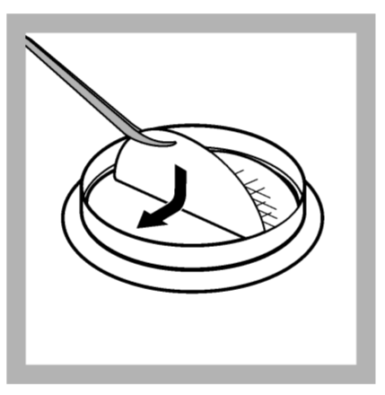 > Place membrane grid-side up. Lower it from one edge to avoid trapping air bubbles between membrane and agar — an air pocket prevents nutrient diffusion and will produce a colony-free zone on the membrane, giving a falsely low count.
| Medium | Organism detected | Incubation temperature | Duration | Colony appearance |
|---|---|---|---|---|
| mEndo Agar LES | Total coliforms | 35 ± 0.5°C | 22–24 hours | Red with golden metallic sheen (typical); dark red, mucoid, or dark centre without sheen (atypical) |
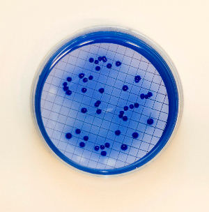
| mFC Agar | Faecal coliforms | 44.5 ± 0.2°C | 22–26 hours | Blue (any shade) — confirmed faecal coliforms |
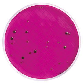 Figure: Coliform colonies in mEndo agar (note green metallic sheen) Membrane filtration
Day 2 (24 hours later):
- After 22 – 24 hours, remove the mEndo agar LES plates from the 35°C incubator and count the colonies that are dark red, mucoid, have a dark center or (more typically) produce a metallic sheen. These are considered to be total coliform colonies.
- From the mEndo agar LES plates, choose two total coliform colonies that are isolated on the membrane and confirm that they are Gram-negative rods and non-spore formers.
- After 22 to 26 hours, remove the mFC agar plates from the 44.5°C incubator and count the colonies that have any blue color. These are considered to be fecal coliform colonies.**
- From the mFC agar plates, choose two fecal coliform colonies that are isolated on the membrane. Confirm that they are Gram-negative rods and non-spore formers.
- Calculate the total and fecal coliform CFU per 100 ml for each sample as described below:
The original density is estimated from the volume of sample filtered (or the volume of dilution and the dilution factor), and the number of colonies counted on the membrane.
As counts are reported per 100 ml of sample (not per ml), the per ml values must be multiplied by a factor of 100.
No. of CFU per 100 ml = [(No. of colonies on the membrane)/(volume filtered)] × 100
If the sample is diluted and the volume of the dilution was filtered, the denominator will be (volume (ml) filtered x dilution)
Colony Interpretation and Calculation
Calculate the total and fecal coliform CFU per 100 ml for each sample as described below:
The original density is estimated from the volume of sample filtered (or the volume of dilution and the dilution factor), and the number of colonies counted on the membrane.
As counts are reported per 100 ml of sample (not per ml), the per ml values must be multiplied by a factor of 100.
No. of CFU per 100 ml = [(No. of colonies on the membrane)/(volume filtered)] × 100
If the sample is diluted and the volume of the dilution was filtered, the denominator will be (volume (ml) filtered x dilution)
Colony counting rules:
- Count only membranes with 20–80 colonies for statistical reliability
- Count all typical AND atypical colonies as potential coliforms; only typical colonies reported as confirmed unless verification performed
- Membranes with >80 colonies: report as TNTC (too numerous to count); retest at higher dilution
- Membranes with <20 colonies: report but note low count; use highest volume result if multiple volumes tested
CFU per 100 mL calculation:
CFU per 100 mL=Colonies countedVolume filtered (mL)×100\text{CFU per 100 mL} = \frac{\text{Colonies counted}}{\text{Volume filtered (mL)}} \times 100CFU per 100 mL=Volume filtered (mL)Colonies counted×100
Worked examples:
Example 1 — Treated tap water:
- Volume filtered: 100 mL
- Colonies on mEndo plate: 3
- Total coliforms = (3 ÷ 100) × 100 = 3 per 100 mL
- Interpretation: Positive — WHO guideline requires absence (0 per 100 mL) → investigate immediately
Example 2 — Borehole water:
- Volume filtered: 10 mL
- Colonies on mFC plate: 12 (blue)
- Faecal coliforms = (12 ÷ 10) × 100 = 120 per 100 mL
- Interpretation: High faecal contamination → water unsafe for drinking without treatment
Example 3 — River water (diluted):
- Volume filtered: 1 mL of 1:10 dilution (= 0.1 mL of original sample)
- Colonies on mFC plate: 35
- Faecal coliforms = [35 ÷ (1 × 0.1)] × 100 = 35,000 per 100 mL
- Interpretation: Severely contaminated → unsuitable for any contact use without treatment
 ### Advantages and Limitations
Advantages:
- Large volume processing — 100 mL or more per membrane; far more sensitive than pour or spread plate (limited to 1 mL)
- Direct colony count — results are precise, not statistical estimates (unlike MPN)
- Faster than MPN — presumptive results in 18–24 hours vs 48–72 hours for MPN three-step
- Field adaptable — portable filtration kits exist for field water testing in remote areas
- Membrane transferability — membrane can be moved between media (from mEndo to confirmation media) without disturbing colonies
- Bacteriostatic agent removal — chlorine and other inhibitory agents are washed through the membrane during filtration; they do not carry over to the incubation medium (unlike MPN, where inhibitory agents may remain in the broth)
Limitations:
- Not suitable for turbid water — particulates block pores; membrane clogs before adequate volume is filtered
- Viruses and mycoplasmas pass through — 0.45 µm pore does not retain organisms smaller than bacteria
- High bacterial loads require dilution — very contaminated samples must be diluted to achieve 20–80 countable colonies
- Higher equipment cost — filtration apparatus, vacuum source, and membrane consumables are more expensive than MPN glassware in resource-limited settings
- False positives from non-coliforms — some non-coliform organisms can produce sheen on mEndo; verification by subculture required for borderline results
Membrane Filtration vs MPN vs Pour Plate
| Feature | Membrane Filtration | MPN | Pour / Spread Plate |
|---|---|---|---|
| Result type | Direct count (CFU/100 mL) | Statistical estimate | Direct count (CFU/mL) |
| Precision | High | Low (wide confidence interval) | High |
| Volume per test | 100 mL+ | 15–55 mL total across tubes | 0.1–1.0 mL |
| Sensitivity (low counts) | High — large volume | High | Low |
| Turbid samples | Not suitable | Suitable | Partially suitable |
| Time to result | 18–24 hrs (presumptive) | 48–72 hrs (three steps) | 24–48 hrs |
| Labour intensity | Moderate | High | Low |
| Equipment cost | Moderate–high | Low | Low |
| Field adaptable | Yes (portable kits) | Difficult | Difficult |
| Inhibitory agents in sample | Washed through | Remain in broth — may suppress growth | Diluted by agar |
| Organism isolation possible? | Yes (pick colonies from membrane) | No (broth only) | Yes |
| Best for | Clear water; large volume; speed | Turbid water; all water types; low resource | Food, urine, general microbiology |
Decision rule for water testing:
- Clear water sample → membrane filtration (faster, more precise)
- Turbid, sediment-laden, or sludge samples → MPN
- Count expected very low, equipment limited → MPN
- Isolation of coliform strains required → membrane filtration (pick colonies) or pour plate with coliform-specific media
Uses Beyond Water Testing
Membrane filtration with different pore sizes serves multiple applications beyond bacteriological water analysis:
- Pharmaceutical sterilization — solutions sensitive to heat (hormones, vitamins, serum, certain antibiotics) cannot be autoclaved; 0.22 µm membrane filtration removes all bacteria and larger particles while preserving heat-labile compounds
- Vaccine preparation — selective pore sizes allow specific virus strains to pass while retaining bacterial contaminants; used in vaccine purification and concentration
- CSF cytology — 0.45 µm membranes used to concentrate cells from cerebrospinal fluid for cytological examination (referenced in Rich 1972)
- Air quality monitoring — membrane filters capture airborne particles and organisms from known volumes of air for counting
- Pharmaceutical environmental monitoring — surface sampling in pharmaceutical clean rooms; air and surface counts against ISO cleanliness standards
- Food and beverage industry — bacterial counts in beverages, dairy products, and brewing where turbidity is minimal
How to Remember
The membrane filter as a biological net: The 0.45 µm membrane works like a net with a fixed mesh size — it catches anything larger (bacteria) and lets everything smaller pass (water, nutrients, viruses). After the catch is made, you bring the net to the feeding ground (agar plate) and let the catch grow into countable colonies.
The two media and their colours — a simple rule:
- mEndo LES at 35°C → metallic golden sheen = total coliforms (including E. coli)
- mFC agar at 44.5°C → blue colonies = faecal coliforms (the clinically dangerous ones)
The temperature difference is the key: standard coliforms grow at 35°C; faecal coliforms (adapted to gut temperature) also grow at 44.5°C. The higher temperature selects for organisms that are genuinely of faecal origin.
The WHO guideline as an exam anchor: E. coli = zero tolerance in drinking water. Not "a few is acceptable." Not "below 10." Zero. Any detection in treated piped water is an immediate action trigger. This zero-tolerance standard is what makes membrane filtration valuable — it can detect single organisms from 100 mL.
When to choose membrane filtration vs MPN:
Clear water, need speed and precision → membrane filtration Turbid water, limited equipment → MPN Need to isolate the coliform strain → membrane filtration (pick the colony)
The cluster connection: Membrane filtration is one technique in the enumeration toolkit. The others:
- Pour plate and spread plate: small volumes, clear samples, food and clinical specimens
- MPN: all water types, turbid samples, statistical estimate
- Serial dilution: required before any plate count when concentration is unknown
- Membrane filtration: water, pharmaceutical, large volume, low count
All methods ultimately serve the same purpose as the growth curve and binary fission: understanding how many bacteria are present and what that means for human health.
References
- World Health Organization. (2022). Guidelines for Drinking-Water Quality (4th ed., incorporating 1st and 2nd addenda). Geneva: WHO. https://www.who.int/publications/i/item/9789240045064
- American Public Health Association (APHA). (2017). Standard Methods for the Examination of Water and Wastewater (23rd ed.). APHA Press. [Method 9222: Membrane Filter Technique for Members of the Coliform Group]
- Forster, B., & Arango Pinedo, C. (2015). Bacteriological Examination of Waters: Membrane Filtration Protocol. American Society for Microbiology.
- Pan American Health Organization (PAHO). Manual for Bacteriological Analysis of Natural Water Supply Sources in Disaster Situations. PAHO.
- Wolochow, H. (1958). The membrane filter technique for estimating numbers of viable bacteria: some observed limitations with certain species. Applied Microbiology, 6(3), 201–206. https://doi.org/10.1128/am.6.3.201-206.1958
- Cheesbrough, M. (2006). District Laboratory Practice in Tropical Countries, Part 2 (2nd ed.). Cambridge University Press.
Frequently Asked Questions
Why is membrane filtration preferred over MPN for most drinking water quality testing?
Why does mEndo agar produce a metallic green sheen on E. coli colonies but not on other organisms?
What is the mFC agar incubation temperature and why is it different from standard incubation?

Tankeshwar Acharya, MSc (Medical Microbiology)
Tankeshwar Acharya is an Assistant Professor in the Department of Microbiology at Patan Academy of Health Sciences (PAHS), Nepal, where he has been teaching and practicing clinical microbiology for over 14 years. He is the founder of Microbe Online, one of the leading free microbiology education resources on the web, covering bacteriology, mycology, parasitology, immunology, and clinical laboratory diagnostics written from direct experience in both the classroom and the diagnostic laboratory.