
Poliovirus and Poliomyelitis: Why <1% of Infections Cause Paralysis
Why most poliovirus infections stay silent, what decides CNS invasion, and how OPV and IPV protect differently.
In the mid-twentieth century, polio paralysed tens of thousands of children every year in the United States alone. Parents kept children away from swimming pools in summer, terrified of an invisible virus they couldn't see coming. Today, the disease is close to global eradication, with fewer than a hundred cases reported annually worldwide. That near-disappearance is one of vaccinology's greatest achievements, and it happened using two completely different vaccines, one swallowed, one injected, built on the same virus but protecting against it in fundamentally different ways.
Here's the biological puzzle that makes poliovirus worth understanding deeply: the vast majority of people infected, over 90%, never know they were infected at all. The paralysis that defined the disease's terrifying reputation occurs in fewer than one in a hundred infections. So what decides whether a given infection stays silently in the gut or crosses into the nervous system and destroys the neurons that control movement?
That question sits at the centre of this article's pathogenesis section, and the answer connects directly to why eradicating poliovirus required two different vaccines rather than one: a virus that can spread silently through a community without causing visible disease needs to be stopped at the gut, not just protected against in the bloodstream.
Poliovirus is the etiologic agent of the paralytic disease known as poliomyelitis. Poliomyelitis is a contagious disease that spreads through person-to-person contact, mainly via the fecal-oral route. The virus affects young children (mainly children under five years of age) and can cause permanent disability.
Poliovirus has been eradicated from most countries, but two countries, Pakistan and Afghanistan, are still endemic.
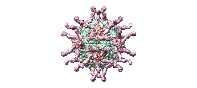 Figure: polio virus
Characteristics of Poliovirus
- Poliovirus is a member of a family of viruses called Picornaviridae. The Picornaviridae includes many other human pathogens, such as Coxsackieviruses, echoviruses, enteroviruses, hepatitis A virus, and rhinoviruses. Poliovirus is one of the smallest viruses.
- Poliovirus is a small and simple virus composed of a shell, or capsid, made of protein. The poliovirus capsid is about 30 nanometers in diameter.
- A single molecule of ribonucleic acid (RNA) is found within the capsid. This RNA carries genetic information to make new virus particles. **It has single-stranded RNA of positive polarity.**Its genome is infective. The infectious nucleic acid of poliovirus can bypass the host range specificity provided by the viral protein-cell receptor interaction. For example, intact poliovirus can grow only in human or primate cells, but purified poliovirus RNA can also enter and replicate in nonprimate cells.
- **Polioviruses have three serotypes.**Based on the nature of surface antigens, polioviruses have three different antigenic determinants (poliovirus type 1, poliovirus type 2, and poliovirus type 3).
- Poliovirus has a narrow range of specificity. It can enter the cells of only humans and other primates.
Pathogenesis
People acquire poliovirus infection from other humans through contact with virus-containing feces or contaminated water. The virus multiplies in the cells of the gastrointestinal tract and lymphatic tissue (tonsils/payers patches). Newly synthesized virus particles are released into the intestine and shed in the feces.
After multiplying in the gastrointestinal tract, poliovirus spreads to the regional lymph nodes and enters the bloodstream, producing a minor (first) viremia. In most people, the immune response clears the virus at this stage and infection remains subclinical. In a minority of cases, viral replication continues and a major (second) viremia develops, with sufficiently high blood titers to breach the blood-brain barrier and enter the spinal cord and brain. Whether major viremia occurs depends on the host's immune response, the infecting serotype, and possibly inoculum size; it is this progression that determines the small fraction of infections that ultimately result in CNS involvement and paralysis.
Direct neural transmission may also occur, e.g., after tonsillectomy. In the CNS, Virus multiplies selectively in the neurons and destroys the anterior horn cells of the spinal cord. The destruction of motor neurons by the virus leads to limb paralysis.
The earliest histological change is degeneration of Nissl bodies (the rough endoplasmic reticulum of neurons), a sign that the infected motor neuron's protein synthesis machinery is already being disrupted before the cell dies. When degeneration becomes irreversible, the necrotic cell lyses or is phagocytosed. Viruses don’t multiply in muscle in vivo. Changes in peripheral nerves and voluntary muscles are secondary to the destruction of nerve cells.
Clinical findings
The incubation period is 7-14 days. The manifestations may range from the asymptomatic stage (>90 % cases) to the most severe paralytic disease (<1%).
- Inapparent infections: Following infection, the majority (91-96%) of cases are asymptomatic.
- **Abortive infection:**About 5% of patients develop minor symptoms such as fever, malaise, sore throat, anorexia, myalgia, and headache.
- Nonparalytic poliomyelitis is seen in 1% of patients who presented with aseptic meningitis.
- Paralytic poliomyelitis is the least common form (<1%) among all the stages and is characterized by descending asymmetric acute flaccid paralysis (AFP). Proximal muscles are affected earlier than the distal muscles; paralysis starts at the hip and proceeds towards the extremities, which leads to the characteristic tripod sign (child sits with flexed hip, both arms are extended towards the back for support). Sites involved can be spinal, bulbospinal, and bulbar. Accordingly, the nature of paralysis varies (e.g., respiratory insufficiency or dysphagia are common in bulbar involvement).
Lab diagnosis
Sample: Poliovirus can be detected in specimens from the throat, feces (stool), and occasionally blood or cerebrospinal fluid (CSF).
Virus Isolation and Detection
Virus isolation from stool specimens is the most sensitive method to diagnose poliovirus infection. Collecting at least two stool specimens 24 hours apart from suspected poliomyelitis patients is recommended to increase the probability of isolating poliovirus. Poliovirus may also be isolated from pharyngeal swabs. Isolation is less likely from blood or CSF. Samples should be collected early during the disease (ideally within 14 days after onset).
Primary monkey kidney cells are the most recommended cell lines. Various methods can identify virus growth.
- Cytopathic effects appear in 3-6 days; described as crenation and degeneration of the entire cell sheet.
- Isolated viruses can be identified and serotyped by neutralization with a specific antiserum.
- Specific gene sequences of the virus can be detected by polymerase chain reaction (PCR) assays.
Serology
For the patients who did not receive polio vaccination, the serological test may help support the diagnosis of paralytic poliomyelitis. An acute serum specimen should be obtained early in the course of the disease, and a convalescent specimen should be obtained at least three weeks later.
A four-fold rise in antibody titer in paired sera confirms the diagnosis. Neutralization and complement fixation tests can be useful to measure the concentration of antibodies.
Prevention
Vaccination is the best way to protect people and stop the transmission of Poliomyelitis. There are two types of vaccine;inactivated poliovirus vaccine (IPV) and oral poliovirus vaccine (OPV).
Two types of vaccine are available against poliomyelitis, inactivated vaccine (IPV, Salk) and a live attenuated oral vaccine (OPV, Sabin). Both vaccine formulations contain all three polio types.
Oral Polio Vaccine (OPV) /Sabin
The oral polio vaccine was the most widely used vaccine for the prevention of poliomyelitis. It is composed of attenuated strains of the three poliovirus types and is administered orally. It has been instrumental in nearly eradicating the virus from the planet. Lately, WHO has recommended the replacement of OPV with inactivated poliovirus vaccine (IPV) as scientist finds evidence of reversion to virulence in OPV strains.
At least two or three doses are considered necessary to ensure adequate immunity. In some countries, even five to six or more doses are given in the primary course. Revaccination is used to varying degrees. A full primary course induces an antibody response against all three types in more than 90% of vaccinated individuals and gives a high degree of protection against disease.
OPV also induces intestinal immunity due to the production of secretory IgA antibodies. This is important for inhibiting virus replication in the gut, diminishing the virus’s possible spread to susceptible contacts.
OPV is almost non-reactogenic and is very safe. However, the attenuated vaccine strain can very rarely revert toward virulence and induce paralytic disease (vaccine-associated paralytic poliomyelitis, VAPP) in about one case per 1–10 million doses. The OPV strains are stabilised with MgCl2; shelf life at 4–8°C is four months and at -20°C is two years. Cold chain failure is an important contributor to OPV's apparent failure in some LMIC settings, since the attenuated live virus is heat-sensitive. If a child's alimentary tract is colonised by another enterovirus at the time of vaccination, establishment of OPV-induced immunity may be blocked. Breastfeeding immediately before or after OPV may also transiently neutralise the vaccine virus.
Inactivated Poliovirus Vaccine (IPV)
Inactivated poliovirus vaccine (IPV) was the first vaccine used against poliomyelitis. It contains all three types of poliovirus inactivated by formaldehyde and is administered parenterally. The use of IPV in the late 1950s was followed by a 90% reduction of poliomyelitis cases when it was replaced in many countries by the more easily administered OPV around 1960. Newer IPVs have higher immunogenic potency, leading to a reintroduction of IPV in many developed and developing countries.
The primary vaccination course with IPV consists of two or three doses, usually followed by revaccination after intervals of about 5–10 years during childhood and adolescence. Some countries use a sequential or combined OPV/IPV schedule. Unlike OPV, IPV does not induce substantial intestinal secretory IgA, meaning IPV-vaccinated individuals are protected from paralysis but may still transiently carry and shed wild poliovirus in the gut if exposed. As wild polio nears eradication globally, WHO now recommends that all countries transition to at least one IPV dose in their routine immunisation schedules to eliminate VAPP risk entirely. Current WHO guidance (2016) supports IPV as the preferred vaccine for countries where wild poliovirus transmission has been interrupted.
How to Remember
Poliovirus is HAV's structural cousin, which explains fecal-oral spread immediately. Both are Picornaviridae, both are small, naked (non-enveloped) positive-sense ssRNA viruses, and both use the same bare-capsid survival strategy that lets them withstand stomach acid and gut bile salts long enough to establish GI infection. If you already remember why HAV spreads through contaminated food and water (naked capsid = acid-resistant = survives the gut), poliovirus spreads the same way for the same reason. No new logic is needed; this is the same mechanism applied to a different family member.
The two-phase viremia is the key to understanding who gets paralysis. Phase one (minor viremia) is a brief, low-level seeding of the bloodstream from gut lymph nodes, most people's immune system mounts an antibody response here and clears the virus before anything else happens; this is the >90% asymptomatic group. Phase two (major viremia) only occurs if the first-phase viremia isn't cleared quickly enough, virus levels rise high enough in the blood to breach the blood-brain barrier and enter the CNS. The determinants of whether major viremia occurs include serotype (type 1 is the most paralytogenic), prior immune status, and possibly inoculum size. Paralysis happens in fewer than 1% of infections because most immune systems win the race during phase one.
OPV protects the gut; IPV protects the bloodstream: the distinction that determines which vaccine wins the eradication race. OPV (swallowed) replicates in the gut, stimulating local secretory IgA production at the mucosal surface, exactly where poliovirus first lands and replicates. A person vaccinated with OPV who encounters wild poliovirus doesn't just avoid paralysis, the gut IgA blocks the virus from establishing itself in the GI tract at all, meaning it can't be shed in stool and can't spread to contacts. IPV (injected) produces excellent serum IgG that prevents major viremia and therefore prevents paralysis, but it doesn't reliably prevent gut replication or fecal shedding. Once wild poliovirus is eliminated from a region, VAPP (vaccine-associated paralytic poliomyelitis, roughly one case per 1-10 million OPV doses) is no longer an acceptable trade-off since there's no longer wild-type transmission to stop. That's why WHO switched the recommendation from OPV to IPV once eradication became achievable.
Where Students Get Confused
"More infection means more paralysis." Not for poliovirus. Over 90% of infections are completely asymptomatic; paralysis occurs in fewer than 1% of all infections. Severity is determined by whether the virus achieves major viremia and reaches the CNS, which depends on host immune response, serotype, and timing, not simply on being infected.
"OPV and IPV protect against polio in the same way." They don't. IPV produces serum IgG that prevents major viremia and paralysis. OPV additionally produces intestinal secretory IgA that blocks gut replication and fecal shedding entirely, meaning OPV-vaccinated contacts also become non-transmitters of the virus. This difference is exactly why OPV was chosen for the global eradication campaign (community transmission protection), and why VAPP risk becomes unacceptable once wild polio is eliminated (the community benefit that justified the risk no longer exists).
"The Sabin and Salk vaccines were just early, cruder versions of the same thing." They represent genuinely different vaccine strategies. Salk (IPV) uses killed virus and induces systemic immunity only. Sabin (OPV) uses live attenuated virus that replicates in the gut and induces both systemic and mucosal immunity. The trade-off between mucosal protection and VAPP risk is a real, ongoing policy decision, not a historical curiosity.
"Poliovirus paralysis is permanent in all cases." Not always. Some motor function can return as inflammation subsides and surviving neurons compensate. However, neurons destroyed by virus lysis cannot regenerate, making permanent deficit common in severe cases, and post-polio syndrome (progressive weakness 25-30 years after the original infection) can occur even in those who partially recovered.
"If a child has a normal stool culture at one point, polio is ruled out." Not reliably. WHO recommends at least two stool specimens collected 24 hours apart to adequately exclude poliovirus, because single-sample sensitivity is insufficient. This is the basis of the standard surveillance protocol for acute flaccid paralysis cases.
Key Exam Facts Table
| Feature | Detail |
|---|---|
| Family / Genus | Picornaviridae / Enterovirus |
| Genome | Positive-sense ssRNA, ~7.5 kb; genome alone is infectious |
| Capsid | Naked (non-enveloped), icosahedral, ~30 nm |
| Serotypes | 3 (Type 1 most paralytogenic, most epidemic; Type 2 eradicated 2015; Type 3 eradicated 2019) |
| Transmission | Fecal-oral (primary); respiratory droplets (minor) |
| Host range | Humans and other primates only (narrow specificity) |
| Receptor | CD155 (poliovirus receptor, PVR) — only on human/primate cells |
| Incubation period | 7–14 days |
| % asymptomatic | >90% |
| % paralytic | <1% |
| Paralysis type | Acute flaccid paralysis (AFP); asymmetric; proximal before distal |
| Classic sign | Tripod sign (flexed hip, extended arms for support) |
| Earliest histological change | Degeneration of Nissl bodies in anterior horn cells |
| OPV (Sabin) immunity | Systemic IgG + intestinal secretory IgA; blocks gut replication and shedding |
| IPV (Salk) immunity | Systemic IgG only; prevents paralysis but doesn't reliably block gut shedding |
| VAPP risk (OPV) | ~1 case per 1–10 million doses |
| Why IPV preferred now | With wild polio near-eradicated, VAPP risk from OPV exceeds residual wild-type risk |
| Gold standard lab diagnosis | Virus isolation from stool (two specimens, 24 hours apart) |
| Cell line for isolation | Primary monkey kidney cells |
| CPE pattern | Crenation and degeneration of entire cell sheet, 3–6 days |
References and Further Reading
- Menant, J. C., & Gandevia, S. C. (2018). Poliomyelitis. Handbook of Clinical Neurology, 159, 337–344. https://doi.org/10.1016/B978-0-444-63916-5.00021-5
- Skern, T. (2010). 100 years poliovirus: from discovery to eradication. A meeting report. Archives of Virology, 155(9), 1371–1381. https://doi.org/10.1007/s00705-010-0778-x
- Alleman, M. M., Wannemuehler, K. A., Weldon, W. C., Kabuayi, J. P., Ekofo, F., Edidi, S., Mulumba, A., Mbule, A., Ntumbannji, R. N., Coulibaly, T., Abiola, N., Mpingulu, M., Sidibe, K., & Oberste, M. S. (2014). Factors contributing to outbreaks of wild poliovirus type 1 infection involving persons aged ≥15 years in the Democratic Republic of the Congo, 2010–2011, informed by a pre-outbreak poliovirus immunity assessment. The Journal of Infectious Diseases, 210(Suppl 1), S62–S73. https://doi.org/10.1093/infdis/jiu282
- Nathanson, N., & Kew, O. M. (2010). From emergence to eradication: the epidemiology of poliomyelitis deconstructed. American Journal of Epidemiology, 172(11), 1213–1229. https://doi.org/10.1093/aje/kwq320
- Chard, A. N., Martinez, M., Matanock, A., & Kassem, A. M. (2021). Estimation of oral poliovirus vaccine effectiveness in Afghanistan, 2010–2020. Vaccine, 39(42), 6250–6255. https://doi.org/10.1016/j.vaccine.2021.09.020
- World Health Organization. (2010). Polio vaccines and polio immunization in the pre-eradication era: WHO position paper–recommendations. Vaccine, 28(43), 6943–6944. https://doi.org/10.1016/j.vaccine.2010.08.023
- Heinsbroek, E., & Ruitenberg, E. J. (2010). The global introduction of inactivated polio vaccine can circumvent the oral polio vaccine paradox. Vaccine, 28(22), 3778–3783. https://doi.org/10.1016/j.vaccine.2010.02.095
- World Health Organization. (2016). Polio vaccines: WHO position paper — March 2016. Weekly Epidemiological Record, 91(12), 145–168. https://apps.who.int/iris/handle/10665/204457
Frequently Asked Questions
Why do most people infected with poliovirus never get paralysed?
What is the difference between OPV and IPV in terms of the immunity they produce?
What is VAPP and why does it matter for the switch from OPV to IPV?
Why is stool the preferred specimen for poliovirus diagnosis over blood or CSF?

Tankeshwar Acharya, MSc (Medical Microbiology)
Tankeshwar Acharya is an Assistant Professor in the Department of Microbiology at Patan Academy of Health Sciences (PAHS), Nepal, where he has been teaching and practicing clinical microbiology for over 14 years. He is the founder of Microbe Online, one of the leading free microbiology education resources on the web, covering bacteriology, mycology, parasitology, immunology, and clinical laboratory diagnostics written from direct experience in both the classroom and the diagnostic laboratory.