
Monospot Test: Principle, Procedure, Results, and When to Use EBV-Specific Tests
The Monospot test detects heterophile antibodies to screen for infectious mononucleosis (EBV). Learn its principle, procedure, false positive and negative causes, age-related limitations, and when EBV-specific antibodies (VCA IgM/IgG, EBNA) are needed instead.
An 18-year-old university student presents with five days of sore throat, fever, and profound fatigue. On examination she has cervical lymphadenopathy, tonsillar exudate, and a palpable spleen tip. Her clinician suspects infectious mononucleosis and orders a Monospot test.
The result returns positive within an hour. Amoxicillin — which would be reasonable for bacterial tonsillitis — is deliberately withheld. Between 80–100% of infectious mononucleosis patients who receive ampicillin or amoxicillin develop a widespread maculopapular rash, an idiosyncratic drug reaction triggered by the EBV-induced immune state rather than a true drug allergy. The patient is advised on rest, hydration, and to avoid contact sports for at least four weeks because of splenomegaly and splenic rupture risk.
This scenario captures the clinical value of the Monospot: rapid, inexpensive confirmation of EBV infection that directly changes management — preventing the ampicillin rash and prompting counselling about splenic rupture.
The monospot test, also known as the Paul-Bunnell test, is a form of the heterophile antibody test used to rapidly screen infectious mononucleosis (mono). Epstein–Barr virus (EBV) causes the disease infectious mononucleosis. Blood, serum, or plasma is taken and is looked for the presence of heterophile antibodies.
When a person is infected with Epstein-Barr virus (EBV), the immune system makes antibodies to fight this infection which is detected in the blood using this test. Monospot testing can usually detect antibodies two to nine weeks after infection.
Paul and Bunnell were the first scientists who suggested hemagglutination of sheep RBC to detect heterophile antibodies associated with infectious mononucleosis.
What Are Heterophile Antibodies?
"Heterophile" (Greek: different species) refers to antibodies that cross-react with antigens from species unrelated to the one that triggered them. In EBV infection, the immune response against the virus also generates cross-reactive IgM antibodies that agglutinate red blood cells from certain animal species — horse, sheep, and ox — despite these animals having no relationship to EBV.
This cross-reactivity is reproducible and diagnostically useful: approximately 85–90% of adolescents and adults with infectious mononucleosis produce detectable heterophile antibodies. They appear within the first two weeks of illness, peak in weeks 2–3, and disappear within 3–6 months.
Why the test is called "Monospot": The Monospot is a commercial refinement of the original Paul-Bunnell test (1932), which detected heterophile antibodies by agglutination of sheep red blood cells. The Monospot uses preserved horse erythrocytes and guinea pig kidney antigen as the absorption reagent, streamlining the test to a single rapid slide read in minutes.
Sample
Serum/Plasma: Serum or plasma may be used with the monospot test, provided an acceptable medical technique collects whole blood. Avoid using grossly hemolyzed sera. The serum should be clear and free of bacterial contamination. Specimens can be stored at 2 to 8°C for 24 hours. For prolonged storage, specimens should be frozen (-20°C).
Principle
A sample of the patient’s serum or plasma is placed on a microscope slide and mixed with guinea pig kidney antigen and preserved horse erythrocytes (these reagents can be purchased from commercial suppliers of kits). Thoroughly mix the reagents and rock the slide for a minute. Suppose heterophil antibodies are present in the serum/plasma and the blood clumps (agglutinates). This result usually indicates a mono infection. It typically is not used to diagnose mono that started more than six months earlier. The results of a monospot test are usually ready within one hour.
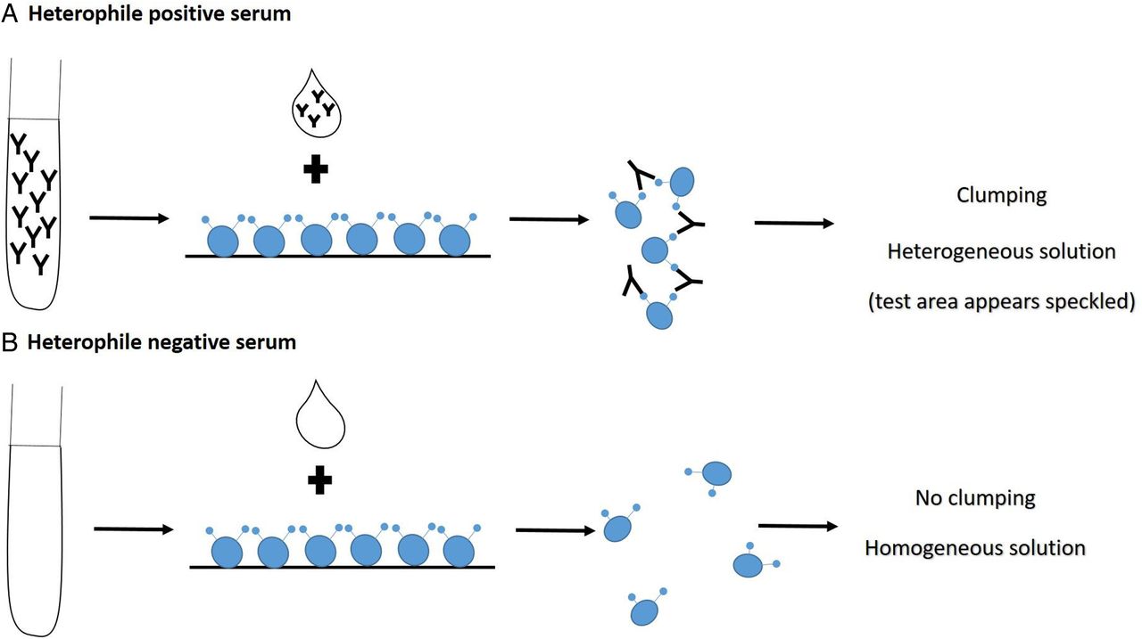 Figure: Heterophile-antibody test
Results:
- Normal (negative): The blood sample does not form clumps (no heterophil antibodies are detected).
- Abnormal (positive): The blood sample clumps (heterophil antibodies are detected). If the blood sample clumps, you probably have mono.
The absorption step — why guinea pig kidney is used:
Normal human serum contains background heterophile antibodies (Forssman antibodies) that non-specifically agglutinate horse red blood cells. Guinea pig kidney antigen absorbs these Forssman antibodies before testing, leaving only the EBV-associated heterophile antibodies. The EBV-specific heterophile antibodies are NOT absorbed by guinea pig kidney — this selectivity is the basis of the test's diagnostic specificity.
This is also why the Monospot card has two circles: the LEFT circle (guinea pig kidney) identifies non-specific Forssman-type agglutination; the RIGHT circle (horse RBCs) detects EBV-specific heterophile antibodies. A true positive shows agglutination on the RIGHT only.
Procedure
- Allow all reagents and patient samples to reach room temperature (20–25°C) before testing.
- Place one drop of guinea pig kidney antigen suspension on the LEFT circle of the reaction card.
- Place one drop of horse erythrocyte suspension on the RIGHT circle.
- Using a clean disposable applicator stick, add one drop of patient serum to both circles.
- Using separate applicator sticks for each circle, mix the contents and spread evenly across each circle.
- Rock the card gently for exactly 2 minutes.
- Read results immediately under a good light source.
Reading results:
| Pattern | Interpretation |
|---|---|
| RIGHT circle agglutinates; LEFT circle does NOT | Positive — EBV heterophile antibodies present |
| Neither circle agglutinates | Negative |
| BOTH circles agglutinate | Equivocal — non-specific Forssman antibodies; follow up with EBV-specific serology |
| LEFT only agglutinates | Equivocal — non-specific; not diagnostic for mono |
Quality control: Run positive and negative controls with each batch. If controls do not perform as expected, results are invalid.
Sensitivity by Age and Timing of Monospot Test
| Patient group | Sensitivity | Clinical implication |
|---|---|---|
| Adolescents and adults (>12 years) | 85–90% | Most reliable age group; standard first-line test |
| Children 4–12 years | ~60–75% | Consider EBV-specific testing if Monospot negative |
| Children <4 years | 25–50% | Not recommended as first-line — use EBV-specific antibodies |
| First week of illness | ~75% | May be seronegative early; repeat at 1–2 weeks if negative |
| Weeks 2–3 | ~90% | Peak heterophile antibody production |
| >6 months after illness | Declining | Not useful retrospectively; VCA IgG more appropriate |
Specificity: ~99% in adolescents and adults — false positives are rare.
Studies have shown that monospot test produces both false-positive and false-negative results. Results should be correlated with clinical and hematological findings.
- False Positive Reactions: The antibodies detected by monospot can be caused by conditions other than infectious mononucleosis. It may occur in persons with: Leukemia or lymphoma Hepatitis Rubella Systemic lupus erythematosus (SLE) Toxoplasmosis
- False Negative Reactions Children, especially those younger than two years, are more likely than adults to have a negative monospot test (25%–50%), even when they have infectious mononucleosis. It takes time to develop antibodies if the test is done within the first few weeks of becoming infected with EBV (approx. 25% false-negative rate in the first week). This may lead to a false-negative result. If the first test does not indicate mono but still has symptoms, the test may be repeated.
When the Monospot is Negative: EBV-Specific Antibody Testing
A negative Monospot does not rule out EBV infection — it rules out detectable heterophile antibodies. In children under 4, early presentations, or strong clinical suspicion with a negative Monospot, EBV-specific antibody testing is needed.
EBV-specific antibody markers:
| Antibody | Timing | Persists | Interpretation |
|---|---|---|---|
| VCA IgM | 1–2 weeks after onset | 4–8 weeks | Active primary EBV infection |
| VCA IgG | 1–2 weeks after onset | Lifelong | Past or current infection |
| EA-D IgG | Acute phase | Months | Active viral replication |
| EBNA IgG | 2–4 months after infection | Lifelong | Critical: ABSENT in acute primary infection |
The diagnostic pattern for primary EBV infection:
VCA IgM positive + VCA IgG positive + EBNA IgG negative = Primary EBV infection confirmed
Why EBNA IgG negativity is the key discriminator: EBNA IgG develops late (2–4 months) and persists for life. If EBNA IgG is already positive in a patient presenting with apparent mono, this is NOT a primary EBV infection — they had EBV before. The current illness may be due to another cause: CMV, toxoplasmosis, HIV primary infection, or other causes of mononucleosis syndrome.
Clinical Context: Infectious Mononucleosis
The Monospot is ordered when infectious mononucleosis is suspected. Recognising the syndrome guides management:
Classic triad: Fever + pharyngitis (often severe, white exudate) + lymphadenopathy (posterior cervical most characteristic)
Additional features:
- Splenomegaly (50%) — contact sports contraindicated ≥4 weeks; splenic rupture risk
- Mild transaminase elevation (80%)
- Periorbital oedema (Hoagland's sign)
- Palatal petechiae
- Maculopapular rash (5% spontaneously; 80–100% if given ampicillin/amoxicillin)
The ampicillin rash: Not a true drug allergy — it is an EBV-immune-mediated reaction. It resolves without desensitisation. The patient can receive penicillin for future unrelated infections — but amoxicillin during the EBV illness will cause the rash again.
Uses of Monospot Test
The monospot test is not recommended for general use. Monospot test may indicate that a person has a typical case of infectious mononucleosis, but does not confirm the presence of EBV infection.
How to Remember
Heterophile = cross-species antibodies — EBV accidentally makes antibodies that agglutinate horse red cells. The "accident" is diagnostically useful because it is reproducible. The Monospot exploits this cross-reactivity.
The two-circle card logic:
- LEFT circle (guinea pig kidney): absorbs non-specific Forssman antibodies — agglutination here = non-specific
- RIGHT circle (horse RBCs): EBV-specific heterophile antibodies cause agglutination here
Positive = RIGHT agglutinates + LEFT does not.
The age rule — "under four, use more": Under 4 years, Monospot sensitivity falls to 25–50%. In young children with suspected EBV, go straight to EBV-specific serology (VCA IgM/IgG, EBNA).
The EBNA rule — the late marker that rules out primary infection: EBNA IgG takes 2–4 months to appear. If EBNA IgG is already positive in a patient with apparent mono → this is NOT primary EBV. Look for another cause of the mononucleosis syndrome.
The ampicillin anchor — the test that prevents a rash: The Monospot is the test that prevents doctors from giving amoxicillin to EBV patients. Confirming mono before treating pharyngitis saves the patient an unnecessary widespread rash and avoids a lifelong false label of penicillin allergy.
References and further readings
- Marshall-Andon T, Heinz P. How to use … the Monospot and other heterophile antibody tests. Archives of Disease in Childhood – Education and Practice2017;102:188-193.
- Luzuriaga K., Sullivan J.L. (2010). Infectious mononucleosis. New England Journal of Medicine, 362(21), 1993–2000. https://doi.org/10.1056/NEJMcp1001116
- Tille, P. M. (2017). Bailey and Scott's Diagnostic Microbiology (14th ed.). Elsevier.
Frequently Asked Questions
Why does the ampicillin/amoxicillin rash occur in infectious mononucleosis, and does it indicate a true penicillin allergy?
What other conditions can cause a positive Monospot test besides EBV infection?
How do EBV-specific antibody tests differentiate primary EBV infection from reactivation or past infection?

Tankeshwar Acharya, MSc (Medical Microbiology)
Tankeshwar Acharya is an Assistant Professor in the Department of Microbiology at Patan Academy of Health Sciences (PAHS), Nepal, where he has been teaching and practicing clinical microbiology for over 14 years. He is the founder of Microbe Online, one of the leading free microbiology education resources on the web, covering bacteriology, mycology, parasitology, immunology, and clinical laboratory diagnostics written from direct experience in both the classroom and the diagnostic laboratory.