
Embryonated Egg Culture for Viruses: Sites, Procedure, and Vaccine Use
Why influenza, herpes, and arboviruses each need a different site inside the egg, and how this method still makes most flu vaccines today.
Every flu season, somewhere around 100-150 million eggs are used in the US alone just to manufacture that year's influenza vaccine. A nursing student administering that vaccine might reasonably be asked by a patient: "I'm allergic to eggs, is this vaccine safe for me?" Answering that question correctly requires actually understanding how the vaccine is made, not just memorizing that eggs are used.
Why eggs at all, when cell culture exists and is used for plenty of other viruses? The answer is practical, not biological: a fertilized chicken egg is a self-contained, sterile, immunologically inert bioreactor. It has no functioning immune system yet, so an inoculated virus replicates undisturbed; it's cheap, widely available, and doesn't require the trained technicians and specialized equipment cell culture demands. For a virus like influenza that needs to be produced at enormous, vaccine-supply scale every single year, that combination of cost and convenience still beats most alternatives, even decades after cell-based methods became available.
But not every virus grows equally well everywhere inside the egg, and that's where this article's real teaching point lives: the egg isn't one uniform environment, it's several different tissues (yolk sac, amniotic sac, allantoic sac, chorioallantoic membrane), each with different cell types and different susceptibility to different viruses. Choosing the right site isn't arbitrary, it's the same principle as choosing the right cell line in tissue culture, match the virus to the tissue it actually infects well.
Embryonated chicken egg is used for the culture of some viruses. The viruses grow in the cells of the embryo and membranes.
Specimens are inoculated into pathogen-free fertilized eggs of 10-11 days and are incubated for 2-9 days before harvesting the viruses. Growth and multiplication of the viruses are indicated by the death of the embryo, or by the formation of typical pocks or lesions on the egg membranes.
The embryonated chicken egg was first used for the cultivation of viruses by Goodpasture (1931) and was later extensively developed by Burnet.
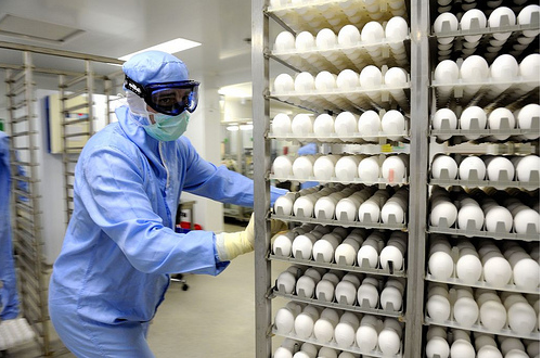 Figure: Handling of eggs in a influenza vaccine plant
Before the development of cell lines for the culture of viruses, egg inoculation was one of the preferred methods of virus cultivation. Until now, egg culture is a preferred method for manufacturing both inactivated influenza vaccine (“flu shot”) and live attenuated (weakened) vaccine (“nasal spray flu vaccine”). Many manufacturers are also producing cell-based influenza vaccine.
Egg Inoculation
Specimens are injected into different sites (different cells) within fertilized eggs. These sites are different for different viruses (specificity of the virus). Inoculation sites are;
- Yolk Sac
- Amniotic Sac
- Chorioallantoic Membrane
- Allantoic Sac
Egg inoculation sites are selected based on the virus yield which in turn depends on the susceptibility and permissiveness of these cells.
The eggs used for cultivation must be sterile and the shell should be intact and healthy. The handling of the eggs should be done in sterilized conditions.
Instead of manual injection by laboratory workers, vaccine manufacturing plants use automated machines to inoculate eggs and to harvest viruses after incubation.
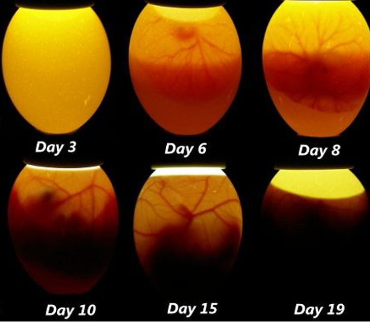 Figure: Candling of embryonated eggs to see chicken embryo inside
| Inoculation method | Name of viruses |
|---|---|
| Allantoic inoculation | Influenza virus, Mumps virus, Newcastle disease virus, Avian adenovirus |
| Amniotic inoculation | Influenza virus, Mumps virus |
| Chorioallantoic membrane inoculation | Herpes simplex virus, Poxvirus, Rous sarcoma irus |
| Yolk sac inoculation | Herpes simplex virus |
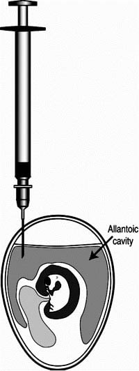 Figure: Allantoic inoculation
How to Remember Sites
Allantoic sac: the "mass production floor." Influenza, mumps, and Newcastle disease virus are all grown here because the allantoic cavity is large (about 10 mL of fluid per egg) and the cells lining it support efficient, high-yield replication, exactly the property you want when manufacturing vaccine doses at a scale of hundreds of millions of eggs per season. Think of the allantoic sac as the high-volume factory floor of the egg.
Amniotic sac: the "first isolation room." It's the preferred site for primary isolation of influenza virus, meaning the first time a clinical sample is being grown, before the virus has been adapted to grow well elsewhere. Newly isolated clinical strains often replicate more reliably here before they're later adapted to the higher-yield allantoic sac for production purposes.
Chorioallantoic membrane (CAM): the "visible scoreboard." Herpes simplex virus and poxviruses are grown on the CAM because they produce visible pock lesions you can actually count, and since each pock comes from a single infectious virus particle, counting pocks is a direct, visual way to quantify how much infectious virus is in a sample, similar in spirit to colony counting on an agar plate. If a question describes "pock counting" as a quantification method, it's testing CAM inoculation.
Yolk sac: the "yolk likes intracellular parasites." Arboviruses like Japanese encephalitis virus, and obligate intracellular bacteria like Rickettsia and Chlamydia, are grown in the yolk sac. A useful anchor: yolk sac cells are particularly good hosts for organisms that need to live and replicate strictly inside cells, which describes both arboviruses and these two obligate intracellular bacterial genera, explaining why this site shows up for bacteria too, not just viruses, in egg culture discussions.
Allantoic Sac
The allantoic cavity is a larger cavity found in fertilized eggs and contains about 10 mL fluid per egg. It is lined with cells and after virus inoculation, the virus replicates in those cells. It is the most convenient method for the propagation of Newcastle disease virus.
An allantoic cavity is also commonly used for manufacturing viral vaccines such as influenza vaccine, yellow fever vaccine, and rabies vaccine.
Procedure of Allantoic Cavity Inoculation
For propagation of influenza virus
- Take pathogen-free fertilized chicken eggs of 11-12 days.
- Locate a non-veined area of the allantoic cavity which is located just below the air sac placing the egg in front of a light source. Mark the area with a pencil
- Make a small nick in the marked area using a jeweler’s scribe.
- Drill a hole at the top of the egg with a Dremel motorized tool. This is done to decrease the pressure of the air sac to prevent the leakage of inoculum.
- Inoculate the eggs using a tuberculin syringe (a 1 ml syringe fitted with a 1/2 inch, 27 gauge needle).
- Pass the needle through the hole in the shell, through the chorioallantoic membrane, and inject the inoculum in the allantoic cavity, which is filled with allantoic fluid.
- Seal these two holes of the shell with melted paraffin.
- Incubate the eggs at 37 degrees C for 48 hours.
During the incubation period, the virus replicates in the cells and virus particles are released by budding into the allantoic fluid.
Harvesting the virus
- Remove the top of the eggshell (the part covering the air sac).
- Pierce the shell membrane and chorioallantoic membrane with a pipette.
- Withdraw about 10 mL of allantoic fluid per egg.
Depending on the virus strain, one or two eggs will produce sufficient virus to produce one 15 microgram dose of vaccine.
Amniotic Sac
It is mainly used for the primary isolation of the influenza virus. Viral growth is measured by the detection of hemagglutinin antigens in amniotic fluid.
Chorio-allantoic Membrane
It is preferred for the Herpes Simplex virus and the pox virus. Viruses produce visible lesions as pocks on the chorioallantoic membrane. Each pock is derived from a single virion so the number of pocks would represent the number of viruses present in the inoculum.
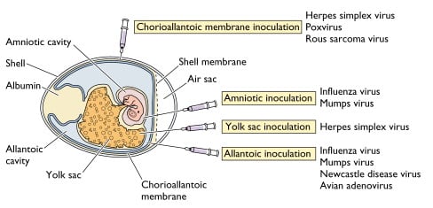 Figure: Cutaway view of an embryonated chicken egg
Yolk Sac
It is a preferred method for arboviruses (e.g. Japanese Encephalitis Virus) and some obligate intracellular bacteria such as Rickettsia and Chlamydia. The growth of encephalitis viruses may result in the death of an embryo.
Advantages of Egg Inoculation
- Egg inoculation is a convenient and easy method to grow various types of viruses (many avian and few mammalian viruses) in massive quantities. In the US alone, 100-150 million eggs are used to make the flu vaccine.
- Different viruses can be injected into an egg at different sites and the egg can be easily observed for viral replication throughout the development of an embryo.
- Eggs have no developed immunologic functions.
- Eggs are free from bacteria and many latent viruses.
- Other advantages include easy availability, less cost, easier maintenance, and less labor.
Disadvantages of Egg Inoculation
Not every virus grows in eggs. Many clinically important viruses, including most enteroviruses and a number of respiratory viruses other than influenza, either don't replicate well in egg tissue or require cell culture for reliable isolation. Egg culture is useful for a specific subset of viruses, not a universal method.
Egg allergy is a genuine clinical consideration for egg-based vaccines. Since egg-grown influenza vaccines may contain trace amounts of egg protein (ovalbumin), this is a real question patients ask, and the answer matters clinically: current guidance from major health bodies generally considers egg-based flu vaccines safe even for most people with egg allergy, including severe allergy, though clinicians should still follow local vaccination guidelines and be prepared to monitor for reactions. Knowing the manufacturing method is what allows you to actually answer a patient's question here, rather than just reassuring them without explanation.
Batch-to-batch and egg-to-egg variability. Biological variation between eggs (and between flocks, seasons, and suppliers) can affect viral yield, requiring quality control testing that adds time and cost to vaccine production.
Antigenic changes can occur during egg adaptation. Influenza virus grown in eggs can acquire mutations that help it adapt to egg tissue, sometimes altering the surface antigens (hemagglutinin) the vaccine is meant to match against circulating human strains. This is one reason cell-based and recombinant influenza vaccines have been developed as alternatives, they avoid this egg-adaptation risk entirely.
Time-consuming relative to some cell culture methods, and dependent on a reliable supply of pathogen-free fertilized eggs, which itself requires dedicated, specialized poultry flocks rather than ordinary commercial eggs.
Where Students Get Confused
"Egg-based flu vaccines are unsafe for people with egg allergy." This is a common patient (and sometimes student) misconception. Current guidance from major health authorities generally supports vaccinating even severely egg-allergic individuals with egg-based influenza vaccines, since modern manufacturing reduces residual egg protein and the actual reaction risk has been found to be very low in studies. This doesn't mean zero caution is needed, standard vaccination safety monitoring still applies, but "contains egg protein" doesn't automatically mean "contraindicated in egg allergy."
"Pock counting on the CAM and plaque counting in cell culture are different concepts." They're not, conceptually. Both rely on the same underlying principle: a single infectious virus particle produces one visible, countable unit of damage (a pock on the CAM, a plaque in a cell monolayer), allowing quantification of infectious virus titer. If you understand plaque assays from cell culture, pock counting is the same idea in a different host system.
"All four egg sites are basically interchangeable, just pick any one." They're not interchangeable, site selection depends on which virus is being grown and what the goal is (primary isolation vs. mass production vs. visible quantification), exactly the logic laid out in the How to Remember section above. Using the wrong site for a given virus can mean poor yield or no detectable growth at all.
References and further readings
- FAO. (n.d.). Inoculation of embryonated eggs by the allantoic cavity route. Food and Agriculture Organization of the United Nations. (Verify current URL in CMS before inserting; FAO occasionally restructures URLs.)
- Centers for Disease Control and Prevention. (2019). How influenza (flu) vaccines are made. CDC. https://www.cdc.gov/flu/vaccine-process/index.html
- Racaniello, V. (2009). Influenza virus growth in eggs. Virology Blog. https://virology.ws/2009/12/10/influenza-virus-growth-in-eggs/
Frequently Asked Questions
Is the flu vaccine unsafe for people with egg allergy?
Why is egg culture still used for flu vaccines when cell culture exists?
Why do different viruses need to be inoculated into different parts of the egg?
What is a "pock" and why does counting them matter?

Tankeshwar Acharya, MSc (Medical Microbiology)
Tankeshwar Acharya is an Assistant Professor in the Department of Microbiology at Patan Academy of Health Sciences (PAHS), Nepal, where he has been teaching and practicing clinical microbiology for over 14 years. He is the founder of Microbe Online, one of the leading free microbiology education resources on the web, covering bacteriology, mycology, parasitology, immunology, and clinical laboratory diagnostics written from direct experience in both the classroom and the diagnostic laboratory.