
Neisseria meningitidis: Properties, Pathogenesis, Virulence Factors, and Lab Diagnosis
Neisseria meningitidis causes life-threatening bacterial meningitis and meningococcemia. Learn its serogroups (A, B, C, W, X, Y), virulence factors (capsule, LOS, fimbriae, IgA protease), clinical features including petechial rash, lab diagnosis (CSF Gram stain, culture, PCR), and vaccines.

On this page
A 19-year-old university student returns to her dormitory after a night out feeling unwell. She develops a headache and fever. By midnight she has a stiff neck. At 2 AM her roommate notices a rash spreading across her body: small red spots that do not blanch when a glass is pressed against them. She is taken to the emergency department. By the time she arrives, the rash has become large, purple, and confluent. She is in septic shock.
Neisseria meningitidis serogroup B is confirmed from blood culture 48 hours later, but the clinical team did not wait for culture. They gave IV benzylpenicillin at the point of clinical recognition, before any diagnostic results. She survives with the loss of two fingertips to peripheral ischemia.
This case illustrates the defining clinical feature of meningococcal disease: speed. From first symptoms to death, the interval can be less than 12 hours. The petechial rash of meningococcemia is the most important physical sign in emergency medicine: a non-blanching rash in a febrile patient is meningococcemia until proven otherwise and demands immediate treatment without waiting for results.
Neisseria meningitidis (Nm), also referred to as meningococci are fastidious, aerobic Gram-negative diplococci with adjacent sides flattened (lens-shape/half-moon-shaped). It is a commensal of the nasopharynx in roughly 10% of individuals (higher, up to 25 to 35%, in adolescents and young adults).
Meningococci cause sepsis and life-threatening meningitis commonly referred to as invasive meningococcal disease (IMD) in susceptible individuals. Historically estimated at up to 1.2 million cases have been reported worldwide, with the African meningitis belt reporting the highest incidence.
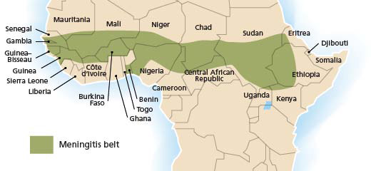 Figure: The African meningitis belt. These sub-Saharan countries are at high epidemic risk for meningococcal meningitis. (Image source: WHO)
Meningococcal-meningitis incidence rates are generally highest in children under five, followed by adolescents. IMD’s case fatality rate (CFR) ranges from 4.1% to 20%.
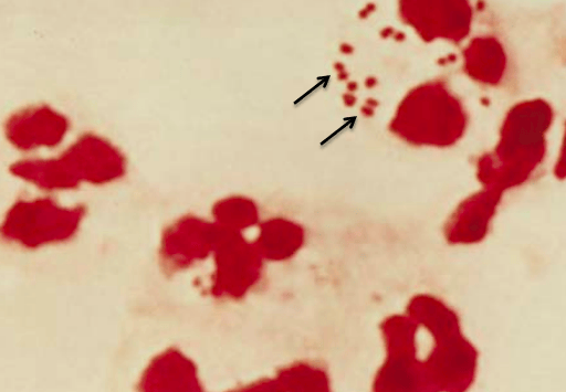 Figure: Gram stain of N. meningitidis in CSF
General Properties of Neisseria meningitidis
- Gram-negative intra-cellular diplococci
- Neisseria meningitidis (meningococcus) has a prominent polysaccharide capsule that enhances virulence through its complement inhibitory and antiphagocytic action. Nonencapsulated N. meningitidis isolates are generally considered nonpathogenic. The capsule is also the immunogen in the meningococcal vaccine.
- Serogroups: Based on the antigenic nature of capsular polysaccharides, N. meningitidis can be typed into 13 serogroups (A-D, X-Z, 29E, W135, H, I, K, and L). Six serogroups (types) of Neisseria meningitidis A, B, C, W, X, and Y cause the majority of cases of IMDs worldwide.
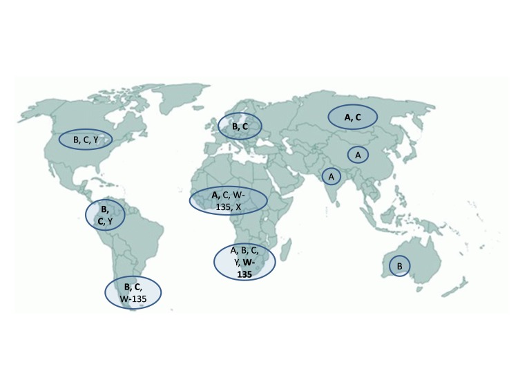 Figure: Distribution of Meningococcal serogroups in the world
Serotype A was the leading cause of epidemic meningitis worldwide, mainly in the African meningitis belt. However, its incidence decreased after the introduction of group A meningococcal polysaccharide–tetanus toxoid conjugate vaccine (PsA-TT; MenAfriVac) in Africa in 2010.
Pathogenesis
Transmission
Neisseria meningitidis lives as normal upper respiratory tract flora in young adults. Carriage is highest in adolescents and young adults (approx 10%), mostly due to their lifestyle. Carriage rates are generally lower in older adults and infants.
The bacteria are transmitted from person to person through carriers’ droplets of respiratory or throat secretions. Smoking, close and prolonged contact (such as kissing, sneezing, or coughing), or living in close quarters with a carrier facilitates the spread of the disease. The reasons for transitioning from asymptomatic carriage to invasive disease are not completely understood.
Virulence Factors of Neisseria meningitidis
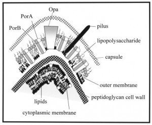 Figure: Surface proteins of Neisseria meningitidis. Source: Sciencedirect.com
1. Polysaccharide Capsule: Primary Virulence Factor
The polysaccharide capsule is the most important virulence determinant of N. meningitidis:
- Antiphagocytic: inhibits complement-mediated opsonization and phagocytosis by neutrophils and macrophages
- Complement inhibition: capsular polysaccharides interfere with C3b deposition, preventing complement-mediated killing
- Basis of serogroup classification: 13 serogroups defined by capsular polysaccharide antigenicity; 6 serogroups (A, B, C, W, X, Y) cause >90% of disease
- Vaccine immunogen: capsular polysaccharides are the antigens in all licensed meningococcal vaccines (MenACWY conjugate; MenB protein-based)
Non-encapsulated N. meningitidis strains are generally considered non-pathogenic, the capsule is essential for invasive disease. However, non-encapsulated strains can colonize the nasopharynx asymptomatically.
2. Lipooligosaccharide (LOS)
As with N. gonorrhoeae, N. meningitidis produces LOS rather than full LPS:
- Carbohydrate lacto-N-neotetraose: a major meningococcal virulence determinant; mimics host cell surface oligosaccharides
- Stimulates TNF-α release: the endotoxin activity of LOS drives the cytokine storm responsible for the rapid circulatory collapse in meningococcemia
- DIC trigger: LOS-induced inflammation activates coagulation cascades → disseminated intravascular coagulation → petechial rash
3. Fimbriae (Pili)
Meningococcal pili mediate:
- Initial attachment to nasopharyngeal epithelial cells
- Microcolony formation facilitating mucosal colonization
- Phase and antigenic variation are similar to gonococcal pili; contributes to immune evasion during carriage
4. IgA1 Protease
All meningococci produce IgA1 protease with the identical function to the gonococcal enzyme:
- Cleaves secretory IgA1 at the hinge region
- Destroys mucosal antibody blocking nasopharyngeal attachment
- Antibodies against IgA1 protease are produced during both carriage and disease, it is a potential vaccine target
5. Outer Membrane Proteins: PorA and PorB
Meningococcal porins have dual roles:
- PorA: major outer membrane protein; highly variable; target for protein-based MenB vaccines
- PorB: serum resistance; inhibits complement deposition; contributes to resistance to killing in the bloodstream
6. Factor H Binding Protein (fHbp)
N. meningitidis expresses a surface protein that binds human complement regulator Factor H:
- Binding Factor H to the bacterial surface prevents complement activation and C3b deposition
- Highly immunogenic and is a key component of protein-based MenB vaccines (Bexsero, Trumenba)
- Represents a rational vaccine target, blocking fHbp would remove complement protection from the bacterium
Virulence Factor Summary
| Virulence factor | Function | Clinical relevance |
|---|---|---|
| Capsule | Antiphagocytic; complement inhibition | Essential for invasive disease; vaccine target |
| LOS | Endotoxin; TNF-α induction; DIC trigger | Drives the rapid circulatory collapse in septicemia |
| Pili | Mucosal attachment; microcolony formation | Colonization and transmission |
| IgA1 protease | Destroys mucosal IgA | Enables nasopharyngeal colonization |
| PorA/PorB | Serum resistance; complement evasion | Allows survival in bloodstream |
| fHbp | Factor H binding; complement evasion | Vaccine target (MenB vaccines) |
Clinical Manifestations
The two main clinical manifestations of invasive meningococcal disease are meningitis and septicemia, with many patients showing features of both. Estimates of the split vary by population and case definition, with meningitis reported in roughly 50 to 80% of cases and septicemia in a substantial minority.
Occasionally, meningococci cause other infections, including pneumonia, pericarditis, conjunctivitis, endophthalmitis, septic arthritis, pelvic disease, or chronic low-grade septicemia.
Meningococcal meningitis
Meningococcal meningitis is a serious infection of the meninges that affects the brain membrane. The liberation of endotoxin (by the bacteria) into the subarachnoid space provokes a marked cytokine-mediated inflammation of the meninges. Early symptoms are fever, malaise, nausea, shivers, tachycardia, and mild headache. As the illness progresses, headache may become more pronounced, accompanied by photophobia, confusion, and vomiting (due to raised intracranial pressure), followed by coma if untreated. In newborns and babies, symptoms such as being slow or inactive, irritable, vomiting, feeding poorly, or the presence of a bulging in the soft spot of the skull (anterior fontanelle) may be an indication rather than the classic symptoms.
Septicemia
A less common but even more severe (often fatal) form of meningococcal disease is meningococcal septicemia, characterized by a hemorrhagic rash and rapid circulatory collapse. Most patients become progressively ill within 24 to 48 hours. However, in a few cases, the disease progresses so rapidly that the patient becomes moribund or dies within a few hours of the onset of infection.
Waterhouse-Friderichsen Syndrome
The most severe form of meningococcal septicemia is Waterhouse-Friderichsen syndrome: bilateral adrenal hemorrhage occurring as a complication of overwhelming meningococcemia:
- Mechanism: Severe DIC and LOS-driven endothelial damage cause hemorrhage into both adrenal glands → acute adrenocortical insufficiency → cardiovascular collapse
- Features: Purpuric rash (rapidly progressing from petechiae to large confluent purpura), hypotension unresponsive to fluids, high fever or hypothermia, shock
- Mortality: Extremely high without immediate treatment; historically approached 100% without antibiotics and supportive care
- Recognition: The glass test: pressing a glass firmly against petechial spots; if spots do not blanch (disappear when compressed) → non-blanching rash → meningococcemia until proven otherwise → immediate medical emergency
Meningococcal Vaccines
Vaccines are the most effective tool for prevention of invasive meningococcal disease:
| Vaccine type | Covers | Examples | Notes |
|---|---|---|---|
| Polysaccharide–protein conjugate | Serogroups A, C, W, Y | MenACWY (Menveo, Nimenrix) | Conjugation to protein carrier gives T-cell dependent response; better immunogenicity in young children |
| Protein-based (serogroup B) | Serogroup B | Bexsero (4CMenB), Trumenba (MenB-FHbp) | Uses PorA, fHbp, NHBA, NadA proteins; serogroup B capsule is identical to human neural cell adhesion molecule, cannot be used as vaccine antigen |
| Polysaccharide (older, non-conjugate) | A, C, W, Y | Menomune | Less immunogenic; not T-cell dependent; poor response <2 years; being replaced by conjugate vaccines |
Why serogroup B vaccine is different: Serogroup B capsular polysaccharide is identical to polysialic acid found on human neural cell adhesion molecules, it is self-antigen and cannot safely be used as a vaccine immunogen. Serogroup B vaccines instead use conserved outer membrane proteins (PorA, fHbp, NHBA, NadA), which is why they are protein-based rather than polysaccharide-based.
Laboratory Diagnosis
Sample
Cerebrospinal fluid (CSF), blood and skin scrapings from petechial rashes from cases, and nasopharyngeal swabs from carriers. Specimens should be collected in sterile containers and transported immediately without any delay.
CSF should never be refrigerated as suspected agents of meningitis (pneumococci, meningococci, and Haemophilus influenzae) are delicate and may die on refrigeration).
Read this post about CSF Collection.
Gram Stain
In Gram-stained smear of centrifuged deposit of specimen (CSF or sterile body fluid), N. meningitidis appear as Gram-negative, coffee-bean-shaped diplococci occurring intracellularly or extracellularly in PMN leukocytes.
Culture
Isolation of N. meningitidis (from blood, CSF, or other normally sterile sites) remains the gold standard as it also provides isolates for strain differentiation and susceptibility testing.
Since meningococci are fastidious, samples from sterile body sites are inoculated on either blood or chocolate agar. The chocolate agar base can be enriched with antibiotics such as vancomycin, colistin, nystatin, and trimethoprim for selective isolation of N. meningitidis. For culture from non-sterile sites such as the nasopharynx, a selective media such as Modified New York City or Modified Thayer Martin medium are required. Culture plates should be incubated for a minimum of 48 hours with a source of 5% CO2.
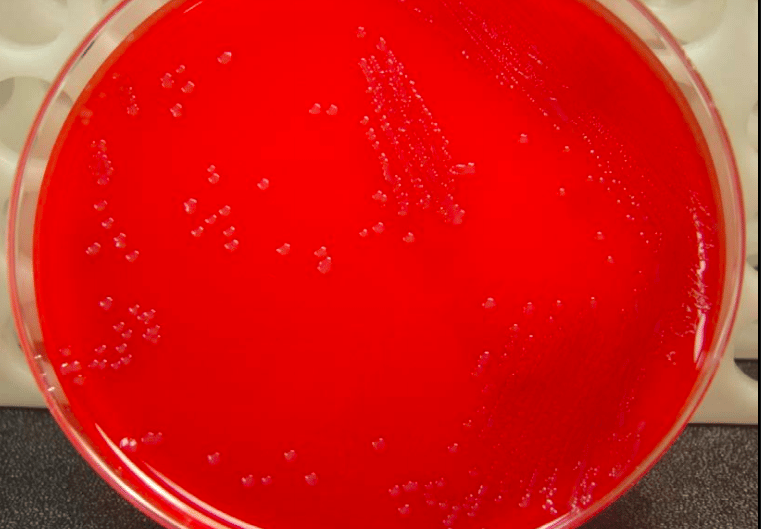 Figure: Neisseria meningitidis in Blood Agar
On Blood agar, young colonies of N. meningitidis are round, smooth, moist, glistening, and convex, with a clearly defined edge whereas actively growing colonies are grey and unpigmented. Older cultures (> 24 hours) become more opaquely grey and sometimes cause the underlying agar to turn dark.
On Modified New York City medium and Thayer Martin medium, Neisseria meningitidis appears as large colorless to bluish-gray mucoid colonies.
Identification
Biochemical tests for the Identification of Neisseria meningitidis:
- Catalase Positive
- Oxidase positive.
- Produce acid from glucose and maltose but not from lactose or sucrose.
- Nitrate Reduction Test: negative
- Produce gamma-glutamyl aminopeptidase
- Resistant to colistin: meningococci are colistin-resistant and grow on selective media containing VCN inhibitor
- DNAse reaction: negative
- Superoxol Test (reaction with 30% hydrogen peroxide): may show weak to a strong reaction.
- Pigmentation: produce pink-brown pigments.
Sero-grouping
Various serogroups of N. meningitidis are differentiated by slide agglutination test using monovalent antisera.
Serological tests
Several serological tests, such as enzyme immunoassay, latex agglutination, and rapid diagnostic tests, are available to detect antibodies against capsular antigens of Neisseria meningitidis. Serological tests help in the retrospective diagnosis of disease. Antibodies are also seen when vaccination is successful and in cases of chronic meningococcemia.
Molecular diagnosis
PCR-based diagnosis provides confirmation of meningococcal disease from blood, CSF, or other normally sterile sites with a validity comparable to that of culture-based diagnosis.
How to Remember
The clinical emergency rule: Non-blanching rash + fever = meningococcemia until proven otherwise. Do not wait for results. Do not wait for a lumbar puncture. Give IV benzylpenicillin immediately if meningococcal disease is suspected clinically.
The six serogroups: A B C W X Y: Africa (meningitis belt) B in Britain/Europe (hardest to vaccinate, identical to human self-antigen) C in Collegiate outbreaks (university students) W at Worldwide events (Hajj outbreaks) X in Africa (emerging) Y in Younger adults and elderly (USA)
Capsule = key distinguisher from gonococcus: Meningococcus HAS capsule → serogroups → vaccine possible Gonococcus has NO capsule → antigenic variation of pili instead → no effective vaccine
The DIC cascade: LOS → TNF-α → endothelial damage → DIC → petechiae → purpura → adrenal hemorrhage (Waterhouse-Friderichsen) → cardiovascular collapse Each step explains the next, the rash is not the infection, it is the body's inflammatory response to LOS destroying the vascular endothelium.
Why CSF should never be refrigerated: Meningococcus, pneumococcus, and H. influenzae: the three classic causes of bacterial meningitis, are all fastidious and cold-sensitive. Refrigerating CSF kills the organisms before culture. Transport immediately to the lab at room temperature or in an incubator at 37°C.
References and Further Readings
- Tille, P. M. (2022). Bailey & Scott's Diagnostic Microbiology (15th ed.). Elsevier.
- Tzeng, Y. L., & Stephens, D. S. (2000). Epidemiology and pathogenesis of Neisseria meningitidis. Microbes and Infection, 2(6), 687–700. https://doi.org/10.1016/S1286-4579(00)00356-2
- Rosenstein, N. E., Perkins, B. A., Stephens, D. S., Popovic, T., & Hughes, J. M. (2001). Meningococcal disease. New England Journal of Medicine, 344(18), 1378–1388. https://doi.org/10.1056/NEJM200105033441807
- Jolley, K. A., Maiden, M. C. J., & Feavers, I. M. (2007). Molecular typing and global epidemiology of Neisseria meningitidis. FEMS Microbiology Reviews, 31(2), 175–191. https://doi.org/10.1111/j.1574-6976.2006.00060.x
- World Health Organization. (2011). Meningococcal vaccines: WHO position paper. Weekly Epidemiological Record, 86(47), 521–540.
- Vipond, C., Wheeler, J. X., Jones, C., Feavers, I. M., & Suker, J. (2005). Characterization of the protein content of a meningococcal outer membrane vesicle vaccine by polyacrylamide gel electrophoresis and mass spectrometry. Human Vaccines, 1(2), 80–84.

Tankeshwar Acharya, MSc (Medical Microbiology)
Tankeshwar Acharya is an Assistant Professor in the Department of Microbiology at Patan Academy of Health Sciences (PAHS), Nepal, where he has been teaching and practicing clinical microbiology for over 14 years. He is the founder of Microbe Online, one of the leading free microbiology education resources on the web, covering bacteriology, mycology, parasitology, immunology, and clinical laboratory diagnostics written from direct experience in both the classroom and the diagnostic laboratory.
Comments
No comments yet. Be the first to share your thoughts.
Leave a comment
All comments are reviewed before they appear.