

A medical student sees their first positive blood culture result: "Gram-positive cocci in clusters." Before any identification test is run, before any culture result comes back, that Gram stain result has already narrowed the differential to Staphylococcus and potentially changed the empiric antibiotic choice.
Staining is not a preliminary step in microbiology. It is often the fastest diagnostic result available and in resource-limited settings where culture may take days or never happen at all, it may be the only result. Understanding which stain to use, why it works, and how to interpret it is among the most important skills in laboratory medicine.
This article covers the major categories of staining techniques used in clinical and general microbiology, with a specimen-to-stain decision guide and links to detailed procedural articles for each technique.
Which Stain to Use: A Quick Reference Guide
The most common question at the bench is not "how does this stain work?" but "which stain should I use for this specimen?" The table below answers that question.
| Specimen or Organism | First-Choice Stain | Alternative | What You Are Looking For |
|---|---|---|---|
| Any bacterial specimen (routine) | Gram stain | Acridine orange (blood culture) | Gram reaction, morphology, arrangement |
| Mycobacterium tuberculosis, M. leprae | Ziehl-Neelsen (ZN) | Auramine-rhodamine | Acid-fast bacilli (red rods on blue background) |
| Mycobacterium (fluorescence lab) | Auramine-rhodamine | ZN for confirmation | Fluorescent yellow-orange rods |
| Corynebacterium diphtheriae | Albert stain | Gram stain | Metachromatic (volutin) granules |
| Fungal elements in clinical specimen | Calcofluor white | KOH mount | Fluorescent hyphae and yeast cells |
| Fungal culture identification | LPCB (lactophenol cotton blue) | Conidial arrangement, hyphal morphology | |
| Malassezia furfur (skin scraping) | KOH mount | Calcofluor white | Spaghetti-and-meatball appearance |
| Malaria and blood parasites | Giemsa stain | Leishman stain | Ring forms, trophozoites, gametocytes |
| Cryptococcus neoformans capsule | India ink (negative stain) | Calcofluor white | Capsule halo around yeast cells |
| Bacterial capsule | Capsule stain (Anthony's) | India ink | Clear capsule zone against dark background |
| Bacterial endospores | Endospore stain (Schaeffer-Fulton) | Gram stain (poorly) | Spore position (terminal, subterminal, central) |
| Bacterial flagella | Flagella stain (Leifson/silver) | Wet mount (motility) | Number and arrangement of flagella |
| Pneumocystis jirovecii in BAL | Toluidine blue | Calcofluor white, GMS | Cyst wall staining (dark purple) |
| Bacteria in blood culture (faint/equivocal) | Acridine orange | Gram stain | Fluorescent orange nucleic acid |
| Chlamydia trachomatis inclusions | Giemsa stain | DFA (direct fluorescent antibody) | Intracellular inclusion bodies |
| Intestinal parasites | Saline wet mount | Iodine wet mount | Motility, ova, trophozoite morphology |
| Giardia, Cryptosporidium (stool) | Modified ZN (cold) | Auramine-rhodamine | Acid-fast oocysts |
| Whipple's disease (tissue biopsy) | PAS stain | PAS-positive macrophages in lamina propria | |
| Actinomyces (sulfur granules) | Gram stain | Gram-positive branching filaments | |
| Nocardia in specimen | Modified ZN (partial acid-fast) | Gram stain | Partially acid-fast branching rods |
| Spirochetes (Treponema, Borrelia) | Dark field microscopy | Silver impregnation | Motile spiral organisms |
| Leishmania in tissue/smear | Giemsa stain | Amastigotes within macrophages |
Why Staining?
Due to a lack of contrast, the structural details of organisms cannot be seen under a light microscope. Hence, we use dyes to stain cells. Dyes bind with cellular constituents, producing color contrast and increasing visibility.
Among the various types of dyes, it is the positively charged (cationic) ones that play a crucial role. Dyes such as methylene blue, crystal violet, safranin, etc, with their positive charge, specifically bind with negatively charged cellular constituents such as nucleic acids and acidic polysaccharides, and even the cell surface of bacteria, enhancing our ability to observe and study these structures.
Based on the types and number of dyes used, staining can be categorized into different types.
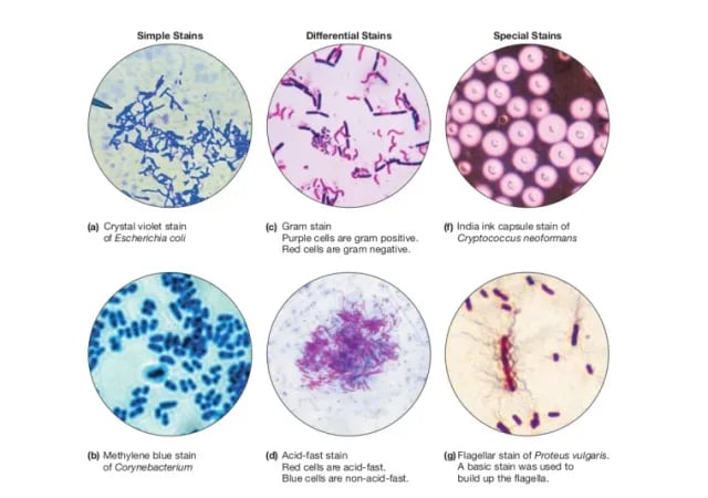 Figure: Types of Microbiological Stains (Image source: Ref-3)
Note: All staining techniques involve direct handling of cultures or clinical specimens. All the procedures below assume you have reviewed and will follow the foundational microbiology laboratory safety rules, which cover PPE, handwashing, aerosol control, and waste disposal for all staining work.
Simple stain
Basic dyes, such as methylene blue or basic fuchsin are used as simple stains. They produce color contrast but impart the same color to all the bacteria in the smear.
Negative staining
A drop of bacterial suspension is mixed with dyes, such as India ink or nigrosin. The background gets stained black whereas the unstained bacterial or yeast capsule stands out in contrast. This is very useful in the demonstration of capsules that do not take up simple stains.
India ink preparation
Negative stains are used when a specimen or a part of it, such as the capsule resists taking up the stain. India Ink preparation is recommended for use in the identification of Cryptococcus neoformans.
Impregnation methods
Bacterial cells and structures that are too thin to be seen under the light microscope are thickened by impregnation of silver salts on their surface to make them visible, e.g., for demonstration of bacterial flagella and spirochetes.
Flagella stain
Demonstrate the presence and arrangement of flagella. Flagellar stains are painstakingly prepared to coat the surface of the flagella with dye or a metal such as silver. The number and arrangements of flagella are critical in identifying species of motile bacteria.
Read more about Flagella Staining
Differential staining
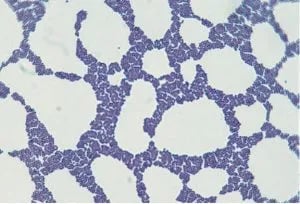 Figure: Staphylococcus in Gram Stain
Here, two stains are used which impart different colors to different bacteria or bacterial structures, which help in differentiating bacteria. The most commonly used differential stains are:
Gram staining
Gram stain is a very important differential staining technique used in the initial characterization and classification of bacteria in microbiology. Gram staining helps to identify bacterial pathogens in specimens and cultures by their Gram reaction (Gram-positive and Gram-negative) and morphology (cocci/rod).
The Gram stain result from a clinical specimen is often the fastest actionable result in the microbiology laboratory; available within 15–30 minutes of specimen receipt, hours before any culture result. A Gram stain showing gram-negative rods in sputum changes empiric antibiotic selection immediately; gram-positive cocci in clusters in a blood culture bottle warrants anti-staphylococcal coverage before susceptibility results are available. Learning to interpret Gram stain patterns clinically, not just morphologically, is one of the most valuable skills in clinical microbiology.
Acid-fast stain (Ziehl-Neelsen technique)
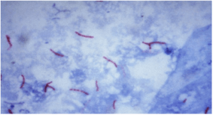 Figure: Acid-fast bacillus
It distinguishes acid-fast bacteria such as Mycobacterium spp from non-acid fast bacteria; which do not stain well by the Gram staining. It is used to stain Mycobacterium species (Mycobacterium tuberculosis, M. leprae, and M. ulcerans). Note that M. leprae is demonstrated in slit-skin smears and biopsy, not by sputum smear. Read more about AFB (Ziehl Neelsen staining technique).
Endospore stain
It demonstrates spore structure in bacteria as well as free spores. Relatively few species of bacteria produce endospores, so a positive result from endospore staining methods is an important clue in bacterial identification. Bacillus spp and Clostridium spp are the main endospores producing bacterial genera.
Read more about: Endospore staining technique.
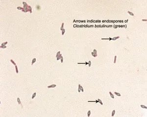 Figure: Spore of Clostridium botulinum Source: ASM
Capsule stain
It helps to demonstrate the presence of capsules in bacteria or yeasts. Streptococcus pneumoniae, Neisseria meningitidis, Haemophilus influenzae, Klebsiella pneumoniae are common capsulated bacteria.
Read more about: Capsule Staining Technique.
Giemsa stain
Giemsa stain is a Romanowsky stain. It is widely used in the microbiology laboratory for the staining of:
- Malaria and other blood parasites
- Chlamydia trachomatis inclusion bodies
- Borrelia species
- Yersinia pestis
- Histoplasma species
- Pneumocystis jirovecii trophic and intracystic forms (cyst wall itself is better shown by toluidine blue, GMS, or calcofluor white
Cytoplasmic inclusion stains
Identifies intracellular deposits of starch, glycogen, polyphosphates, hydroxybutyrate, and other substances. E.g. Albert staining is used to stain the volutin or metachromatic granules of C. diphtheriae.
Fluorescence Staining
Fluorescence staining uses dyes (fluorochromes) that absorb ultraviolet or near-UV light and emit visible light at a longer wavelength; producing a bright, glowing signal against a dark background. Fluorescence staining requires a fluorescence microscope equipped with the appropriate excitation filter.
Advantages over conventional staining:
- Higher sensitivity: organisms are easier to detect at low density
- Faster screening: scanning slides under low power is quicker than under oil immersion
- Particularly valuable for specimens with few organisms (e.g., CSF in TB meningitis)
Key fluorescent stains in clinical microbiology:
- Auramine-rhodamine fluorochrome stain for mycobacteria; more sensitive than ZN for direct smear examination of sputum. WHO recommends it as the preferred smear method where fluorescence microscopy is available. Positive results are confirmed by ZN staining.
- Acridine orange: binds nucleic acid; used to detect bacteria in blood cultures when the Gram stain is equivocal, and for screening cell-wall-deficient organisms such as mycoplasmas.
- Calcofluor white: binds chitin in fungal cell walls; highly sensitive for detecting fungi in direct specimens (BAL, skin scrapings, corneal scrapings). Requires UV fluorescence microscopy.
Lactophenol cotton blue (LPCB) wet mount
LPCB mount is the most widely used method of staining and observing fungi. Read more about LPCB mount.
Key Exam Facts in One Table
| Stain | Category | Principle | Key Use | Organism/Structure |
|---|---|---|---|---|
| Gram stain | Differential | Crystal violet-iodine complex retained by thick peptidoglycan | Bacterial identification | GP (blue-purple) vs GN (pink-red) |
| Ziehl-Neelsen | Differential | Mycolic acid retains carbol fuchsin after acid-alcohol decolorization | Mycobacteria detection | M. tuberculosis, M. leprae |
| Albert stain | Differential | Toluidine blue + malachite green; metachromatic granules stain dark | C. diphtheriae ID | Metachromatic (volutin) granules |
| Endospore stain | Differential | Malachite green driven into spore by heat; safranin counterstain | Spore detection | Bacillus, Clostridium |
| Capsule stain | Special | Negative staining of capsule against stained background | Capsule detection | S. pneumoniae, K. pneumoniae |
| India ink | Negative | Background stained; capsule unstained | Cryptococcus ID | Capsule halo |
| Giemsa stain | Special (Romanowsky) | Azure-eosin complex differentiates cell components | Blood parasites, intracellular organisms | Malaria, Leishmania, Chlamydia |
| LPCB | Special | Phenol kills organisms; cotton blue stains chitin | Fungal culture ID | Conidial arrangement, hyphal morphology |
| Calcofluor white | Fluorescent | Binds chitin/cellulose; fluoresces under UV | Fungal detection in specimens | Hyphae, yeast, Pneumocystis |
| Auramine-rhodamine | Fluorescent | Fluorochrome binds mycolic acid | Mycobacteria (more sensitive than ZN) | Mycobacterium spp |
| Acridine orange | Fluorescent | Intercalates nucleic acid; fluoresces orange | Bacteria in blood cultures | All bacteria, mycoplasmas |
| PAS stain | Special | Periodic acid oxidizes polysaccharides → Schiff reagent → magenta | Fungal wall, glycogen, Tropheryma | Whipple's (Tropheryma whipplei) disease, fungi in tissue |
| Toluidine blue | Special | Metachromatic staining of cyst wall | Pneumocystis cysts in BAL | Pneumocystis jirovecii |
| Saline wet mount | Direct | No stain; phase contrast or reduced light | Motility, parasite morphology | Intestinal parasites, trichomonas |
How to Remember: Staining Categories
A simple framework for exam recall:
"One dye, two dyes, or special":
- Simple stain = one basic dye, one color, no differentiation (e.g., methylene blue)
- Differential stain = two dyes, two colors, distinguishes organisms or structures (Gram stain, ZN stain, endospore stain, Albert stain)
- Special stain = designed for a specific structure or organism (capsule, flagella, India ink, Giemsa, LPCB, Calcofluor white)
The "GAZCEF" is an acronym-based mnemonic used in microbiology to recall the major types of stains.
- Gram: bacteria (gram-positive vs gram-negative)
- Albert: C. diphtheriae metachromatic granules
- Ziehl-Neelsen: mycobacteria (acid-fast bacilli)
- Capsule stain: capsulated organisms
- Endospore stain: Bacillus, Clostridium spores
- Flagella stain: motile bacteria, flagellar arrangement
References and further readings
- Procop GW, Koneman EW. Koneman's Color Atlas and Textbook of Diagnostic Microbiology. 7th ed. Wolters Kluwer; 2017.
- Tille PM. Bailey & Scott's Diagnostic Microbiology. 15th ed. Elsevier; 2022.
- Willey JM, Sherwood LM, Woolverton CJ. Prescott's Microbiology. 11th ed. McGraw-Hill; 2023.
- Murray PR, Rosenthal KS, Pfaller MA. Medical Microbiology. 9th ed. Elsevier; 2020.
- World Health Organization. Laboratory Biosafety Manual. 4th ed. Geneva: WHO; 2020.

Comments
No comments yet. Be the first to share your thoughts.
Leave a comment
All comments are reviewed before they appear.