
What Is a Virus? Structure, Genome Types, and Common Viral Diseases

On this page
A medical student encounters dozens of individual virus articles across this site: rabies, hepatitis B, dengue, HIV, each with its own structure, genome, and disease story. What ties all of them together, and why does it matter to learn the general pattern before studying any one virus in detail?
The answer is that almost every clinically important fact about a specific virus traces back to a handful of general properties covered on this page: what kind of genome it carries (which predicts mutation rate and vaccine durability, covered in depth in the Baltimore classification article), whether it has an envelope (which predicts how it spreads and what disinfects it), and what shape and symmetry its capsid takes (which predicts how stable it is outside the body). Once you understand these general patterns, you stop memorizing each new virus from scratch and start recognizing which family of behavior it belongs to before you have even learned its name.
This page is a properties-and-classification overview, not a full diagnostic manual. For the hands-on diagnostic methods (cytopathic effect recognition, neutralization testing, PCR-based detection), each has its own dedicated article linked from the Lab Diagnosis section below.
What is a virus?
Viruses are genetic elements that cannot replicate independently of a living host cell. The host cell provides the energy and metabolic intermediates needed to replicate the viral genome and synthesize viral proteins. Because viruses need suitable living cells to multiply, they are called obligate intracellular parasites.
Viruses exist in either an extracellular or an intracellular form. The extracellular form is called a virus particle or virion: a microscopic particle containing nucleic acid surrounded by a protein coat. A virion is metabolically inert. It cannot generate energy or carry out biosynthesis. Its role is to carry the viral genome safely from one host cell to another.
The entire virus, including nucleic acid, capsid, envelope, and glycoprotein spikes, is called the virion, or virus particle.
Once a virus enters a new host cell, its intracellular state begins and replication starts. Using the host cell's structural and metabolic components, the virus makes new copies of its genome and its coat proteins, then assembles new virions. Progeny viruses eventually leave the host cell either by budding or by lysis of the cell.
Size and Shape of Viruses
Virions come in many sizes and shapes. Most viruses are smaller than prokaryotic cells, ranging from 0.02 to 0.3 μm (20 to 300 nm). Because of this small size, viruses pass through bacterial filters and cannot be seen with a light microscope. The common unit for measuring viruses is the nanometer, one-thousandth of a micrometer.
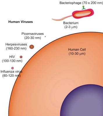 Figure: Viruses and cell size comparison
- Smallpox virus, one of the largest viruses, is about 200 nm in diameter (about the size of the smallest bacterial cells).
- Poliovirus, one of the smallest viruses, is only 28 nm in diameter (about the size of a ribosome).
Most animal viruses are roughly spherical, with some exceptions:
- Rabies virus: bullet-shaped
- Ebola virus: filamentous
- Poxvirus: brick-shaped
- Adenovirus: space-vehicle-shaped
Viral Structure
The structures of virions are diverse, varying widely in size, shape, and chemical composition.
Nucleocapsid
A virus is a nucleic acid genome surrounded by a protein shell called a capsid. Together, the genome and capsid are the nucleocapsid.
The capsid is built from individual protein molecules called capsomers. A few viruses have only one kind of capsid protein, but most have several. Capsomers are arranged in a precise, highly repetitive pattern around the nucleic acid, and the capsomer is the smallest morphological unit seen with the electron microscope.
A single virion can contain a large number of capsomers. The information needed to fold and assemble the proteins into capsomers is contained within the proteins themselves, so the process is called self-assembly. The nucleocapsid is the complete complex of nucleic acid and protein packaged in the virion.
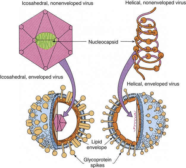 Figure: Enveloped and non-enveloped virions (Modified from Murray PR, Drew WL, Kobayashi GS, et al, editors: Medical microbiology, St Louis, 1990, Mosby.)
Virus Symmetry
Viral nucleocapsids are built in highly symmetric ways. Three kinds of symmetry are recognized: helical, icosahedral, and complex.
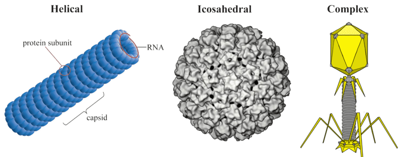 Figure: Virus symmetry
- Helical symmetry: rod-shaped viruses have helical symmetry. Examples: tobacco mosaic virus (TMV), measles, mumps, influenza, rabies.
- Icosahedral symmetry: the icosahedron is the most efficient arrangement of subunits in a closed shell. Roughly spherical viruses have icosahedral symmetry. An icosahedron has 20 faces, each an equilateral triangle. The simplest icosahedral capsids are built from 60 identical subunits; larger ones are built from multiples of 60 (described by the triangulation number, T). Most viruses have icosahedral symmetry, for example poliovirus and adenovirus.
- Complex symmetry: some viruses have complex or uncertain symmetry. Smallpox virus has one of the most complex virion structures, with many different proteins and lipoproteins. Bacteriophages are structurally the most complicated, with icosahedral heads and helical tails.
Viral Genome
Although viruses are acellular, they carry a genome encoding the information needed for replication. Viral genomes are smaller than those of most cells. The largest known viral genome, that of Mimivirus, is 1.18 Mbp of double-stranded DNA, larger than some cellular genomes. At the other extreme, some viruses have fewer than five genes.
Viral genomes are made of DNA or RNA (in contrast, all cells have double-stranded DNA genomes). They are classified first by whether the genome is DNA or RNA, then by whether it is single-stranded (ss) or double-stranded (ds), and whether it is linear or circular.
Viruses are unusual in that their genomes can take forms not found in cellular life, including single-stranded DNA and double-stranded RNA:
- Double-stranded RNA genome: families Reoviridae (rotavirus, Colorado tick fever virus) and Birnaviridae.
- Single-stranded DNA genome: family Parvoviridae (parvovirus B19).
- Segmented genome: the RNA genome can exist in several separate pieces. Influenza virus and rotavirus have segmented RNA genomes.
- Ploidy: almost all viruses are haploid, carrying a single copy of the genome. The major exception is the retrovirus family, which is diploid (two copies of the RNA genome).
 Figure: Central dogma concept
Most viral genomes are linear, though some are circular. DNA viruses follow the central dogma of molecular biology; RNA viruses are the exception. Regardless of genome type, every virus must produce messenger RNA (mRNA), which the host cell's ribosomes then translate.
Virus Envelope
Some viruses are naked (non-enveloped), whereas others have a lipid-containing layer around the nucleocapsid called an envelope.
The envelope is a lipid bilayer derived from the host cell's membranes, with viral proteins (usually glycoproteins, encoded by viral genes) embedded in it. Glycoprotein spikes project from the surface and act as attachment proteins or as enzymes (for example, neuraminidase). A virus acquires its envelope by budding through a host membrane, which is a gentler exit that often does not kill the cell immediately. This is also why enveloped viruses are fragile outside the body.
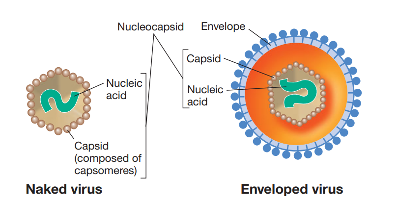 Figure: Naked vs Enveloped virus
Because the envelope makes first contact with the host cell, it controls the specificity of infection. The envelope proteins are essential for attachment: the virus binds specific receptors on the host cell membrane through its glycoprotein spikes, and the specificity of that interaction determines which hosts and which cells the virus can infect.
Enveloped virions are sensitive to lipid solvents such as ether and chloroform, which destroy their ability to infect cells. Naked viruses are not affected by lipid solvents.
Enzymes in Virions
Most virions carry no enzymes of their own, but some contain one or more virus-specific enzymes that act during infection and replication. The common ones are summarized below.
| Enzyme | Virions carrying it | Function |
|---|---|---|
| Lysozyme | Bacteriophages | Makes a small hole in the bacterial cell wall so the phage can inject its nucleic acid into the host cytoplasm. Lysozyme made late in infection also helps lyse the cell wall to release progeny phages. |
| RNA-dependent DNA polymerase (reverse transcriptase) | Retroviruses | Transcribes the viral RNA into a DNA intermediate |
| RNA-dependent RNA polymerase | RNA viruses | Synthesizes RNA from an RNA template, a reaction host cells cannot perform, which is why RNA viruses must carry this enzyme themselves |
| Neuraminidase | Certain animal viruses (e.g. influenza) | Cleaves sialic acid (neuraminic acid) residues from host cell surface glycoproteins, releasing newly budded virions and preventing them from clumping together |
Viral Diseases
Clinically important viruses and the diseases they cause are listed below. Each linked virus has its own dedicated article with full structure, pathogenesis, and diagnosis.
| Virus | Disease or condition |
|---|---|
| Herpes simplex virus types 1 and 2 | Painful vesicles on the face and genitals |
| Varicella-zoster virus | Varicella (chickenpox), typically in children, and zoster (shingles) |
| Cytomegalovirus | Congenital malformations |
| Epstein-Barr virus | Infectious mononucleosis |
| Human herpesvirus 8 | Kaposi's sarcoma |
| Hepatitis B virus | Viral hepatitis |
| Poxvirus | Smallpox |
| Adenovirus | Upper and lower respiratory tract infection (commonly pharyngitis and pneumonia) |
| Papillomavirus | Skin and mucosal papillomas; some strains cause carcinoma of the cervix |
| Parvovirus B19 | Slapped-cheek syndrome |
| Measles virus | Measles |
| Mumps virus | Mumps |
| Rubella virus | Rubella |
| Rabies virus | Rabies (fatal encephalitis) |
| Hepatitis C virus | Chronic hepatitis; predisposes to hepatocellular carcinoma |
| Human T-cell lymphotropic virus | T-cell leukemia and tropical spastic paraparesis |
| Human immunodeficiency virus | AIDS |
| Poliovirus | Polio (aseptic meningitis and paralysis) |
| Rhinovirus | Common cold |
| Rotavirus | Gastroenteritis in young children |
| Hepatitis A virus | Hepatitis |
| Norovirus | Gastroenteritis, especially in adults |
| Hepatitis E virus | Hepatitis (fecal-oral route) |
Lab Diagnosis of Viral Infections
Diagnosing a viral infection generally relies on one or more of the following, often combined for confirmation:
- Microscopy: direct examination of specimens can reveal characteristic inclusion bodies or multinucleated giant cells (for example, the Tzanck smear for herpesvirus giant cells), or detect virus particles directly by electron microscopy.
- Cytopathic effect (CPE) in cell culture: growing the virus in cell culture and observing characteristic patterns of cell damage is useful for provisional identification, especially where molecular testing is not readily available. See the dedicated CPE article for the full range of patterns and their associated viruses.
- Antigen detection: tests such as ELISA detect viral antigens directly in blood or tissue, for example HBsAg for hepatitis B or p24 antigen for HIV.
- Serology (antibody detection): a four-fold or greater rise in antibody titer between acute and convalescent samples, or the presence of IgM antibody, indicates current infection. See the hepatitis B serology interpretation article for a worked example of how such a panel is read.
- Neutralization testing: confirms whether antibody in serum can actually block viral infectivity, used both for diagnosis and for confirming protective immunity (such as rabies vaccination titers). See the dedicated neutralization test article for the mechanism and applications.
- Nucleic acid detection (PCR): the most sensitive and rapid method for many viral infections, amplifying and detecting specific viral sequences. See the dedicated PCR article for the method and its applications, including viral load monitoring in HIV.
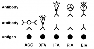 Figure: Serological methods used in viral diagnosis
How to Remember
Shape often predicts function, not just appearance. Helical symmetry (rod-shaped, like rabies, measles, influenza) tends to appear in enveloped RNA viruses, where flexibility matters for budding through a host membrane. Icosahedral symmetry, the most efficient way to pack identical subunits into a closed shell, is the default for viruses that need a rigid, stable, protective container. That is why most viruses, including many that must survive outside cells for a while, are built this way. Complex symmetry (smallpox, bacteriophages) appears where the virus needs to do something mechanically unusual, such as injecting DNA through a bacterial cell wall. A simple icosahedron or helix does not have the right shape for that job.
Envelope or no envelope is a transmission-strategy fork, not a random feature. An envelope makes a virus fragile outside the body (sensitive to ether, chloroform, drying, heat) but lets it bud out of a cell relatively gently, often without killing the cell immediately. A naked (non-enveloped) virus is tougher and better suited to surviving outside the body (water, surfaces, the gut), but it usually has to lyse the host cell to escape, because it has no membrane to bud through. This single trade-off explains a great deal of clinical microbiology. Enveloped viruses spread mainly by close contact, blood, or respiratory droplets and do not survive well on surfaces. Naked viruses (such as hepatitis A, poliovirus, rotavirus) are the ones associated with fecal-oral spread and outbreak-causing environmental persistence.
Genome type predicts almost everything else. This is covered in full mechanistic depth in the Baltimore classification article, but the short version worth anchoring here: RNA genomes generally mutate faster (no proofreading), DNA genomes are generally more stable, and only specific genome types (retroviruses, hepadnaviruses) require reverse transcriptase. That is exactly why that one drug class works only against that specific subset of viruses.
Key exam facts
| Property | What it predicts |
|---|---|
| Helical symmetry | Often enveloped, flexible budding viruses (TMV, measles, influenza, rabies) |
| Icosahedral symmetry | Stable, efficient packing; most viruses, including many naked viruses (poliovirus, adenovirus) |
| Complex symmetry | Unusual structural or mechanical needs (smallpox, bacteriophages) |
| Enveloped | Fragile outside host; sensitive to lipid solvents, heat, drying; spreads by close contact, blood, droplets |
| Naked (non-enveloped) | Resistant to lipid solvents; survives well outside host; spreads by fecal-oral route and fomites |
| dsDNA genome | Generally more stable, lower mutation rate; often uses host DNA polymerase |
| ssRNA/dsRNA genome | Generally faster mutation rate (no proofreading by viral RNA polymerase) |
| Retrovirus/hepadnavirus genome | Requires reverse transcriptase; only these are vulnerable to RT-inhibitor drugs |
| Smallest viral genomes | Fewer than 5 genes in some viruses |
| Largest viral genome | Mimivirus, 1.18 Mbp dsDNA, larger than some cellular genomes |
Where Students Get Confused
- "All viruses leave the cell the same way." They do not, and this follows directly from the envelope distinction. Enveloped viruses generally bud through the host membrane, a gentler exit that does not necessarily kill the cell. Naked viruses usually have to lyse the cell to escape, because they have no membrane to bud through. This is part of why naked viruses are more often linked to rapid, destructive cytopathic effects. See the CPE article for specific examples.
- "A virus without its own polymerase cannot replicate at all." Most viruses do need a polymerase to copy their genome or make mRNA, but where that enzyme comes from varies. Many DNA viruses borrow the host's polymerase entirely. RNA viruses and retroviruses must carry their own, because host cells have no enzyme that can use RNA as a template. That is exactly why those viruses package these enzymes inside the virion.
- "RNA polymerase" and "reverse transcriptase" blur together in an enzyme table. RNA-dependent RNA polymerase (used by RNA viruses) makes RNA from an RNA template. RNA-dependent DNA polymerase, that is, reverse transcriptase (used by retroviruses), makes DNA from an RNA template. Both solve the same underlying problem (host cells cannot do either), but they make different products and are used by different virus groups.
Frequently Asked Questions
Why do some viruses have an envelope and others don't?
Does virus shape (symmetry) actually matter clinically, or is it just classification trivia?
Why do RNA viruses need to carry their own polymerase, but many DNA viruses don't?
What's the difference between detecting a virus by PCR versus by cytopathic effect (CPE)?
References
- Levinson W, Chin-Hong P, Joyce EA, Nussbaum J, Schwartz B. Review of Medical Microbiology and Immunology. 17th ed. McGraw-Hill Education; 2022.
- Madigan MT, Bender KS, Buckley DH, Sattley WM, Stahl DA. Brock Biology of Microorganisms. 16th ed. Pearson; 2021.
- Louten J. Virus structure and classification. In: Essential Human Virology. Academic Press; 2016. p. 19-29. DOI: 10.1016/B978-0-12-800947-5.00002-8

Tankeshwar Acharya, MSc (Medical Microbiology)
Tankeshwar Acharya is an Assistant Professor in the Department of Microbiology at Patan Academy of Health Sciences (PAHS), Nepal, where he has been teaching and practicing clinical microbiology for over 14 years. He is the founder of Microbe Online, one of the leading free microbiology education resources on the web, covering bacteriology, mycology, parasitology, immunology, and clinical laboratory diagnostics written from direct experience in both the classroom and the diagnostic laboratory.
Comments
No comments yet. Be the first to share your thoughts.
Leave a comment
All comments are reviewed before they appear.