
Decarboxylase Test (Lysine, Ornithine, Arginine): Principle, Procedure, and How to Read the Pattern
How the decarboxylase test identifies Enterobacteriaceae using lysine, ornithine, and arginine. Why the control tube is essential, how arginine dihydrolase differs from true decarboxylation, and the result patterns that separate Salmonella, Klebsiella, and others.
The two amines at the center of this test have names that give the game away. Lysine decarboxylation yields cadaverine; ornithine yields putrescine. These are the same foul-smelling compounds that form in decomposing flesh, which is exactly where they were first characterized. A bacterium that is decarboxylase-positive is running, in miniature, the same chemistry that putrefies a corpse: stripping the carboxyl group off an amino acid and releasing a stinking, alkaline amine.
That alkalinity is the whole basis of the test. In the diagnostic lab, this is one of the most useful ways to sort otherwise-similar Enterobacteriaceae. Whether an organism decarboxylates lysine, ornithine, or arginine, and in which combination, helps separate Salmonella from Shigella, Klebsiella from Enterobacter, and Proteus from the rest. This article covers how the reaction works, why the control tube is non-negotiable, how the arginine dihydrolase pathway differs from true decarboxylation, and how to read the result pattern.
Principle
Decarboxylases are substrate-specific enzymes that remove the carboxyl (–COOH) group from an amino acid, releasing carbon dioxide and producing an alkaline amine. Each enzyme is specific for one amino acid, and three are routinely tested to identify Enterobacteriaceae: lysine, ornithine, and arginine.
The reactions and their products are:
- Lysine → cadaverine (by lysine decarboxylase, LDC)
- Ornithine → putrescine (by ornithine decarboxylase, ODC)
- Arginine → agmatine (by arginine decarboxylase) but arginine is more often handled by a different enzyme, see below.
Decarboxylation only proceeds in an acidic, anaerobic environment. The medium (Møller decarboxylase base) contains glucose, so the organism first ferments the glucose and acidifies the broth. That drop in pH turns the indicator yellow and switches on the decarboxylase enzymes. If the organism then decarboxylates the amino acid, the alkaline amine produced reverses the pH, and the indicator (bromcresol purple) turns from yellow back to purple. A layer of sterile oil seals the tube, keeping conditions anaerobic and preventing false alkalinization at the surface.
So a positive tube goes purple → yellow → purple, and you read the final purple. This is why the control tube matters (below): it proves the organism acidified the broth in the first place.
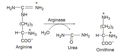 Arginine: two different pathways
Arginine is a source of confusion because two unrelated enzyme systems act on it, and both end up raising the pH:
- Arginine dihydrolase (ADH) — the pathway usually detected in the routine tube. Arginine is converted to citrulline (losing an –NH₂ group), then to ornithine, and the ornithine is decarboxylated to putrescine. Net result: alkaline, purple tube.
- Arginine decarboxylase — a true single-step decarboxylation of arginine to agmatine. Less commonly the operative route in the organisms tested.
For identification purposes the routine "arginine" tube is read as arginine dihydrolase (ADH) positive or negative, and standard identification tables label it ADH, not arginine decarboxylase. The important teaching point: citrulline and ornithine here are intermediates, not the reported amine end-product.
Requirements
Medium: Møller decarboxylase base broth, dispensed as four tubes per organism: one each with L-lysine, L-ornithine, and L-arginine (each ~1%), plus a control tube with no amino acid. The base contains peptone, glucose (~0.5%), the pH indicators bromcresol purple and cresol red, and pyridoxal (a cofactor for the decarboxylases).
Note on methods: the Møller method (described here) is the reference method and requires the oil overlay and the control tube. The older Falkow method uses a different base and a shorter incubation; Møller is preferred for non-fermenters and is the standard in most identification schemes.
Procedure
- Lightly inoculate each of the four tubes (three amino acids + control) from a pure culture. For glucose-fermenters, a light inoculum into each tube; for non-fermenters, a heavy suspension is used and inoculated into base broths.
- Overlay each tube with ~4 mm of sterile mineral/paraffin oil to maintain anaerobic conditions. This is essential: without it, surface oxidation causes false alkaline (false-positive) reactions.
- Incubate at 35–37°C.
- Read the control tube first, then the test tubes, daily for up to 4 days.
Reading the result pattern
Always read the control tube first:
- The control (no amino acid) should turn and stay yellow — proof that the organism fermented the glucose and acidified the broth. Only then are the decarboxylase enzymes switched on and the test valid.
- If the control is purple (alkaline), the test is invalid: the organism did not acidify the medium, and any purple in the test tubes cannot be trusted.
Then read each test tube against the control:
| Test tube color | Compared to control | Interpretation |
|---|---|---|
| Purple (or reverted to purple) | Control yellow | Positive — amino acid decarboxylated (alkaline amine raised pH) |
| Yellow | Control yellow | Negative — no decarboxylation; broth stayed acidic |
| Any color | Control purple | Invalid — organism did not acidify; repeat/troubleshoot |
A positive tube is often described as passing through yellow early (from glucose fermentation) and reverting to purple as decarboxylation takes over. Read at the endpoint.
QC Control
| Reaction | Positive control | Negative control |
|---|---|---|
| Lysine decarboxylase (LDC) | Klebsiella pneumoniae | Enterobacter cloacae |
| Ornithine decarboxylase (ODC) | Enterobacter cloacae | Klebsiella pneumoniae |
| Arginine dihydrolase (ADH) | Enterobacter cloacae | Klebsiella pneumoniae |
Uses of decarboxylation Tests
The three reactions are read together as a pattern (often written LDC / ADH / ODC) and combined with the rest of the biochemical panel. Key discriminations:
- Lysine decarboxylase (LDC): helps separate Salmonella (positive) from Shigella (negative). LDC-positive organisms include E. coli (variable), most Salmonella (except S. Paratyphi A, which is LDC-negative), Klebsiella pneumoniae, Serratia marcescens, and Vibrio cholerae. Proteus, Morganella, and Providencia are LDC-negative.
- Ornithine decarboxylase (ODC): helps separate Klebsiella (ODC-negative) from Enterobacter (ODC-positive), and is positive in Proteus mirabilis but negative in Proteus vulgaris — a useful Proteus split.
- Arginine dihydrolase (ADH): aids differentiation among enterics and non-fermenters; Pseudomonas aeruginosa is ADH-positive, a useful non-fermenter clue.
Relationship to LIA (Lysine Iron Agar): LIA detects lysine decarboxylation and deamination in a single slant/butt tube and is often used alongside or instead of the Møller lysine tube in enteric workups. For the combined lysine reading in that medium, see the LIA article. This article owns the decarboxylase concept; LIA owns the LIA-specific reading.
Organisms Identification Table
| Organism | LDC | ADH | ODC |
|---|---|---|---|
| Escherichia coli | +/− | +/− | +/− |
| Klebsiella pneumoniae | + | − | − |
| Enterobacter cloacae | − | + | + |
| Salmonella Typhi | + | +/− | − |
| Salmonella Paratyphi A | − | +/− | + |
| Serratia marcescens | + | − | + |
| Proteus vulgaris | − | − | − |
| Proteus mirabilis | − | − | + |
| Pseudomonas aeruginosa | − | + | − |
| Vibrio cholerae | + | − | − |
How to Remember
Cadaverine and putrescine — the smell of decay. Lysine gives cadaverine (think cadaver), ornithine gives putrescine (think putrid). A positive decarboxylase reaction is corpse chemistry: the same amines that make rotting flesh reek. If you can name the smell, you can name the product.
Purple is positive, but only if the control is yellow. The reaction runs acid-then-alkaline, so you are reading a reversal back to purple. The control tube is the honesty check: no yellow control, no valid test. Yellow control + purple test = real positive.
Oil keeps it honest. Decarboxylases need anaerobic, acidic conditions. The oil overlay is not optional packaging; without it the surface goes alkaline on its own and fakes a positive.
Arginine is the trickster: dihydrolase, not decarboxylase. The routine arginine tube usually reports arginine dihydrolase (ADH). Citrulline and ornithine are stops along the way, not the answer you report.
Key exam facts in one table
| Question | Answer | The reason behind it |
|---|---|---|
| What does the test detect? | Decarboxylation of lysine, ornithine, arginine | Enzyme removes –COOH, releases CO₂ + alkaline amine |
| Lysine product | Cadaverine | By lysine decarboxylase (LDC) |
| Ornithine product | Putrescine | By ornithine decarboxylase (ODC) |
| Arginine (routine) | Arginine dihydrolase (ADH) pathway | Arginine → citrulline → ornithine → putrescine |
| Indicator | Bromcresol purple (+ cresol red) | Purple alkaline, yellow acidic |
| Why glucose is in the medium | To acidify the broth first | Acid pH switches the decarboxylases on |
| Color sequence of a positive | Purple → yellow → purple | Ferment (acid) then decarboxylate (alkaline) |
| Why the oil overlay | Maintains anaerobic conditions | Prevents false surface alkalinization |
| Why the control tube | Confirms the broth was acidified | Purple control = invalid test |
| Positive result | Purple test tube (control yellow) | Alkaline amine raised the pH |
| Negative result | Yellow test tube (control yellow) | No decarboxylation; stayed acidic |
| LDC key split | Salmonella (+) vs Shigella (−) | High-yield enteric discrimination |
| LDC exception | S. Paratyphi A is LDC-negative | Important Salmonella exception |
| ODC key split | Enterobacter (+) vs Klebsiella (−); P. mirabilis (+) vs P. vulgaris (−) | Useful genus/species splits |
| Reference method | Møller (vs older Falkow) | Møller preferred, esp. for non-fermenters |
| Relationship to LIA | LIA reads lysine decarboxylation + deamination | This article owns the concept; LIA owns its reading |
Where students get confused
Thinking the arginine tube measures arginine decarboxylase. In routine identification it almost always reports arginine dihydrolase (ADH), a different enzyme system that reaches the same alkaline endpoint by a longer route (arginine → citrulline → ornithine → putrescine). Standard tables label it ADH. Citrulline and ornithine are intermediates, not the reported product.
Ignoring the control tube. The single most common error. A purple test tube means nothing unless the amino-acid-free control is yellow, proving the organism first acidified the broth. If the control is purple, the whole set is invalid.
Forgetting the oil overlay, or reading only the surface. Decarboxylation needs anaerobic, acidic conditions. Without the oil seal, the surface oxidizes and turns alkaline on its own, mimicking a positive. Always overlay, and read the body of the tube.
Expecting a simple purple-or-yellow with no time course. A true positive typically goes yellow first (glucose fermentation) then reverts to purple (decarboxylation). Reading too early can call a positive as negative. Read daily up to four days.
Confusing decarboxylation with deamination (the "opposite" reaction). Decarboxylation removes the carboxyl group and produces an alkaline amine (purple). Deamination (as in the phenylalanine deaminase / PDA test) removes the amino group and is detected differently (green with ferric chloride). They are opposite ends of amino-acid metabolism. See the PDA article, which owns deamination.
Reading a single reaction as an ID. LDC, ODC, and ADH are read as a pattern together and combined with the rest of the panel, never as a standalone identification.
References
- Leber AL, editor. Clinical Microbiology Procedures Handbook. 4th ed. Washington, DC: ASM Press; 2016. doi:10.1128/9781555818814
- Procop GW, Church DL, Hall GS, Janda WM, Koneman EW, Schreckenberger PC, Woods GL. Koneman's Color Atlas and Textbook of Diagnostic Microbiology. 7th ed. Philadelphia: Wolters Kluwer; 2017.
- Tille PM. Bailey and Scott's Diagnostic Microbiology. 15th ed. St. Louis: Elsevier; 2022.
Frequently Asked Questions
What is the difference between the decarboxylase test and the arginine dihydrolase test?
Why is a control tube necessary in the decarboxylase test?
Why must the tubes be overlaid with oil?
Why does a positive decarboxylase tube change color twice?
What do cadaverine and putrescine have to do with this test?
Which key organisms does lysine decarboxylase help identify?

Tankeshwar Acharya, MSc (Medical Microbiology)
Tankeshwar Acharya is an Assistant Professor in the Department of Microbiology at Patan Academy of Health Sciences (PAHS), Nepal, where he has been teaching and practicing clinical microbiology for over 14 years. He is the founder of Microbe Online, one of the leading free microbiology education resources on the web, covering bacteriology, mycology, parasitology, immunology, and clinical laboratory diagnostics written from direct experience in both the classroom and the diagnostic laboratory.