
Catalase Test: The 3-Second Test That Separates Staph from Strep, and Five Ways It Lies
Bubbles in 3 seconds means Staphylococcus. But red blood cells bubble, nichrome loops bubble, and enterococci grown on blood agar bubble weakly. Learn what the catalase test actually detects, why streptococci cannot make the enzyme, and how to tell a true positive from the four things that imitate one.

On this page
The boy whose neutrophils could not finish the job
A three-year-old is admitted for the fourth time in eighteen months. Lymph node abscess at two. Staphylococcus aureus osteomyelitis at two and a half. Liver abscess growing Serratia marcescens at three. Now a pneumonia, and the culture is growing Burkholderia cepacia.
His neutrophil count is normal. His neutrophils phagocytose bacteria normally. Under the microscope they engulf everything you feed them.
They just cannot kill it.
The diagnosis is chronic granulomatous disease. His NADPH oxidase is broken, so his phagocytes cannot produce the respiratory burst, and the hydrogen peroxide they need to kill ingested bacteria is never made.
Except that some bacteria make hydrogen peroxide themselves, as a by-product of their own metabolism. A phagocyte with no working oxidase can still borrow that peroxide and kill the organism with it.
Unless the organism destroys its own peroxide first.
Which is what catalase does. And so the child gets infected, again and again, by Staphylococcus aureus, Serratia, Nocardia, Burkholderia, Aspergillus. Every one of them catalase positive.
The catalase test takes three seconds and costs nothing. What it detects is an enzyme that decides, in some patients, who lives.
Catalase Test Overview
The catalase test asks whether an organism can destroy hydrogen peroxide. Add 3% H₂O₂ to a colony, and if catalase is present the peroxide is torn apart into water and oxygen fast enough to see. The oxygen escapes as bubbles. No enzyme, no bubbles.
It is the first test run on any Gram-positive coccus, because it splits the two genera that account for most Gram-positive infection: Staphylococcus bubbles, Streptococcus does not.
Living with oxygen forces this problem on a bacterium. Aerobic respiration leaks superoxide (O₂·⁻) and hydrogen peroxide as by-products, and both will destroy DNA, protein, and membrane lipid if left alone. Superoxide dismutase converts superoxide into hydrogen peroxide. Catalase then converts hydrogen peroxide into water and oxygen. The two enzymes work in series, and an organism that has both can live in air.
An organism that has neither must avoid air entirely. That is what strict anaerobes do, and it is why they are catalase negative.
An organism with superoxide dismutase but no catalase sits in between, and understanding that middle category is what separates a student who has memorized this test from one who understands it.
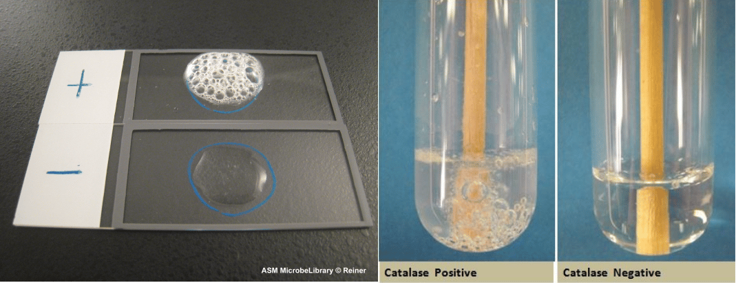 Figure: Slide and tube catalase test methods
Principle
2H₂O₂→ 2H₂O+ O₂ (gas bubbles)
Catalase mediates the breakdown of hydrogen peroxide (H₂O₂) into oxygen and water. To find out if a particular bacterial isolate can produce catalase enzyme, a small inoculum of a bacterial isolate is mixed into hydrogen peroxide solution (3%). It is observed for the rapid elaboration of oxygen bubbles. The lack of catalase is evident by a lack of or weak bubble production.
Catalase-positive bacteria include strict aerobes as well as facultative anaerobes. They all can respire using oxygen as a terminal electron acceptor.
Catalase-negative bacteria include strict anaerobes, which never encounter oxygen, and aerotolerant organisms such as the streptococci, which encounter it constantly and survive anyway.
Why streptococci cannot make catalase, even though they grow in air
A streptococcal colony on a blood agar plate has been sitting in room air for eighteen hours. It is thriving. It has been generating hydrogen peroxide the entire time. Some streptococci, notably S. pneumoniae, generate so much of it that they kill neighboring bacteria with it.
So why has the peroxide not killed them?
Because catalase is not the only way to dispose of hydrogen peroxide. Streptococci carry NADH peroxidase, which reduces H₂O₂ to water without ever releasing oxygen gas. No oxygen, no bubbles. The organism is protected, and the test reads negative. Aerotolerance and catalase are two different things, and streptococci have the first without the second.
But there is a deeper reason, and it explains a result you will eventually see on the bench and mistrust.
Catalase is a heme enzyme. Its active site is built around a heme group. Streptococci, enterococci, and lactococci are heme auxotrophs: they lack the biosynthetic pathway to make heme at all. They cannot build the enzyme, not because they have no gene for it, but because they have no cofactor to put in it.
Hand them heme, and some of them will build it.
Enterococcus faecalis grown on a medium containing heme, which is exactly what blood agar is, can express a functional, heme-dependent catalase. It will bubble. Weakly, but it will bubble. This is the origin of the "pseudocatalase" reaction described in every textbook, and the textbooks generally do not explain it. The organism has not changed. Its medium has.
The practical rule follows from the biology: for any Gram-positive coccus, test from a non-blood-containing medium such as nutrient agar or trypticase soy agar. Do it from blood agar and you risk a bubble that came from the plate, from the red cells, or from an enterococcus that borrowed a cofactor.
Percentage of H₂O₂ used in catalase test
| Percentage | Purpose |
|---|---|
| 3% H₂O₂ | Routine testing of aerobes |
| 15% H₂O₂ | Identification of anaerobic bacteria. |
| 30% H₂O₂ | In the superoxol catalase (used for the presumptive speciation of certain Neisseria species) |
Procedure
Slide Catalase Test
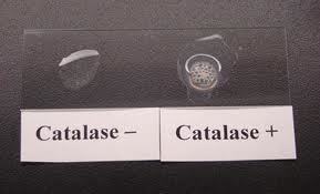 Figure: Catalase Test: Positive and Negative results
- Transfer a small amount of bacterial colony to a surface of a clean, dry glass slide using a loop or sterile wooden stick. Be sure the colony is visible to the naked eye on the slide.
- Place a drop of 3% H₂O₂ onto the slide and mix.
- A positive result is the rapid evolution of oxygen (within 5-10 seconds), as evidenced by bubbling.
- A negative result is no bubbles or only a few scattered bubbles.*
- Dispose of your slide in the biohazard glass disposal container.
Tube Catalase Test
- Add 4 to 5 drops of 3% H₂O₂ to a test tube
- Using a wooden applicator stick, collect a small amount of organism from a well-isolated 18 to 24-hour colony and place it into the test tube (Note: Be careful not to pick up any agar (especially if using Blood Agar).
- Place the tube against a dark background and observe for immediate bubble formation at the end of the wooden applicator stick.
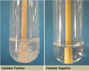 Figure: Catalase Positive and Catalase Negative Reactions
Pseudocatalase
Rare strains of staphylococci may be catalase-negative, and some enterococci produce a “pseudocatalase” and are weakly positive with H₂O₂. "Pseudocatalase" is used loosely in the literature to cover two distinct situations, and it is worth separating them.
True pseudocatalase is a manganese-dependent, non-heme enzyme found in some Lactobacillus and a few other Gram-positive organisms. It degrades hydrogen peroxide and is genuinely not catalase.
Heme-dependent catalase in enterococci is not pseudocatalase at all. Enterococcus faecalis carries a functional catalase gene but cannot synthesize heme. Grown on a heme-containing medium such as blood agar, it acquires the cofactor and expresses real catalase, giving a weak but true positive. Grown on nutrient agar, the same organism is catalase negative.
In both cases the practical consequence is identical: a weak, delayed bubble from a Gram-positive coccus is not a reason to call it Staphylococcus. Repeat from a medium without blood.
Results
- Catalase positive reaction: Evident by immediate effervescence (bubble formation)
- Catalase negative reaction: No bubble formation (effervescence) or a few bubbles after 20 seconds.
| Organism | Gram Stain | Catalase Result | Clinical Significance |
|---|---|---|---|
| Staphylococcus aureus | +ve cocci | Positive | Differentiates from Streptococcus |
| Staphylococcus epidermidis | +ve cocci | Positive | Common skin commensal |
| Micrococcus spp. | +ve cocci | Strongly positive | Differentiates from Staphylococcus |
| Bacillus spp. | +ve rods | Positive | Differentiates from Clostridium |
| Neisseria gonorrhoeae | −ve cocci | Positive (explosive in 30% superoxol | Explosive bubbling in 30% H₂O₂ (superoxol) separates it from N. meningitidis and N. lactamica |
| Haemophilus influenzae | −ve rods | Positive | Fastidious GNR identification |
| Brucella spp. | −ve rods | Positive | Zoonotic pathogen ID |
| Helicobacter pylori | −ve rods | Positive | Gastric ulcer pathogen |
| Streptococcus pyogenes | +ve cocci | Negative | Key distinction from Staphylococcus |
| Streptococcus pneumoniae | +ve cocci | Negative | Key distinction from Staphylococcus |
| Enterococcus spp. | +ve cocci | Negative (rarely pseudocatalase+) | Occasional weak false positive |
| Clostridium perfringens | +ve rods | Negative | Differentiates from Bacillus |
| Escherichia coli | −ve rods | Positive | All Enterobacteriaceae are catalase positive; the test does not differentiate within the family |
| Listeria monocytogenes | +ve rods | Positive | Catalase positive with tumbling motility; separates it from Streptococcus in CSF and blood |
| Mycobacterium tuberculosis | Acid-fast | Positive, heat-labile at 68°C | Catalase-peroxidase (KatG) activates isoniazid; katG mutants are catalase negative and INH resistant |
| Shigella dysenteriae (certain serotypes) | −ve rods | Negative | The exception. Every other member of the Enterobacteriaceae is catalase positive |
Note: Some bacteria possess enzymes other than catalase that can decompose peroxide, a few tiny bubbles forming after 20-30 seconds is not considered a positive test.
Quality Control
Each new lot or shipment of the reagent should be tested with positive and negative control before using them.
A. Staphylococcus aureus ATCC 25923- catalase-positive
B. Streptococcus pyogenes ATCC 19615- catalase-negative
S. aureus secretes catalase and superoxide dismutase, which inhibit organism destruction by the myeloperoxidase system of phagocytic cells.
Five ways the catalase test lies to you
Three of these give you a false positive. Two give you a false negative. Knowing which is which is the difference between calling a Streptococcus a Staphylococcus and the reverse.
- The nichrome loop. False positive.
Do not use a metal loop or needle with H₂O₂; it will give a false positive and degrade the metal. Instead, use a platinum loop or wooden stick to perform this test. Many metals (especially iron-containing ones, common in nichrome loops) can non-enzymatically catalyze the breakdown of hydrogen peroxide into oxygen and water. This chemical reaction, not mediated by bacterial catalase, would produce bubbles and lead to a false-positive result. Platinum loops are chemically inert and do not react with H₂O₂, making them safe for use, as are wooden or plastic sticks. - Red blood cell carryover from blood agar. False positive.
If using colonies from a blood agar plate, be careful not to scrape up any of the blood agar as red blood cells are catalase-positive. The presence of any contaminating agar (carryover of red blood cells) could give a false positive catalase reaction. Red blood cells, like many eukaryotic cells, naturally contain the catalase enzyme to protect themselves from oxidative damage. If red blood cells from the blood agar medium are accidentally transferred along with the bacterial colony to the hydrogen peroxide solution, their intrinsic catalase will react with the H2O2, producing bubbles. This reaction would be mistakenly attributed to the bacterial isolate, resulting in a false-positive catalase test. - Enterococci grown on blood agar. False positive. A weak bubble from a Gram-positive coccus on a blood plate. The organism acquired heme from the medium and built a real catalase. See the pseudocatalase section above
- A culture older than 24 hours. False negative.
Catalase enzyme is present in viable cultures; do not test colonies older than 24 hours. Older cultures may give false-negative results. Bacterial enzyme production, including catalase, is highest during the logarithmic (log) phase of growth when cells are metabolically active and rapidly dividing. As cultures age beyond 24 hours and enter the stationary or decline phase, their metabolic activity decreases, and enzyme production can significantly diminish. This reduction in catalase activity in older, less viable cultures can lead to insufficient bubble production, resulting in a false-negative test, even if the organism inherently produces catalase. - A rare catalase-negative Staphylococcus aureus. False negative. These strains exist. They are uncommon, and they are the reason the catalase test is a screening test and not a confirmatory one. A Gram-positive coccus in clusters that is catalase negative should go to coagulase testing, not be dismissed as a Streptococcus.
NOTE: While performing catalase test, test from a medium whose composition you know. Nutrient agar and trypticase soy agar are ideal. Blood agar is the medium to avoid, for the reasons given above.
Mueller-Hinton agar is not a catalase medium and should not be used for this test, but the reason is simpler than interference: an MHA plate is a susceptibility plate. Colonies on it have been growing in the presence of antimicrobial disks, they are frequently older than 24 hours by the time the plate is read, and they were never intended for identification work. Subculture and test from a fresh plate.
Uses
- The catalase test is primarily used to distinguish among Gram-positive cocci: members of the genus Staphylococcus are catalase-positive, and members of the genera Streptococcus and Enterococcus are catalase-negative.
- Catalase test is used to differentiate Clostridium (catalase-negative for the clinically common species such as C. perfringens) from Bacillus species (catalase-positive). A few aerotolerant Clostridium species are an exception and can be weakly catalase-positive.
- A semiquantitative catalase test is used for the identification of Mycobacterium tuberculosis.
- Catalase test is helpful to separate among the fastidious Gram-negative rods.
- Catalase test can be used as an aid to the identification of Enterobacteriaceae. Members of the Enterobacteriaceae family are catalase positive.
- Shigella dysenteriae types 1, 3, 4, 6, 9, 11, and 12 and S. boydii type 12 are catalase-negative, whereas other species of Shigella, EIEC, and STEC are catalase-positive.
- Neisseria gonorrhoeae produces immediate, vigorous, explosive bubbling when tested with 30% hydrogen peroxide. This reagent is called superoxol, and the test is called the superoxol test. Other Neisseria species, including N. meningitidis, bubble weakly or not at all under the same conditions. All Neisseria are catalase positive in the routine 3% test; superoxol separates them.
Catalase, isoniazid, and why a drug-resistant tuberculosis isolate stops bubbling
Two catalase tests are used for mycobacteria, and each answers a different question.
The semiquantitative catalase test measures how much oxygen is produced, by reading the height of the bubble column in a tube. M. tuberculosis produces a low column, under 45 mm. Most non-tuberculous mycobacteria produce more.
The 68°C heat-stable catalase test asks whether the enzyme survives heating. M. tuberculosis catalase is heat-labile and is destroyed at 68°C. Most non-tuberculous mycobacteria carry a heat-stable catalase. Heat the suspension, add peroxide, and if it still bubbles the organism is probably not M. tuberculosis.
Then the part that matters clinically.
The mycobacterial catalase is KatG, a catalase-peroxidase. Isoniazid, the backbone of first-line TB therapy, is not an active drug when it enters the cell. It is a prodrug. KatG activates it. Without KatG, isoniazid is inert.
So an M. tuberculosis isolate that loses or mutates katG becomes isoniazid resistant, and it becomes catalase negative at the same time, by the same mutation. A large proportion (commonly cited around 50 to 70 percent, varying by region) of all isoniazid-resistant clinical isolates carry a katG mutation.
The organism paid for its resistance by giving up an enzyme that protects it from oxidative killing. It is a genuine trade, and it is visible in a test tube.
How to remember
Cats Need PLACESS to poop
The catalase-positive organisms that infect a child with chronic granulomatous disease. This is the list examiners want, and the hook at the top of this article is where it comes from.
- N — Nocardia
- P — Pseudomonas
- L — Listeria
- A — Aspergillus
- C — Candida
- E — E. coli
- S — Staphylococcus aureus
- S — Serratia
Add Burkholderia cepacia, which the mnemonic omits and which is one of the most characteristic CGD pathogens of all. Aspergillus and Candida are the fungi, not bacteria.
The bench rule, in one line
Bubbles, cluster, catalase positive: think Staphylococcus. No bubbles, chains, catalase negative: think Streptococcus.
Then stop. Catalase gets you to the genus and no further. A Staphylococcus is not S. aureus until coagulase says so.
The question to ask yourself
Before you look anything up: streptococci grow in air, generate hydrogen peroxide, and have no catalase. Why are they still alive?
If you can answer that in one sentence, you understand what this test does and does not detect. If you cannot, reread the section on heme auxotrophy. Every confusing result in this article, including the enterococcal weak positive, follows from it.
Key exam facts in one table
| Question | Answer | The reason behind it |
|---|---|---|
| What is the reaction? | 2H₂O₂ → 2H₂O + O₂ | Oxygen escapes as visible bubbles |
| What reagent, routine test? | 3% hydrogen peroxide | Enough to drive a visible reaction, gentle enough not to lyse the colony |
| 15% H₂O₂ is used for what? | Anaerobes | Their catalase activity is low; more substrate is needed to see the reaction |
| 30% H₂O₂ is used for what? | The superoxol test, to separate N. gonorrhoeae from other Neisseria | N. gonorrhoeae bubbles explosively; N. meningitidis barely does |
| Positive result | Immediate effervescence, within 3 to 10 seconds | Catalase is abundant and turns over extremely fast |
| Negative result | No bubbles, or a few bubbles after 20 seconds | Late scattered bubbles come from other peroxidases, not catalase |
| The core separation | Staphylococcus positive; Streptococcus and Enterococcus negative | The first test on any Gram-positive coccus |
| The second separation | Bacillus positive; Clostridium negative | Both are Gram-positive rods; catalase splits them |
| Why are streptococci negative? | Catalase is a heme enzyme, and streptococci are heme auxotrophs | They cannot build the cofactor, so they cannot build the enzyme |
| Then how do they survive in air? | NADH peroxidase reduces H₂O₂ to water, releasing no oxygen | Protected, but no bubbles, so the test reads negative |
| Why do enterococci on blood agar bubble weakly? | Blood agar supplies heme; E. faecalis has a catalase gene and builds a real enzyme | Not "pseudocatalase." Repeat from nutrient agar |
| What is true pseudocatalase? | A manganese-dependent, non-heme peroxide-degrading enzyme, seen in some Lactobacillus | Genuinely not catalase |
| Why not a nichrome loop? | Iron and nickel decompose H₂O₂ non-enzymatically | False positive, with no organism involved |
| Why not blood agar? | Red blood cells contain catalase | False positive, from the plate |
| Why not a culture over 24 hours old? | Catalase production peaks in log phase and falls in stationary phase | False negative |
| Rare catalase-negative S. aureus? | They exist | False negative. Never call species from catalase alone; coagulase is required |
| Which Enterobacteriaceae is catalase negative? | Certain Shigella dysenteriae serotypes (1, 3, 4, 6, 9, 11, 12) and S. boydii type 12 | Every other member of the family is positive |
| Positive QC organism | Staphylococcus aureus ATCC 25923 | Vigorous and unambiguous |
| Negative QC organism | Streptococcus pyogenes ATCC 19615 | True negative, no peroxidase artifact |
| Catalase in TB? | KatG, a catalase-peroxidase, heat-labile at 68°C | KatG activates isoniazid. katG mutants are catalase negative and INH resistant |
| Catalase in immunology? | Catalase-positive organisms infect patients with chronic granulomatous disease | The classic list: N-PLACESS plus Burkholderia cepacia |
| Catalase as a virulence factor | Destroys phagocyte-derived H₂O₂, resisting oxidative killing | Works with superoxide dismutase; S. aureus has both |
Where students get confused
"Streptococci are catalase negative because they only ferment." They are not. Streptococci grow on an aerobic plate, respire to some degree, and generate hydrogen peroxide continuously. They are catalase negative because catalase is a heme enzyme and streptococci cannot synthesize heme. They survive the peroxide using NADH peroxidase instead, which produces water and no oxygen, and therefore no bubbles. Aerotolerance and catalase are two separate things.
A weak bubble from a Gram-positive coccus on blood agar. Three different things produce this, and they are not equivalent. It could be red blood cell carryover from the plate. It could be an Enterococcus faecalis that has acquired heme from the medium and expressed a genuine catalase. It could be a true Staphylococcus with a light inoculum. The only way to resolve it is to repeat the test from a plate with no blood in it. This single habit prevents more misidentifications than any other on this list.
Reading the direction of each error wrong. Students reliably remember that nichrome loops, blood agar, and old cultures are all forbidden, and reliably forget which way each one pushes. Metal and blood give you a false positive. Age gives you a false negative. If you remember nothing else: the plate and the loop make bubbles appear, time makes them disappear.
Calling it Staphylococcus aureus on catalase alone. Catalase separates Staphylococcus from Streptococcus. It does not identify a species. Gram-positive cocci in clusters that are catalase positive are staphylococci, and they may be S. epidermidis, S. saprophyticus, or any of forty others. Coagulase is what names S. aureus. Catalase-negative S. aureus strains exist, and a student who stops at catalase will miss them entirely.
Confusing catalase with oxidase. Both are rapid, both are enzyme tests, both are done early. Nothing else is shared. Catalase uses 3% hydrogen peroxide, is applied to Gram-positive cocci, and a nichrome loop is unacceptable. Oxidase uses TMPD, is applied to Gram-negative rods, and a nichrome loop is also unacceptable. Note that the loop rule is the one thing they have in common, which is precisely why students transpose everything else.
"Anaerobes are catalase negative." Mostly, and not always. Some aerotolerant Clostridium species and several Bacteroides strains produce catalase. The 15% peroxide concentration exists because anaerobic catalase activity is low, not because it is absent. The rule is a useful default, not a law.
Thinking the semiquantitative catalase test is a lab curiosity. The mycobacterial catalase-peroxidase, KatG, is what converts isoniazid from an inert prodrug into an active one. A katG mutation makes the organism catalase negative and isoniazid resistant simultaneously. The test tube result and the drug resistance are the same molecular event.
Treating the CGD catalase hypothesis as settled. The clinical association is solid: patients with chronic granulomatous disease get recurrent infections with catalase-positive organisms. The mechanism taught to explain it, that catalase-negative organisms hand over their own peroxide and are killed by it, is elegant and probably incomplete. Catalase-deficient S. aureus remains virulent in CGD mouse models. Answer the exam question with the dogma. Know that it is dogma.
References and further readings
- Reiner K. Catalase test protocol. American Society for Microbiology, 2010 (updated 2013).
- Leber AL, editor. Clinical Microbiology Procedures Handbook. 4th ed. Washington, DC: ASM Press; 2016. doi:10.1128/9781555818814
- Tille PM. Bailey and Scott's Diagnostic Microbiology. 15th ed. St. Louis: Elsevier; 2022.
- Procop GW, Church DL, Hall GS, Janda WM, Koneman EW, Schreckenberger PC, Woods GL. Koneman's Color Atlas and Textbook of Diagnostic Microbiology. 7th ed. Philadelphia: Wolters Kluwer; 2017.
- Mandell GL. Catalase, superoxide dismutase, and virulence of Staphylococcus aureus. In vitro and in vivo studies with emphasis on staphylococcal-leukocyte interaction. J Clin Invest. 1975;55(3):561-566. doi:10.1172/JCI107963
- Frankenberg L, Brugna M, Hederstedt L. Enterococcus faecalis heme-dependent catalase. J Bacteriol. 2002;184(22):6351-6356. doi:10.1128/JB.184.22.6351-6356.2002
- Zhang Y, Heym B, Allen B, Young D, Cole S. The catalase-peroxidase gene and isoniazid resistance of Mycobacterium tuberculosis. Nature. 1992;358(6387):591-593. doi:10.1038/358591a0
- Messina CG, Reeves EP, Roes J, Segal AW. Catalase negative Staphylococcus aureus retain virulence in mouse model of chronic granulomatous disease. FEBS Lett. 2002;518(1-3):107-110. doi:10.1016/s0014-5793(02)02658-3
Frequently Asked Questions
What is the principle of the catalase test?
Why is the catalase test important in clinical microbiology?
It separates Staphylococcus (catalase-positive) from Streptococcus and Enterococcus (catalase-negative), guiding further identification. It also helps identify Mycobacterium tuberculosis and differentiate Bacillus from Clostridium.
What causes a false positive in the catalase test?
What causes a false negative in the catalase test?
What is the difference between the slide and tube catalase test?
Why should you not use a metal loop in the catalase test?
Are all Staphylococcus species catalase positive?
Are all Staphylococcus species catalase positive?
Almost all Staphylococcus species are catalase positive, distinguishing them from Streptococcus and Enterococcus. Rare catalase-negative staphylococcal strains exist, so results should be interpreted with other tests.
What is pseudocatalase and which bacteria produce it?
Pseudocatalase is a cytochrome-based mechanism in some Enterococcus and Lactobacillus strains that weakly decomposes H₂O₂, producing delayed weak bubbling after 20-30 seconds unlike the immediate vigorous bubbling of true catalase-positive organisms.

Tankeshwar Acharya, MSc (Medical Microbiology)
Tankeshwar Acharya is an Assistant Professor in the Department of Microbiology at Patan Academy of Health Sciences (PAHS), Nepal, where he has been teaching and practicing clinical microbiology for over 14 years. He is the founder of Microbe Online, one of the leading free microbiology education resources on the web, covering bacteriology, mycology, parasitology, immunology, and clinical laboratory diagnostics written from direct experience in both the classroom and the diagnostic laboratory.
Comments
No comments yet. Be the first to share your thoughts.
Leave a comment
All comments are reviewed before they appear.