
Phenylalanine Deaminase Test: The 5-Minute Green That Flags Proteus, Morganella, and Providencia
Add ferric chloride to a phenylalanine slant and a green color within five minutes means the organism is one of the Proteeae: Proteus, Morganella, or Providencia. The catch is that the green fades fast, so a delayed reading turns a positive into a false negative. Here is the deamination mechanism, why PDA pairs with urease to screen for Proteus, and how to read it before it disappears.
The green that appears, and then vanishes before you write it down
A urine culture grows a swarming Gram-negative rod that has spread as a thin film across the whole blood agar plate, refusing to form discrete colonies. Swarming is a strong clue for Proteus, but a clue is not an identification, and Proteus has to be separated from the other enteric rods that cause urinary infection.
The technician inoculates a phenylalanine slant, incubates it overnight, and adds a few drops of ferric chloride. The slant flushes green almost at once.
Green is positive. It means the organism deaminated phenylalanine to phenylpyruvic acid, and among the Enterobacteriaceae only three genera do that strongly: Proteus, Morganella, and Providencia, together called the Proteeae tribe. The swarming rod is one of them.
But here is the part that catches people. That green is unstable. It begins to fade within a minute or two of adding the reagent and can be gone in five. A technician who adds the ferric chloride, sets the tube down, answers the phone, and comes back to read it will find a straw-colored slant and record a false negative on a genuine Proteus.
This is a read-it-now test. The reaction is fast, the color is diagnostic, and the window is short. This article covers the deamination chemistry behind the green, why phenylalanine deaminase pairs with urease to pin down Proteus quickly, and how to read the result before it disappears.
Principle
The phenylalanine deaminase test, also called the phenylpyruvic acid (PPA) test, detects an organism's ability to produce the enzyme phenylalanine deaminase. This enzyme removes the amine group from the amino acid phenylalanine, an oxidative deamination, producing phenylpyruvic acid (PPA) and ammonia. When ferric chloride is then added, the phenylpyruvic acid reacts with the ferric ions to form a green complex. That green is the readout: it is not the enzyme or the ammonia you are seeing, but the phenylpyruvic acid the enzyme left behind, made visible by iron.
Its main use is narrow and useful: among the Enterobacteriaceae, a strongly positive test points to the Proteeae tribe, Proteus, Morganella, and Providencia.
Phenylalanine agar, also known as phenylalanine deaminase medium which contains DL-phenylalanine and nutrients is used as a test medium.
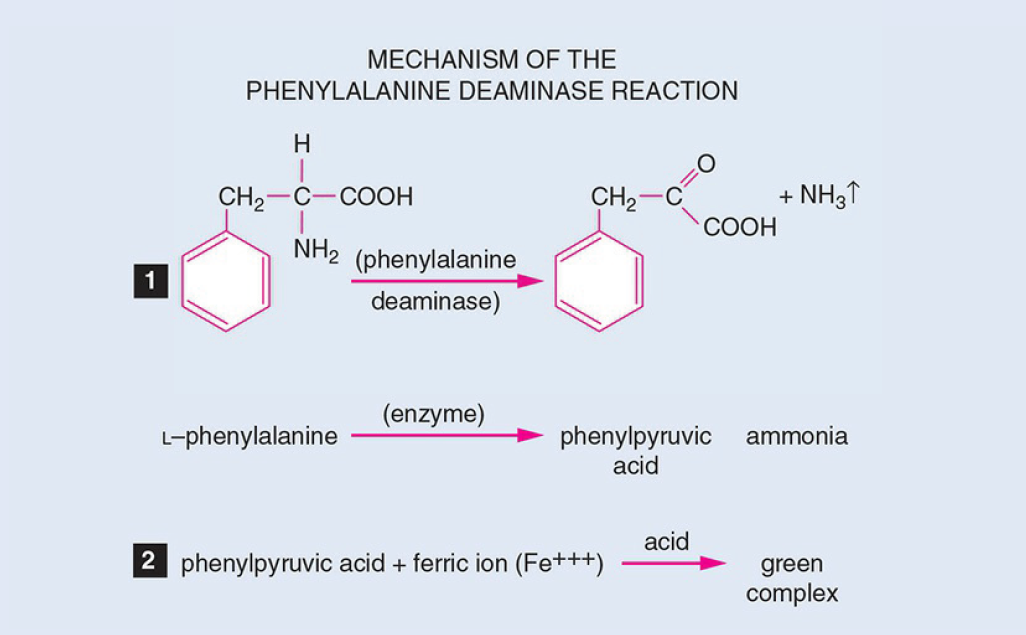 Figure: Phenylalanine Deaminase Test
Oxidative deamination and why only the Proteeae do it strongly
Most bacteria that use amino acids do so by decarboxylation, removing the carboxyl (COOH) group, which is the reaction behind the lysine, ornithine, and arginine decarboxylase tests. The phenylalanine deaminase test detects the opposite chemistry: deamination, removing the amino (NH₂) group instead.
When phenylalanine deaminase acts on phenylalanine, it strips off the amino group as ammonia and leaves behind an α-keto acid, phenylpyruvic acid. This is oxidative deamination: it requires oxygen, which is why the test is done on a slant with a large aerobic surface and read from that surface, not from an anaerobic butt.
Phenylpyruvic acid is colorless on its own. To see it, you add ferric chloride. The ferric ion chelates the α-keto acid and forms a green coordination complex. No phenylpyruvic acid, no green. So the test is really a two-part question: did the organism make the enzyme, and did the enzyme leave behind enough phenylpyruvic acid for the iron to find.
The reason this test is so tightly linked to three genera is that strong, constitutive phenylalanine deaminase activity is characteristic of the Proteeae and uncommon elsewhere in the family. E. coli, Klebsiella, Salmonella, Shigella, and the rest are negative. So a green slant, in the context of a Gram-negative enteric rod, is close to a genus-level statement: this is Proteus, Morganella, or Providencia.
Media and Reagents
Phenylalanine agar medium is prepared and poured as a slant into a tube. The composition of the medium is as follows:
- DL-Phenylalanine: 2 gm
- Yeast extract: 3 gm
- Sodium chloride: 5 gm
- Disodium phosphate: 1 gm
- Agar: 12 gm
- Distilled water: 1 L
- pH: 7.3
Yeast extract serves as carbon and nitrogen source. Meat extracts or protein hydrolysates cannot be used because of the varying natural content of phenylalanine.
Reagent
- Ferric chloride: 12 gm
- Concentrated HCl: 2.5 mL
- Distilled water: 100 mL
Procedure
- Take or prepare a phenylalanine deaminase agar medium
- Inoculate the phenylalanine slant (with a loop on the surface) with a test organism. Note: If you are using the test medium i.e. phenylalanine agar for the first time use positive (Proteus vulgaris) and negative control (Escherichia coli) to check the efficacy of the test medium.
- Incubate at 37°C overnight (18 to 24 hours).
- Add 4 to 5 drops of 10% aqueous ferric chloride (FeCl₃) directly onto the slant, then rotate the tube to wash the reagent over the surface growth. Read within 1 to 5 minutes. The green color develops almost immediately and then fades, so read and record at once. A tube read even ten minutes after adding the reagent can look straw-colored and give a false negative on a truly positive organism. If in doubt, repeat rather than trust a late reading.
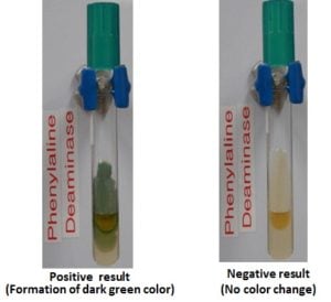 Figure: Phenylalanine deaminase test results(Source: microbugz)
Results
- Positive test: Production of green color (Phenylpyruvic acid thus formed reacts with ferric chloride producing a green colored compound thus turning the medium dark green). Proteus sp., Morganella sp., Providenica sp. give positive PPA test.
- Negative: No color change (medium remains straw/yellow color; no PPA to react with ferric chloride).
Uses of Phenylalanine Deaminase Test
Phenylalanine deaminase test is used to differentiate members of the genera Proteus, Morganella, and Providencia (+ve) from other members of Enterobacteriaceae which give negative results.
Why PDA and urease are run together
Phenylalanine deaminase rarely works alone. In practice it is paired with the urease test, and the pairing is what makes it powerful.
The Proteeae, Proteus, Morganella, and Providencia, are the organisms in the Enterobacteriaceae that are strongly positive for both phenylalanine deaminase and urease. Very few other enterics are. So running the two tests together gives a fast, two-reaction screen:
| Result | Points to |
|---|---|
| PDA positive and urease positive | Proteeae: Proteus, Morganella, Providencia |
| PDA negative, urease positive | Non-Proteeae urease producer (e.g. Klebsiella, some Enterobacter, Yersinia) |
| PDA negative, urease negative | Most other Enterobacteriaceae (E. coli, Salmonella, Shigella) |
This is why a rapid urea-PDA disk was developed: it reads both enzymes from a single inoculation. The combination narrows a Gram-negative enteric rod to the Proteeae in minutes, before the slower IMViC and sugar-fermentation battery. From there, further tests separate the three genera and the individual species. For the swarming urinary isolate in the opening, PDA positive plus urease positive is most of the way to naming it Proteus.
The historical note is worth keeping for context: Henriksen first showed in 1950 that Proteus deaminates phenylalanine, and Ewing and colleagues built that observation into the slant medium in 1957. Ederer and colleagues later developed the rapid urea-PDA disk in 1971. The pairing of the two enzymes has been the point from the beginning.
Phenylalanine deaminase by organism
Positive (the Proteeae):
- Proteus vulgaris, Proteus mirabilis
- Morganella morganii
- Providencia species (P. rettgeri, P. stuartii, P. alcalifaciens)
Negative (the rest of the Enterobacteriaceae):
- Escherichia coli
- Klebsiella species
- Salmonella and Shigella species
- Enterobacter and Citrobacter species
- Serratia species
The clean split is what makes the test useful: positive means Proteeae, negative means almost anything else in the family. There is little middle ground, which is unusual and convenient.
Quality Control
- Positive control: Proteus mirabilis ATCC 12453 or Proteus vulgaris ATCC 13315 — green within 5 minutes
- Negative control: Escherichia coli ATCC 25922 — no color change (straw)
Run controls with each new batch of medium or reagent. Ferric chloride reagent degrades and can give false negatives if old, so the positive control is what confirms the reagent still works.
How to remember
Green flags the Proteeae.
A green slant on phenylalanine agar is nearly a genus-level answer: this is Proteus, Morganella, or Providencia, the three-genus Proteeae tribe. Almost nothing else in the Enterobacteriaceae turns it green. When you see the green, think Proteeae and reach for the urease result to confirm.
PDA plus urease equals Proteus, fast.
The two enzymes travel together in the Proteeae. Positive for both, in a Gram-negative enteric rod, is your rapid ticket to the Proteeae before any of the slower tests come back. Picture them as a pair of keys that only these three genera carry.
Read it before it fades.
The green is a sprinter, not a marathoner. It shows up in seconds and it is gone in minutes. The discipline this test demands is simple and easy to forget: add the reagent, look now, record now. Ask yourself as you drop the ferric chloride, am I watching this tube right now? If not, you are about to lose the result.
Deamination, not decarboxylation.
Most amino-acid tests in this cluster remove the carboxyl group (the decarboxylases). This one removes the amino group and leaves an acid behind. Deaminase takes the amine: the name tells you which end of the molecule is lost.
Key exam facts in one table
| Question | Answer | The reason behind it |
|---|---|---|
| What does the test detect? | Phenylalanine deaminase enzyme | Removes the amino group from phenylalanine |
| What reaction? | Oxidative deamination | Requires oxygen; done and read on the slant surface |
| Products | Phenylpyruvic acid + ammonia | Phenylpyruvic acid is the one detected |
| Reagent | 10% ferric chloride | Chelates phenylpyruvic acid into a green complex |
| Positive result | Green on the slant surface | Phenylpyruvic acid present |
| Negative result | Straw/yellow, no change | No phenylpyruvic acid to react |
| Reading window | Within 1 to 5 minutes | The green fades fast; late reading gives false negative |
| Which genera are positive? | The Proteeae: Proteus, Morganella, Providencia | Strong constitutive deaminase activity is characteristic of this tribe |
| Which are negative? | Most Enterobacteriaceae (E. coli, Klebsiella, Salmonella, Shigella, etc.) | They lack strong phenylalanine deaminase |
| Why paired with urease? | Proteeae are positive for both | PDA + urease is a rapid two-reaction Proteeae screen |
| PDA + / urease + | Proteeae | Narrows to Proteus/Morganella/Providencia in minutes |
| PDA − / urease + | Non-Proteeae urease producer | Klebsiella, some Enterobacter, Yersinia |
| Deamination vs decarboxylation | Deaminase removes NH₂; decarboxylase removes COOH | The name tells you which group is lost |
| Why yeast extract, not meat extract, in the medium? | Meat extract has variable natural phenylalanine | Would confound the substrate; yeast extract is defined |
| Why a slant, not a butt? | The reaction is aerobic | Needs the oxygenated surface |
| QC positive | Proteus mirabilis ATCC 12453 | Green within 5 minutes |
| QC negative | E. coli ATCC 25922 | Straw, no change |
| Rapid method | Urea-PDA disk (Ederer, 1971) | Reads both enzymes from one inoculation |
Where students get confused
Reading the tube too late. The defining error and the main cause of false negatives. The green develops within a minute and fades within about five. Add the ferric chloride, read immediately, and record at once. A tube left to sit will read straw-colored and be called negative on a genuine Proteus. If you were distracted, repeat rather than trust the late reading.
Confusing deamination with decarboxylation. This test removes the amino group (NH₂); the lysine, ornithine, and arginine decarboxylase tests remove the carboxyl group (COOH). They detect opposite chemistry on amino acids and are easy to mix up. The word deaminase points to the amine being removed.
Thinking the green is the enzyme or the ammonia. The green is neither. It is the ferric chloride reacting with phenylpyruvic acid, the α-keto acid the enzyme left behind. The ammonia is real but invisible here. If there is no phenylpyruvic acid, there is no green, regardless of how much ammonia was released.
Treating PDA as a standalone identification. On its own, PDA positive tells you the organism is one of three genera. It does not separate Proteus from Morganella from Providencia, and it does not name a species. It is a tribe-level screen, most powerful when paired with urease and followed by further tests. Reading too much into a single green slant is a classic overreach.
Using meat-extract medium. The medium deliberately uses yeast extract, not meat extract or protein hydrolysate, because those contain variable natural phenylalanine that confounds the defined substrate. Substituting the medium can invalidate the result.
Reading a degraded reagent as negative. Ferric chloride reagent deteriorates over time. An old reagent can fail to develop green even on a positive organism. The positive control run with each batch is what tells you the reagent still works; skip it and a reagent failure looks like a negative organism.
Forgetting the reaction is aerobic. Phenylalanine deamination needs oxygen. Inoculate and read the slant surface, which is aerobic. Do not stab the butt expecting a reaction there.
References
- Henriksen SD. A comparison of the phenylpyruvic acid reaction and the urease test in the differentiation of Proteus from other enteric organisms. J Bacteriol. 1950;60(2):225-231. doi:10.1128/jb.60.2.225-231.1950
- Ewing WH, Davis BR, Reavis RW. Phenylalanine and malonate media and their use in enteric bacteriology. Public Health Lab. 1957;15:153-167.
- Ederer GM, Clark M. Motility-indole-lysine-sulfide medium and a urea-phenylalanine disk for the identification of enteric bacteria. Appl Microbiol. 1971;21(3):545-547. doi:10.1128/am.21.3.545-547.1971
- Procop GW, Church DL, Hall GS, Janda WM, Koneman EW, Schreckenberger PC, Woods GL. Koneman's Color Atlas and Textbook of Diagnostic Microbiology. 7th ed. Philadelphia: Wolters Kluwer; 2017.
- Tille PM. Bailey and Scott's Diagnostic Microbiology. 15th ed. St. Louis: Elsevier; 2022.
Frequently Asked Questions
What does a positive phenylalanine deaminase test indicate?
Why does the green color fade so quickly in the PDA test?
What is the difference between deamination and decarboxylation?
Why is the phenylalanine deaminase test run together with the urease test?
Which organisms are phenylalanine deaminase positive?
What is the green color in the PDA test actually made of?
Why must the phenylalanine test use yeast extract rather than meat extract?

Tankeshwar Acharya, MSc (Medical Microbiology)
Tankeshwar Acharya is an Assistant Professor in the Department of Microbiology at Patan Academy of Health Sciences (PAHS), Nepal, where he has been teaching and practicing clinical microbiology for over 14 years. He is the founder of Microbe Online, one of the leading free microbiology education resources on the web, covering bacteriology, mycology, parasitology, immunology, and clinical laboratory diagnostics written from direct experience in both the classroom and the diagnostic laboratory.