
Lactophenol Cotton Blue (LPCB) Mount: Principle, Procedure, and Fungal Identification
LPCB is the standard mounting medium for microscopic fungal identification — staining chitin in cell walls to reveal hyphae, conidia, and spore arrangements. Learn the tease mount procedure, what to look for, and how to identify common fungi.
A patient with poorly controlled diabetes develops a rapidly spreading black necrotic lesion involving the palate and periorbital region. CT scan shows sinus involvement. An urgent biopsy is taken. In the mycology laboratory, the tissue is cultured on Sabouraud dextrose agar — and within 48 hours, a rapidly growing fluffy grey-white mould appears. An LPCB mount is prepared.
Under the microscope: broad, non-septate (aseptate) hyphae with irregular branching at wide angles. No structures visible from fruiting heads yet, but the combination of tissue presentation, growth rate, and hyphal morphology is already pointing to Mucorales — mucormycosis, one of the most lethal fungal infections in immunocompromised patients. Treatment decisions cannot wait for species confirmation.
LPCB is the workhorse of the mycology bench — the fastest way to examine fungal morphology in a wet preparation. Understanding what you are looking at under oil immersion is as important as knowing how to prepare the mount.
The lactophenol cotton blue (LPCB) wet mount preparation is the most widely used method of staining and observing fungi and is simple to prepare. The preparation has the following constituents;
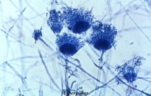 Figure: Fig: Aspergillus spp in LPCB Mount
- Phenol kills fungus.
- Lactic acid acts as a clearing agent and helps preserve the fungal structures.
- Cotton blue is an aniline dye that stains the chitin in the fungal cell walls which adds colour to the fungal preparation thereby enhancing and contrasting the structures.
- Glycerol is a viscous substance that prevents drying of the prepared slide specimen.
Why LPCB is a mounting medium, not a fixative: LPCB preserves and stains fungal structures in their native three-dimensional arrangement — it does not fix or kill organisms immediately on contact (though phenol does kill over time). This is why the preparation should be examined promptly after preparation, and why it is essential to work under a biological safety cabinet with gloves throughout.
Lactophenol cotton blue solution is a mounting medium and staining agent used in the preparation of slides for microscopic examination of fungi. Fungal elements are stained intensely blue. This operation should always be performed under a biological safety hood and gloves should always be worn.
Procedure
Standard Tease Mount
- Place a drop of 70% alcohol on a clean microscope slide. Material from cultures of filamentous fungi should be removed using a stiff inoculating wire, not the loop used for manipulations with bacteria or yeasts.
- Flame the wire by holding it upright in the hottest part of the Bunsen flame, just above the blue cone, until the whole length of the wire glows red hot. You must ensure that the inoculating wire has cooled before placing it in a fungal culture – it should have cooled sufficiently after approximately ten seconds.
- Remove the cap from the culture tube (but do not put it on the bench). Kill any contaminating microorganisms by flaming the neck of the tube.
- Remove a small amount of the culture. It is often useful to take a little of the agar medium together with the fungus. In any case, the material should be disturbed as little as possible when being transferred to the slide.
- Flame the neck of the tube once more and replace the cap.
- Immerse the fungal material in the drop of 70% alcohol. This drives out the air trapped between the hyphae.
- Tease out the material very gently with mounted needles*. Do not forget to sterilize the inoculating wire and the needles after use by heating to red heat in a Bunsen flame
- Before the alcohol dries out add one or at most two drops of the stain. A common fault is to add too much stain to the preparation.
- Holding the coverslip between your index finger and thumb, touch one edge of the drop of stain with the edge of the coverslip.
- Lower the coverslip gently onto the slide, trying to avoid air bubbles. Your preparation is now ready for examination.
Teasing the colony often disrupts the delicate fruiting structures of the filamentous molds, making it difficult in some instances to observe the characteristic spore arrangements or hyphal attachments necessary for definitive identification. In such cases, a transparency tape mount or microslide culture may be required.
Cellophane Tape Preparation
The cellophane or transparency tape method for microscopic examination of fungi is simple to perform, inexpensive and, rapid. It preserves the conidial arrangements of the more delicate filamentous molds and allows one to make an accurate identification.
- Place a drop of lactophenol aniline blue stain on a microscope slide.
- Press the sticky side of unfrosted, clear cellophane tape gently but firmly to the surface of the colony, picking up a portion of the aerial mycelium.
- Stick one end of the tape to the surface of the slide adjacent to the drop of stain.
- Stretch the tape over the stain, gently lowering it so that the mycelium becomes permeated with stain.
- Pull the tape taught and stick the opposite end to the glass, avoiding as much as possible the trapping of air bubbles.
- Your preparation is now ready for examination.
Examination
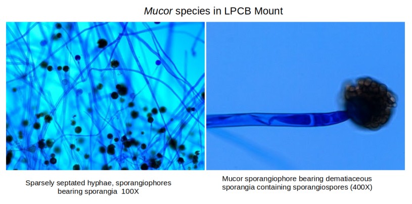 Figure: Mucor species in LPCB mount (Image source)
- Make the initial examination using a low-power objective lens. The thinner parts of the preparation, generally around the edges of the mounting material, will yield the best images.
- Switch to a higher power 40X objective for a more detailed examination of spores and other structures.
What to Look For: Key Morphological Features Under LPCB
LPCB stains chitin in fungal cell walls blue, revealing the following structures:
Hyphae:
- Septate hyphae — hyphae divided by cross-walls (septa) into compartments. Seen in most medically important moulds: Aspergillus, Fusarium, Dermatophytes. Septa are often visible as faint lines perpendicular to the hyphal wall.
- Aseptate (non-septate) hyphae — hyphae without cross-walls; broad, ribbon-like, occasionally collapsing. Characteristic of Mucorales (Mucor, Rhizopus, Lichtheimia). Width is typically 6–25 μm — dramatically wider than septate mould hyphae (2–4 μm).
- Pseudohyphae — chains of elongated yeast cells that resemble hyphae but have constrictions at junctions. Characteristic of Candida albicans.
Key organisms and their LPCB appearance:
| Organism | Hyphae | Key Structure | Distinguishing Feature |
|---|---|---|---|
| Aspergillus fumigatus | Septate, 2–4 μm | Conidiophore with vesicle bearing phialides on upper 2/3 | Blue-grey conidia; phialides on upper portion only (uniseriate) |
| Aspergillus niger | Septate | Large vesicle with biseriate phialides covering entire surface | Black conidia head; radiate arrangement |
| Aspergillus flavus | Septate | Large irregular vesicle; biseriate phialides | Yellow-green conidia; rough conidiophore |
| Mucor / Rhizopus | Aseptate, broad | Sporangiophore bearing spherical sporangium | Large round sporangia; sporangiospores inside; Rhizopus has rhizoids |
| Fusarium spp. | Septate | Sickle-shaped (banana-shaped) macroconidia | Characteristic curved macroconidia with foot cell |
| Penicillium spp. | Septate | Brush-like (penicillus) conidiophore | Branching phialides resembling a paintbrush |
| Trichophyton spp. | Septate | Thin-walled pencil-shaped macroconidia; microconidia | Macroconidia with smooth walls |
| Microsporum canis | Septate | Thick-walled spindle-shaped macroconidia with rough walls | Echinulate (spiky) macroconidia |
| Epidermophyton floccosum | Septate | Club-shaped smooth macroconidia in clusters | No microconidia — only dermatophyte without microconidia |
| Candida albicans | Pseudohyphae | Blastoconidia along pseudohyphae; terminal chlamydospores | On cornmeal-Tween 80 agar: chlamydospores confirm C. albicans |
| Cryptococcus neoformans | None (yeast only) | Round yeast with narrow-based budding | Capsule not visible on LPCB — use India ink for capsule |
The three most important patterns to recognise immediately:
- Broad aseptate hyphae → Mucorales → urgent clinical situation in immunocompromised patients
- Septate hyphae with acute-angle branching + blue-grey conidia → Aspergillus fumigatus
- Sickle-shaped macroconidia → Fusarium → important to distinguish from Aspergillus as treatment differs
LPCB vs Calcofluor White — When to Use Each
Both LPCB and calcofluor white stain fungal cell walls, but they have different applications:
| Feature | LPCB | Calcofluor White |
|---|---|---|
| Microscope required | Ordinary bright-field microscope | Fluorescence microscope |
| Appearance | Blue fungal elements on clear background | Bright fluorescent white/blue-white on dark background |
| Sensitivity | Moderate — good for culture preparations | Higher — detects sparse fungal elements in direct specimens |
| Best use | Culture identification — morphological detail | Direct clinical specimens (skin scrapings, BAL, corneal scrapings) |
| Preparation type | Wet mount from culture | Direct specimen examination |
| Background interference | Minimal on culture preparations | Can have autofluorescent artefacts in tissue |
| Skill required | Standard microscopy | Fluorescence microscopy experience |
Practical guidance:
- Culture identification → LPCB (standard choice — bright-field, full morphological detail)
- Direct specimen (skin scraping, BAL, nail, hair) → Calcofluor white if fluorescence microscope available; KOH mount if not
- Both can be used together for difficult cases where standard bright-field and fluorescence complement each other
For calcofluor white staining procedure, see: Calcofluor White Staining
LPCB for Intestinal Parasite Detection
The existing reference (Parija et al., 2003) notes a use that many microbiologists overlook: LPCB wet mounts of stool specimens can detect Cryptosporidium, Cyclospora, and Isospora oocysts.
Appearance on LPCB:
- Cryptosporidium parvum oocysts — small (4–6 μm), round, pink-tinged or pale against blue background; internal sporozoites occasionally visible
- Cyclospora cayetanensis oocysts — larger (8–10 μm), round, wrinkled appearance; appear as two clusters of sporocysts when sporulated
- Isospora belli oocysts — elongated oval (20–33 μm × 10–19 μm); most easily identified by size and shape
Important limitation: LPCB is not the preferred method for parasite detection — modified Ziehl-Neelsen (cold acid-fast stain) is the standard for Cryptosporidium and Cyclospora, and saline wet mount for intestinal parasites broadly. LPCB can be used when these dedicated methods are unavailable, but sensitivity is lower.
Troubleshooting LPCB Preparations
| Problem | Likely Cause | Action |
|---|---|---|
| Air bubbles under coverslip | Coverslip lowered too quickly; too much stain | Lower coverslip at an angle slowly; use only 1–2 drops of stain |
| Fruiting structures disrupted or absent | Over-teasing with needles; disturbed colony during transfer | Use minimal mechanical force; cellophane tape method preserves conidial arrangements better for delicate moulds |
| Hyphae appear colourless/unstained | Too little LPCB; stain too old | Add slightly more LPCB; replace if precipitate visible |
| Preparation drying out | Too little glycerol in stain; too much alcohol used | Ensure 70% alcohol evaporates before adding LPCB; use fresh stain with adequate glycerol |
| Cannot distinguish septate from aseptate | Low-power examination only | Switch to 40x objective; look for faint perpendicular lines (septa) within hyphal walls |
| Yeast cells not visible | Yeast may be sparse in the preparation | Prepare a second mount using a heavier inoculum from a yeast colony |
The transparency tape method is specifically recommended when fruiting structures have been disrupted by the tease mount — always attempt the tape method first for Aspergillus, Penicillium, and Fusarium where conidial arrangement is the key identification feature.
How to Remember: LPCB
The four components — "Please Let Cotton Grow":
- Phenol — kills
- Lactic acid — clears and preserves
- Cotton blue — stains chitin blue
- Glycerol — prevents drying
The broad vs narrow hyphal rule:
- Broad + aseptate = Mucorales (Mucor, Rhizopus) — emergency in diabetics and immunocompromised patients
- Narrow + septate = everything else (Aspergillus, Fusarium, dermatophytes)
This single distinction is the most urgent clinical differentiation in the mycology lab.
The three conidiophore shapes to know:
- Aspergillus = flask on a stick (conidiophore + vesicle + phialides)
- Penicillium = paintbrush (branching phialides)
- Fusarium = sickle/banana (curved macroconidia)
Composition of LPCB mount
| Ingredients | Amount |
|---|---|
| Lactic acid | 20 mL |
| Glycerol (or glycerine) | 40 mL |
| Phenol crystals or phenol concentrate | 20 g 20 mL |
| Distilled water | 20 mL |
| Aniline blue or 1% aqueous solution (This is analogous to cotton blue.) | 0.05 g 2 mL |
Preparation of LPCB mount
LPCB can be purchased from commercial suppliers or prepared in-house by mixing the above-mentioned ingredients as mentioned in the steps below.
- Add 20 mL of lactic acid in a beaker.
- Add 40 mL of glycerol or glycerine.
- Add 20 mL of distilled water
- Add 22 g. of phenol crystals (22 ml of melted phenol).
- Add 0.05 g. aniline blue.
- Heat and stir the solution to dissolve the stain. Do not boil or go over 100°C.
- Mix well and let the solution cool. You can store the solution at room temperature and dispensed it with a pipette when needed.
Key Exam Facts in One Table
| Feature | Detail |
|---|---|
| Full name | Lactophenol cotton blue (LPCB) |
| Type | Mounting medium and staining agent |
| What it stains | Chitin in fungal cell walls → blue |
| Phenol role | Kills fungal organisms |
| Lactic acid role | Clearing agent; preserves fungal morphology |
| Cotton blue (aniline blue) role | Stains chitin blue for contrast |
| Glycerol role | Prevents drying of preparation |
| Broad aseptate hyphae | Mucorales (Mucor, Rhizopus, Lichtheimia) |
| Narrow septate hyphae | Aspergillus, Fusarium, dermatophytes, most other moulds |
| Aspergillus fumigatus | Conidiophore + vesicle + phialides on upper 2/3 + blue-grey conidia |
| Fusarium | Sickle-shaped (banana-shaped) macroconidia |
| Epidermophyton | Smooth club-shaped macroconidia; NO microconidia |
| Tease mount vs tape method | Tease mount for most; cellophane tape preserves conidial arrangement for delicate moulds |
| LPCB vs calcofluor white | LPCB for culture ID (bright-field); calcofluor for direct specimens (fluorescence) |
| Safety requirement | Biological safety cabinet + gloves — phenol is toxic |
References
- Winn, W. C., & Koneman, E. W. (2006). Koneman’s Color Atlas and Textbook of Diagnostic Microbiology (Color Atlas & Textbook of Diagnostic Microbiology). Lippincott Williams & Wilkins
- Parija, S. C., Shivaprakash, M. R., & Jayakeerthi, S. R. (2003). Evaluation of lacto-phenol cotton blue (LPCB) for detection of Cryptosporidium, Cyclospora and Isospora in the wet mount preparation of stool. Acta tropica, 85(3), 349–354. https://doi.org/10.1016/s0001-706x(02)00265-6
- Khubnani, H., Sivarajan, K., & Khubnani, A. H. (1998). Application of lactophenol cotton blue for identification and preservation of intestinal parasites in faecal wet mounts. Indian journal of pathology & microbiology, 41(2), 157–162.
- Larone DH. Larone's Medically Important Fungi: A Guide to Identification. 6th ed. ASM Press; 2018.
- Murray PR, Rosenthal KS, Pfaller MA. Medical Microbiology. 9th ed. Elsevier; 2020.
Frequently Asked Questions
What does each component of LPCB do?
What is the most urgent morphological distinction to make on an LPCB mount?
Why should the cellophane tape method be used instead of the tease mount for some fungi?

Tankeshwar Acharya, MSc (Medical Microbiology)
Tankeshwar Acharya is an Assistant Professor in the Department of Microbiology at Patan Academy of Health Sciences (PAHS), Nepal, where he has been teaching and practicing clinical microbiology for over 14 years. He is the founder of Microbe Online, one of the leading free microbiology education resources on the web, covering bacteriology, mycology, parasitology, immunology, and clinical laboratory diagnostics written from direct experience in both the classroom and the diagnostic laboratory.