
KOH Mount: Principle, Procedure, Results, Uses
KOH preparation: principle, procedure, 10% concentration rationale, organism-specific findings (dermatophytes, Candida, Mucorales, Coccidioides), troubleshooting artifacts vs true fungal elements, and comparison with calcofluor white.

On this page
Potassium hydroxide (KOH) preparation is one of the most common direct methods for detecting fungi in clinical specimens. Strictly, it is a clearing agent rather than a stain: it dissolves the surrounding tissue so the fungus becomes visible, unlike true stains such as PAS, GMS, or calcofluor white, which bind the fungus and make it stand out by color or fluorescence.
Treatment of skin scrapings, nails, or hairs with potassium hydroxide (KOH) dissolves tissue material, leaving the alkali-resistant fungi intact. Microscopic examination of KOH preparation reveals the presence of fungal structure and aids in diagnosing mycoses.
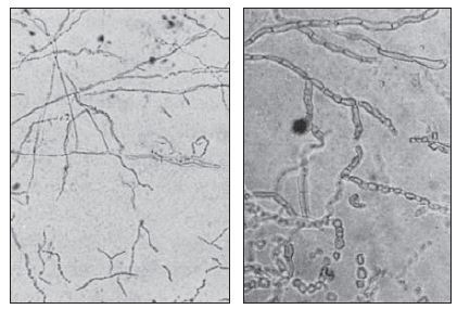 Figure: Image 1:Left: Fungal hyphae in a (KOH) preparation of skin scales as seen with the 10x objective. Right: Hyphae and arthroconidia as seen with the 40x objective.(Image source-Reference-1)
KOH is a strong alkali. When specimen such as skin, hair, nails or sputum is mixed with 10% w/v KOH, it softens, digests and clears the tissues (e.g., keratin present in skins) surrounding the fungi so that the hyphae and conidia (spores) of fungi can be seen under a microscope.
Why 10%
The chosen KOH concentration must balance two competing needs: strong enough to dissolve keratin within a practical timeframe, but not so concentrated that it damages or distorts the fungal elements you're trying to see.
| Concentration | Use case | Clearing time |
|---|---|---|
| 10% KOH | Standard for skin scrapings, vaginal/oral swabs | 5–10 minutes |
| 20% KOH | Thicker keratinized specimens: nails, thick skin scales | 20–30 minutes, or with gentle heating |
| 40% KOH | Rarely used. Useful for very thick nail clippings | Extended clearing, often with heat |
For nail specimens, gentle warming (not boiling) over a flame or on a slide warmer significantly accelerates keratin digestion. Without heat, thick nail material may take hours to clear adequately, during which fungal elements can also start to degrade.
KOH Mount Overview
| Method | Potassium hydroxide preparation (KOH mount) |
|---|---|
| Use | Clearing of specimens to make fungi more readily visible |
| Time required | 5 min; if clearing is not complete, an additional 5-10 min is necessary. |
| Advantages | Rapid detection of fungal elements |
| Disadvantages | Requires experience because background artifacts are often confusing; clearing of some specimens may require an extended time. |
Procedure of KOH Preparation
- Place a drop of KOH solution on a slide.
- Transfer the specimen (small pieces) to the drop of KOH, and cover with glass. Place the slide in a petri dish, or another container with a lid, together with a damp piece of filter paper or cotton wool to prevent the preparation from drying.Note: To assist in clearing, hairs should not be more than 5 mm long, and skin scales, crusts, and nail snips should not be more than 2 mm across.
- As soon as the specimen has cleared, examine it microscopically using the 10X and 40X objectives with the condenser iris diaphragm closed sufficiently to give a good contrast. If too intense a light source is used, the contrast will not be adequate, and the unstained fungi will not be seen.
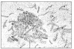 Figure: Malassezia furfur yeast cells and hyphae in KOH blue–black ink preparation. (Image source: Reference 1)
Uses
The KOH test is one of the main methods of diagnosing fungal infections in diagnostic laboratories. It is used as a primary screening tool. This wet-mount procedure helps to visualize fungal elements but may not necessarily identify the species of the fungi.
Complete Organism-Specific Findings Table
Suspected condition | Specimen | Diagnostic characteristics |
|---|---|---|
Dermatophyte infection (ringworm) | Skin scrapings, nail clippings, hair | Hyaline, septate, branching hyphae; arthroconidia in chronic infections |
Candida infection | Skin scrapings, vaginal/oral swabs | Budding yeast cells (blastoconidia) together with pseudohyphae and true hyphae |
Aspergillus infection | Sputum, BAL fluid, sinus aspirate | Septate hyphae with acute-angle (~45°) dichotomous branching |
Mucormycosis | Exudates, tissue, sinus aspirate | Broad, ribbon-like, non-septate hyphae with wide-angle (~90°) branching; urgent finding, notify clinician immediately |
Blastomyces dermatitidis infection | Pus, sputum, skin specimens | Large yeast cells (8–15 μm) with characteristic broad-based bud and thick, double-contoured wall |
Chromoblastomycosis | Scrapings from crusted skin lesions | Muriform cells ("copper pennies"); clusters of dark brown, thick-walled cells with internal septation, 4–10 μm |
Coccidioidomycosis | Sputum, pus, tissue | Large spherules (up to ~80 µm, occasionally larger) filled with endospores; unique among fungi, do NOT confuse with yeast budding |
Sporothrix schenckii infection | Pus, exudate from lesion | Small, elongated "cigar-shaped" yeast cells (rarely seen directly so culture usually required for confirmation) |
Tinea versicolor (Malassezia furfur) | Skin scrapings | Short, curved hyphae together with clusters of round yeast cells, the classic "spaghetti and meatballs" appearance |
KOH preparation is recommended in the following suspected conditions (this is not the exclusive list);
Note: To identify the fungal isolate, specimens must be cultured in either general purpose fungal culture media such as Sabouraud Dextrose Agar (SDA) or specific media based on the type of anticipated isolate.
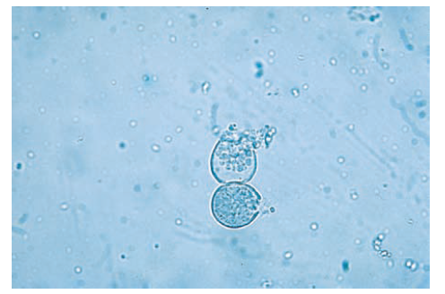 Figure: KOH mount showing two spherules of Coccidioides spp. filled with endospores.
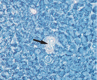 Figure: Potassium hydroxide preparation examined in phase-contrast microscopy showed a large budding yeast cell with a distinct broad base bud (Blastomyces dermatitidis).
Troubleshooting: Distinguishing True Fungal Elements from Artifacts
This is the skill that genuinely separates an experienced reader from a beginner, and it is the single biggest source of both false positives and missed diagnoses in KOH microscopy.
| What you might see | True fungal element or artifact? | How to tell the difference |
|---|---|---|
| Long, thin, branching, refractile structures | Could be either | True hyphae have consistent width throughout and show true branching (not just crossing); refractile (light-bending) appearance |
| Cotton or textile fibres | Artifact | Fibers are usually much wider than hyphae, show NO branching, have sharp/clean broken ends, and are often birefringent under polarized light |
| Cell border edges (mosaic/honeycomb pattern) | Artifact, "mosaic fungus" | This is the most common false-positive trap. Cellular outlines of normal skin cells can resemble pseudohyphae. True hyphae cross OVER cell borders; cell border artifacts follow the cell boundary exactly and do not cross into adjacent cells |
| Round, refractile droplets | Usually artifact (oil droplets, air bubbles) | Air bubbles have a distinct dark ring/halo and are perfectly spherical with sharp black edges; true yeast cells have a more subtle cell wall outline and may show budding |
| Crystals (KOH or specimen-related) | Artifact | Sharp geometric edges, no internal structure, often appear at the edge of the preparation where evaporation has concentrated the KOH |
| Elastic and collagen fibres (especially in nail specimens) | Artifact | Wavy, ribbon-like, but lack the consistent tubular diameter and septation of true hyphae |
The single most reliable rule: True fungal hyphae almost always show septation (cross-walls, except in Mucorales) and consistent diameter along their length. When in doubt, look for definitive structures such as clear branching points, septa, or budding with a visible bud scar, rather than relying on a single ambiguous structure.
When still uncertain: Add a drop of calcofluor white and re-examine under a fluorescence microscope. Chitin-specific fluorescent binding eliminates almost all the ambiguity above, since artifacts (fibers, cell borders, crystals) generally do not contain chitin and will not fluoresce the same way.
If you want to know about other fungal staining methods check this article: Fungal Staining Methods and Uses
Procedure to make 100 ml of KOH 10% w/v solution
- Weigh 10 g potassium hydroxide (KOH) pellets.
- Transfer the chemical to a screw-cap bottle.
- Add 50 ml distilled water, and mix until the chemical is completely dissolved. Add remaining distilled water and make the volume 100 ml.
- Label the bottle and mark it as corrosive. Store it at room temperature. The reagent is stable for up to 2 years.
Caution: Potassium hydroxide is a highly corrosive deliquescent chemical therefore handle it with great care and ensure the stock bottle of chemical is tightly stoppered after use.
Modification in KOH Preparation method
- Use of dimethylsulphoxide-KOH reagent: Adding dimethylsulphoxide (DMSO) to KOH enables specimens to be examined immediately or after only a few minutes.
- KOH with blue-black fountain pen ink added: The ink is not specific for fungi as it stains cells and other components in the skin. The addition of ink is recommended when Malassezia furfur is suspected.
How Remember KOH Preparation
KOH preparation's theory is simple (dissolve keratin, see what's left). The genuine struggle is entirely practical: telling a real hypha from a skin cell border or cotton fiber under time pressure with a queue of specimens waiting. The troubleshooting table above addresses this directly.
One sentence that captures the entire clinical relevance
"KOH doesn't identify the fungus; it just clears away everything that isn't one, so the real skill is knowing what 'fungus' actually looks like versus everything else trying to fool you."
Key exam facts
| Question | Answer |
|---|---|
| What does KOH chemically do to the specimen? | Dissolves keratin and other tissue material via alkaline hydrolysis |
| What concentration of KOH is standard for skin scrapings? | 10% w/v |
| What concentration is used for thick nail specimens? | 20% (sometimes with gentle heat) |
| What does "spaghetti and meatballs" describe? | The short hyphae and round yeast clusters of Malassezia in tinea versicolor |
| What is unique about Coccidioides on KOH? | Forms large spherules filled with endospores, not a yeast or hyphal form |
| What feature distinguishes Mucor hyphae from Aspergillus on KOH? | Mucor: non-septate, wide-angle branching. Aspergillus: septate, acute-angle branching |
| What is the most common false-positive artifact? | Skin cell border outlines mimicking pseudohyphae ("mosaic fungus") |
| How can DMSO improve the KOH procedure? | Allows immediate or near-immediate examination instead of waiting for clearing |
| Why is blue-black ink added for suspected Malassezia? | Enhances visibility of the organism's characteristic short hyphae and yeast clusters (non-specific stain) |
References and Further Readings:
- Cheesbrough, M. (2006). District Laboratory Practice in Tropical Countries, Part 2 (2nd ed.). Cambridge University Press.
- Tille, P. M. (2022). Bailey & Scott's Diagnostic Microbiology (15th ed.). Elsevier.
- Levitt, J. O., Levitt, B. H., Akhavan, A., & Yanofsky, H. (2010). The sensitivity and specificity of potassium hydroxide smear and fungal culture relative to clinical assessment in the evaluation of tinea pedis: a pooled analysis. Dermatology Research and Practice, 2010, 764843. https://doi.org/10.1155/2010/764843
- Leber, A. L. (Ed.). (2016). Clinical Microbiology Procedures Handbook (4th ed.). ASM Press.
Frequently Asked Questions
Is KOH a stain?
Why is 10% KOH used for skin but 20% for nails?
What is the most common false-positive in KOH microscopy?
How can I tell true hyphae from a cotton fiber?
Does a positive KOH mount identify the fungal species?

Tankeshwar Acharya, MSc (Medical Microbiology)
Tankeshwar Acharya is an Assistant Professor in the Department of Microbiology at Patan Academy of Health Sciences (PAHS), Nepal, where he has been teaching and practicing clinical microbiology for over 14 years. He is the founder of Microbe Online, one of the leading free microbiology education resources on the web, covering bacteriology, mycology, parasitology, immunology, and clinical laboratory diagnostics written from direct experience in both the classroom and the diagnostic laboratory.
Comments
No comments yet. Be the first to share your thoughts.
Leave a comment
All comments are reviewed before they appear.