
Infective Stages of Parasites: Complete Reference Table with Examples
The infective stage of every major human parasite; sporozoites, cysts, larvae, oocysts, and more — with route of entry, organism examples, and why knowing the infective stage matters for prevention.
A single mosquito bite can transmit malaria because the infective stage (the sporozoite) concentrates specifically in the mosquito's salivary glands, ready to be injected the moment the mosquito feeds. A single swallowed cyst of Entamoeba histolytica, invisible without a microscope, can establish amoebic dysentery within days. A child walking barefoot on contaminated soil can be infected by hookworm larvae actively burrowing through intact skin in under a minute.
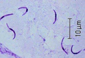 Figure: Sporozoites of Malarial Parasites
Every parasitic infection begins with a single, specific developmental form — the infective stage — without which transmission cannot occur. Knowing exactly what that stage is, where it is found, and how it enters the body is not academic trivia. It is the foundation of every prevention strategy in parasitology: footwear prevents hookworm because it blocks skin-penetrating larvae; cooking meat thoroughly prevents toxoplasmosis because heat destroys tissue cysts; boiling water prevents giardiasis because heat destroys cysts. Prevention always targets the infective stage.
Organisms that live on or inside a host organism deriving nutrients from the host and often causing harm, can cause parasitic infections. Parasites can belong to various biological groups, including protozoa, ectoparasites (such as ticks and lice), and helminths (worms). These infections can affect humans and animals, and they are prevalent in different parts of the world, specifically in areas with poor sanitation and limited access to healthcare.
Parasitic infections go through various stages as they progress through their life cycle, often involving both the host organism (where the parasite resides) and the environment. The stages include infestation, entry into the host, colonization/reproduction, development, migration, evading the host immune response, release of infective forms, transmission to a new host, or survival in the environment.
The infective stage of a parasitic organism's life cycle refers to the developmental stage at which the organism can infect a host and initiate a new cycle of its life. The specific characteristics of the infective stage vary among different parasites, and understanding these stages is crucial for comprehending the transmission dynamics and implementing effective control measures.
Why Infective Stage Knowledge Matters for Prevention
Every major parasite prevention strategy exists because someone identified the exact infective stage and designed an intervention to block it:
| Infective stage type | Why it matters | Prevention strategy it justifies |
|---|---|---|
| Sporozoite (mosquito-injected) | Cannot be filtered from water or food — only vector contact matters | Bed nets, insecticide spraying, repellents |
| Cyst (ingested) | Resistant to chlorination at standard doses; survives in water | Boiling water, filtration (not chlorination alone) |
| Filariform larva (skin-penetrating) | Active penetration — does not require ingestion | Footwear, avoiding contact with contaminated soil |
| Tissue cyst (in undercooked meat) | Destroyed by adequate cooking temperature | Cooking meat to safe internal temperature |
| Egg (ingested, environmentally resistant) | Survives in soil for years; resistant to many disinfectants | Hand hygiene, sanitation infrastructure, vegetable washing |
| Cercariae (water-penetrating) | Active skin penetration in freshwater — swimming/wading sufficient | Avoiding contact with contaminated freshwater |
A clinician who knows the infective stage of a suspected parasite can immediately generate the right exposure history questions — and the right prevention counselling — without needing to look anything up.
Complete Reference: Infective Stages by Organism
| Organism | Disease | Infective stage | Route of entry |
|---|---|---|---|
| Plasmodium spp. | Malaria | Sporozoite (in mosquito salivary glands) | Mosquito bite (inoculation) |
| Entamoeba histolytica | Amoebiasis | Mature quadrinucleate cyst | Ingestion (feco-oral) |
| Giardia lamblia | Giardiasis | Cyst | Ingestion (feco-oral, contaminated water) |
| Leishmania donovani (and other Leishmania spp.) | Leishmaniasis | Metacyclic promastigote | Sandfly bite (inoculation) |
| Trypanosoma brucei | African sleeping sickness | Metacyclic trypomastigote | Tsetse fly bite (inoculation) |
| Trypanosoma cruzi | Chagas disease | Metacyclic trypomastigote (in triatomine faeces) | Contamination of bite wound/mucosa (not direct bite) |
| Hookworm (Ancylostoma duodenale, Necator americanus) | Hookworm infection | Filariform (L3) larva | Active skin penetration (or ingestion for A. duodenale) |
| Strongyloides stercoralis | Strongyloidiasis | Filariform (L3) larva | Active skin penetration |
| Toxoplasma gondii | Toxoplasmosis | Tachyzoite (transplacental); Bradyzoite in tissue cyst (undercooked meat); Sporozoite in oocyst (cat faeces, environmental) | Three distinct routes — see Toxoplasma gondii: Properties, Life Cycle, and Diagnosis for full detail |
| Ascaris lumbricoides | Ascariasis | Mature fertilised egg (containing rhabditiform larva) | Ingestion (feco-oral) |
| Trichuris trichiura | Trichuriasis | Mature embryonated egg | Ingestion (feco-oral) |
| Wuchereria bancrofti | Lymphatic filariasis | Third-stage larva (L3) | Mosquito bite (inoculation) |
| Taenia solium | Taeniasis (intestinal) / Cysticercosis (tissue) | Cysticercus (pork → taeniasis) OR Egg (→ cysticercosis) | Ingestion — two infective stages, two diseases; see Taenia solium: Life Cycle, Pathogenesis and Lab Diagnosis for full mechanism |
| Taenia saginata | Taeniasis | Cysticercus (in beef) | Ingestion |
| Echinococcus granulosus | Hydatid disease | Egg | Ingestion (contact with dog faeces) |
| Enterobius vermicularis | Enterobiasis (pinworm) | Egg (embryonated, on perianal skin) | Ingestion (autoinfection, fomites) — NOT typically via stool |
| Schistosoma spp. | Schistosomiasis | Cercaria (free-swimming) | Active skin penetration in fresh water |
| Trichomonas vaginalis | Trichomoniasis | Trophozoite (no cyst stage exists) | Direct sexual contact |
| Cryptosporidium parvum | Cryptosporidiosis | Oocyst | Ingestion (water, feco-oral) |
| Naegleria fowleri | Primary amoebic meningoencephalitis | Trophozoite (free-living, in warm freshwater) | Nasal mucosa (via nose during swimming) |
| Balantidium coli | Balantidiasis | Cyst | Ingestion (feco-oral, pig reservoir) |
| Hymenolepis nana | Hymenolepiasis | Egg (directly infective — no intermediate host required) | Ingestion (autoinfection common) |
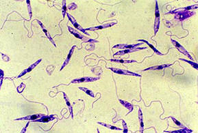 Figure: Promastigote form of Leishmania
Where Students Actually Get Confused
1. The infective stage and the diagnostic stage are not always the same. In malaria, the sporozoite is the infective stage (injected by the mosquito), but the ring-stage trophozoite seen on a blood smear is the diagnostic stage — sporozoites are never seen on a peripheral blood smear because they exist only briefly before invading liver cells.
2. Taenia solium has two distinct infective stages causing two different diseases. Ingesting cysticerci in undercooked pork causes intestinal taeniasis (the adult tapeworm). Ingesting eggs (via contaminated hands, autoinfection, or contaminated food) causes cysticercosis, including potentially fatal neurocysticercosis. Same organism, two infective stages, two completely different clinical pictures.
3. Not all helminth eggs are immediately infective once passed. Ascaris and Trichuris eggs require a period of maturation in soil (typically 2–4 weeks) before becoming infective — freshly passed eggs are not yet capable of causing infection. In contrast, Enterobius eggs are infective almost immediately after being laid on the perianal skin (within hours), and Hymenolepis nana eggs are directly infective without requiring environmental maturation at all.
4. Cysts and oocysts are biologically distinct structures. A cyst (e.g., Entamoeba, Giardia) is the resistant resting stage of a single trophozoite, formed by the organism itself. An oocyst (e.g., Cryptosporidium, Toxoplasma, Cyclospora) is the product of sexual reproduction (a zygote with a resistant wall), typically associated with coccidian parasites and often containing multiple sporozoites internally.
5. Filariform and rhabditiform larvae are not interchangeable terms. Rhabditiform larvae (the form found in fresh stool for hookworm and Strongyloides) are non-infective, feeding, free-living larvae. Filariform larvae (L3 stage) are the infective, non-feeding form capable of skin penetration. Finding rhabditiform larvae in a stool sample indicates infection but is not itself the infective stage — it's a diagnostic finding pointing to the cycle that will eventually produce infective filariform larvae in the environment.
Key Exam Facts in One Table
| Fact | Detail | Memory hook |
|---|---|---|
| Plasmodium infective stage | Sporozoite (mosquito salivary glands) | Injected directly — no ingestion |
| Entamoeba histolytica infective stage | Mature quadrinucleate cyst | 4 nuclei = mature = infective |
| Leishmania infective stage | Metacyclic promastigote | Sandfly-injected |
| Hookworm infective stage | Filariform (L3) larva | Active skin penetration |
| Toxoplasma: 3 infective stages | Tachyzoite (transplacental); bradyzoite (meat); sporozoite in oocyst (cat faeces) | Three routes, three stages |
| Taenia solium: 2 infective stages | Cysticercus (pork → taeniasis); egg (→ cysticercosis) | Same organism, two diseases |
| Schistosoma infective stage | Cercaria | Free-swimming, skin-penetrating |
| Trichomonas vaginalis | Trophozoite only — no cyst stage | Direct contact required (fragile organism) |
| Ascaris/Trichuris eggs | Require 2–4 weeks soil maturation | NOT immediately infective when passed |
| Enterobius eggs | Infective within hours of being laid | Fast — enables rapid autoinfection |
| Hymenolepis nana eggs | Directly infective, no intermediate host needed | Simplest helminth life cycle |
| Cyst vs oocyst | Cyst = single trophozoite resting stage; Oocyst = product of sexual reproduction | Oocyst = coccidian parasites |
| Rhabditiform vs filariform larva | Rhabditiform = non-infective, feeding; Filariform = infective, non-feeding | Diagnostic stage ≠ infective stage |
| Wuchereria bancrofti infective stage | Third-stage larva (L3) | Mosquito-transmitted |
Self-Check Questions
- A patient develops neurocysticercosis after years of eating well-cooked pork. The patient insists they have never eaten undercooked meat. Explain how this infection occurred and identify the infective stage responsible.
- Why is the sporozoite of Plasmodium never seen on a routine blood smear, despite being the infective stage of the parasite?
- A pregnant woman asks whether she is at higher risk of toxoplasmosis from gardening or from eating a rare steak. Explain the two different infective stages involved and the prevention advice for each.
- What is the key biological difference between a protozoan cyst and a coccidian oocyst?
- A stool sample shows rhabditiform larvae. Is this the infective stage of the parasite? Explain your answer.
- Why must Ascaris lumbricoides eggs spend time in soil before becoming infective, while Enterobius vermicularis eggs are infective almost immediately?
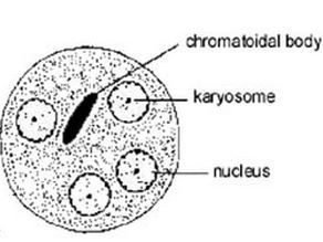 Figure: Mature Quadrinucleate Cyst of Entamoeba histolytica
Answers
- Neurocysticercosis results from ingesting Taenia solium eggs, not cysticerci in pork. The patient likely acquired the eggs through contaminated food, water, or hands — possibly from a household contact who has intestinal taeniasis (from eating undercooked pork themselves) and is shedding eggs. This illustrates the critical distinction: cysticerci (in pork) cause intestinal taeniasis when ingested, while eggs (via fecal-oral contamination) cause cysticercosis/neurocysticercosis — two entirely different infective stages and disease outcomes from the same organism.
- Sporozoites exist only transiently in the bloodstream — within 30 to 60 minutes of the mosquito bite, they invade hepatocytes and are no longer present in circulating blood. A peripheral blood smear, by definition, can only show parasite stages present in the blood at the time of sampling. The diagnostic stages visible on a blood smear (ring forms, trophozoites, schizonts, gametocytes) all occur after the sporozoite has already left the bloodstream and completed liver-stage development.
- Gardening risk involves sporozoites within oocysts shed in cat faeces and present in contaminated soil; eating rare steak risk involves bradyzoites within tissue cysts in undercooked meat. Prevention advice differs accordingly: for the gardening/cat-litter route, wearing gloves and thorough hand-washing after soil contact is key; for the meat route, cooking meat to a safe internal temperature (which destroys bradyzoites) is the relevant intervention. Both routes carry the same risk of congenital toxoplasmosis if infection occurs during pregnancy.
- A cyst is the resistant resting stage formed by a single protozoan trophozoite (e.g., Entamoeba, Giardia) through encystation — it is not the product of sexual reproduction. An oocyst is specifically the product of sexual reproduction in coccidian parasites (e.g., Cryptosporidium, Toxoplasma, Cyclospora) — it is essentially a resistant zygote, often containing multiple sporozoites formed through subsequent asexual division (sporogony) within the oocyst wall.
- No — rhabditiform larvae are not the infective stage. They are the non-infective, free-living, feeding larval form found in fresh stool (for hookworm and Strongyloides). The infective stage is the filariform (L3) larva, which develops from the rhabditiform form after a period in the environment (typically several days) and becomes capable of skin penetration. Finding rhabditiform larvae in stool confirms active intestinal infection but does not represent the form responsible for new infections.
- Ascaris and Trichuris eggs require an obligatory period of embryonic development in soil (typically 2–4 weeks under favourable temperature and moisture conditions) before the larva inside reaches an infective state — eggs passed in fresh stool are not yet capable of causing infection. Enterobius eggs, by contrast, are deposited on the perianal skin already containing a nearly fully developed embryo, requiring only a few hours of further development to become infective — this rapid timeline is precisely what enables the efficient autoinfection cycle characteristic of pinworm transmission.
References
- Rogers, W. P., & Sommerville, R. I. (1963). The infective stage of nematode parasites and its significance in parasitism. Advances in Parasitology, 1, 109–177. https://doi.org/10.1016/s0065-308x(08)60503-5
- Siński, E. (2003). Environmental contamination with protozoan parasite infective stages: biology and risk assessment. Acta Microbiologica Polonica, 52(Suppl), 97–107.
- Koprivnikar, J., Thieltges, D. W., & Johnson, P. T. J. (2023). Consumption of trematode parasite infectious stages: from conceptual synthesis to future research agenda. Journal of Helminthology, 97, e33. https://doi.org/10.1017/S0022149X23000111
- Garcia, L. S. (2016). Diagnostic Medical Parasitology (6th ed.). ASM Press.
- Markell, E. K., John, D. T., & Krotoski, W. A. (1999). Markell and Voge's Medical Parasitology (8th ed.). W.B. Saunders.
- World Health Organization. (2023). Soil-transmitted helminth infections. WHO Fact Sheet. https://www.who.int/news-room/fact-sheets/detail/soil-transmitted-helminth-infections

Tankeshwar Acharya, MSc (Medical Microbiology)
Tankeshwar Acharya is an Assistant Professor in the Department of Microbiology at Patan Academy of Health Sciences (PAHS), Nepal, where he has been teaching and practicing clinical microbiology for over 14 years. He is the founder of Microbe Online, one of the leading free microbiology education resources on the web, covering bacteriology, mycology, parasitology, immunology, and clinical laboratory diagnostics written from direct experience in both the classroom and the diagnostic laboratory.