
Buffy Coat: Definition, Composition, Preparation, and Diagnostic Uses
What is the buffy coat, what does it contain, and how is it used to diagnose malaria, filariasis, leishmaniasis, and trypanosomiasis? Step-by-step preparation with organism layer positions explained.
A laboratory technician in a rural health post receives a blood sample from a febrile patient. The rapid malaria RDT is negative, but clinical suspicion remains high. Rather than accepting the result and sending the patient home, the technician centrifuges the EDTA blood in a narrow tube, withdraws a few microliters from the thin pale layer sitting just above the packed red cells, and makes a smear. Under the microscope: ring-form trophozoites, present but too few to be reliably detected on a standard thick smear.
That thin pale layer is the buffy coat — and concentrating it is one of the simplest, cheapest ways to improve the sensitivity of parasite detection when the standard methods fall short. Understanding what the buffy coat is, why it forms where it does, and which parasites concentrate in which zone is fundamental to clinical parasitology laboratory practice.
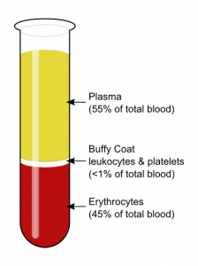 Figure: Fig: Schematic picture of a fractioned whole blood sample
Composition of the Buffy Coat
When anticoagulated whole blood is centrifuged, it separates into three layers based on the density of its components:
| Layer | Component | Proportion of whole blood | Density |
|---|---|---|---|
| Top (supernatant) | Plasma | ~55% | ~1.025 g/mL (least dense) |
| Middle (thin band) | Buffy coat — WBCs + platelets | <1% | 1.050–1.077 g/mL |
| Bottom (pellet) | Erythrocytes (RBCs) | ~45% | ~1.093 g/mL (most dense) |
The name "buffy coat" comes from its characteristic buff (pale yellow-grey) colour, reflecting the mix of white cells and platelets it contains.
What the Buffy Coat Actually Contains
The buffy coat is not a uniform layer — it has internal structure. Different cell types have slightly different densities and separate into sublayers:
| Sublayer (top → bottom) | Cell type | Approximate density |
|---|---|---|
| Uppermost (plasma interface) | Platelets | ~1.050 g/mL |
| Upper middle | Lymphocytes, monocytes | ~1.060–1.067 g/mL |
| Lower middle | Granulocytes (neutrophils, eosinophils, basophils) | ~1.070–1.077 g/mL |
| Bottom (RBC interface) | Granulocytes + reticulocytes | ~1.077+ g/mL |
Why this matters for parasite diagnosis: Different blood parasites have different densities and concentrate in different sublayers. Plasmodium-infected RBCs are less dense than normal RBCs and accumulate near the RBC–buffy coat interface. Trypanosomes and microfilariae are motile and concentrate in the plasma just above the buffy coat. This is why the layer from which you sample — and the small zone above or below it — determines which parasite you find.
Why Mammalian RBCs Don't Contribute to the Buffy Coat
Mammalian erythrocytes are anucleate (no nucleus, no DNA). They cannot be used for genomic DNA extraction. The buffy coat, concentrated with WBCs (which are fully nucleated), is therefore the standard source of gDNA from peripheral blood — useful for molecular diagnostics, forensic analysis, and genetic testing when large quantities of DNA are needed from relatively small blood volumes.
How the Buffy Coat Forms: The Density Principle
The formation of the buffy coat is explained entirely by differential density centrifugation. When force is applied by centrifugation, particles in a fluid migrate to positions where their density equals that of the surrounding medium — denser particles sink further, less dense particles rise.
Applied to blood:
- RBCs (~1.093 g/mL) are densest → sink to the bottom
- Plasma (~1.025 g/mL) is least dense → stays at the top
- WBCs and platelets (1.050–1.077 g/mL) are intermediate → collect in the middle band
This is the same principle exploited by the QBC (Quantitative Buffy Coat) system, where a plastic float of specific gravity 1.055 is inserted to physically expand the buffy coat layer, making it easier to examine under fluorescence microscopy. The float's density places it between plasma and packed RBCs — exactly where the buffy coat forms.
The parasitology implication: Parasites and parasite-infected cells also have characteristic densities. Plasmodium-infected RBCs are slightly less dense than normal RBCs (the parasite metabolises haemoglobin, reducing cell density), so they concentrate just above the main RBC pellet — at the bottom of the buffy coat layer. This physical property is what makes buffy coat concentration useful for malaria detection.
Uses of the Buffy Coat
1. Parasite Detection and Concentration (Diagnostic Microbiology)
The most clinically important use on this site. The buffy coat concentrates blood parasites that would be missed or underdetected on a standard thick or thin smear, particularly in low-parasitaemia infections.
Different parasites, different layers — critical for correct sampling:
| Parasite | Disease | Position after centrifugation | Why |
|---|---|---|---|
| Plasmodium spp. | Malaria | Just above packed RBCs (bottom of buffy coat) | Infected RBCs slightly less dense than normal RBCs |
| Trypanosoma spp. | African sleeping sickness; Chagas disease | Plasma just above the buffy coat | Motile; less dense than cells |
| Microfilariae (Wuchereria, Brugia) | Lymphatic filariasis | Plasma just above the buffy coat | Motile; larger than cells, remain in plasma |
| Leishmania donovani amastigotes | Visceral leishmaniasis | Within buffy coat (monocyte/macrophage layer) | Obligate intracellular — inside monocytes which settle in buffy coat |
| Histoplasma capsulatum | Histoplasmosis (disseminated) | Within buffy coat (macrophage layer) | Intracellular in macrophages |
Practical sampling note: When withdrawing material for a parasite smear, take the buffy coat layer AND a small amount of plasma immediately above it — this ensures you capture trypanosomes and microfilariae which sit above the main WBC band. This is explicitly noted in WHO malaria microscopy guidelines.
2. Quantitative Buffy Coat (QBC) System
The QBC method (Becton Dickinson) is an enhanced application of the buffy coat principle. A pre-coated acridine orange capillary tube concentrates the buffy coat under a plastic float and examines it under fluorescence microscopy. The acridine orange stains parasite nucleic acids, making parasites visible as fluorescent objects against a dark background.
→ For full QBC procedure and results interpretation, see: Quantitative Buffy Coat (QBC) Test
3. Genomic DNA Extraction
Because mammalian RBCs lack nuclei, the buffy coat (rich in nucleated WBCs) is the standard source of genomic DNA from peripheral blood. Applications include:
- Molecular diagnostics (PCR for pathogens)
- Genetic testing and pharmacogenomics
- Forensic DNA analysis
- Biobanking
A single 5 mL EDTA blood tube yields enough buffy coat for multiple high-quality gDNA extractions.
4. Blood Banking — Platelet Concentrates
In blood transfusion medicine, the buffy coat method is one of two standard approaches for preparing platelet concentrates. Whole blood units are centrifuged; the buffy coat (containing platelets) is pooled from multiple donors and re-centrifuged to produce a therapeutic platelet unit. This is the standard platelet preparation method used in many European blood banking systems.
5. Viral Culture and Immunological Studies
Peripheral blood mononuclear cells (PBMCs) — lymphocytes and monocytes — isolated from the buffy coat are used in:
- Viral culture (HIV, CMV, EBV)
- Lymphocyte stimulation assays
- Flow cytometry and immunophenotyping
- Research applications requiring large numbers of viable immune cells
Where Students Actually Get Confused
1. "The buffy coat contains mostly lymphocytes." Not quite. In normal blood, neutrophils are the most abundant white blood cell (~50–70% of WBCs), and they dominate the buffy coat numerically. Lymphocytes (~20–40%) are the second most abundant. The clinical utility of the buffy coat for intracellular parasite detection (Leishmania, Histoplasma) relates specifically to the monocyte/macrophage fraction — which is a small but functionally critical component.
2. "Trypanosomes and microfilariae are in the buffy coat layer." They are in the plasma just above the buffy coat — not in the buffy coat itself. This is a consistently examined distinction. When preparing a smear for trypanosome or microfilariae detection, withdraw the buffy coat and a small amount of the plasma immediately above it. Sampling only the buffy coat layer may miss motile organisms that have migrated upward into the plasma.
3. "Buffy coat preparation and QBC are the same thing." The QBC is a commercial, enhanced version of the buffy coat principle — it uses a pre-coated tube, a specific float, and fluorescence microscopy. A standard buffy coat preparation uses a plain centrifuge tube and conventional Giemsa or Field's staining. Both exploit the same density centrifugation principle, but QBC is more sensitive and more expensive. A basic buffy coat smear can be done in any laboratory with a centrifuge and a microscope.
4. "Fixing the buffy coat smear with methanol before it is completely dry." Same rule as the thin blood smear: allow the preparation to air-dry completely before fixing with methanol or ethanol. Fixing a wet smear causes cell lysis and distortion, making parasite identification impossible. The preparation step notes say "allow to air-dry" — this step must not be rushed.
5. "Buffy coat = blood culture." Buffy coat preparations are not blood cultures. They are concentrated smears for direct microscopic examination. Some protocols use buffy coat as the inoculum for culture media (e.g., NNN medium for Leishmania), but the buffy coat smear itself is a direct diagnostic test, not a culture.
Key Exam Facts in One Table
| Fact | Detail | Memory hook |
|---|---|---|
| Definition | Thin pale layer between plasma and RBCs after centrifugation | Named for buff (pale yellow-grey) colour |
| Composition | WBCs + platelets (<1% of whole blood volume) | Leukocytes concentrate here |
| Plasma proportion | ~55% of whole blood | Top layer, least dense |
| RBC proportion | ~45% of whole blood | Bottom layer, most dense |
| Buffy coat proportion | <1% of whole blood | Middle band, intermediate density |
| Why RBCs don't yield DNA | Mammalian RBCs are anucleate | No nucleus = no genomic DNA |
| Buffy coat DNA source | WBCs (nucleated cells) | Standard source for gDNA extraction |
| Centrifugation speed | 1,000g × 15 minutes (standard preparation) | Lower speed than QBC (12,000g) |
| Anticoagulant used | EDTA (prevents clotting) | Same as for full blood count |
| Staining after preparation | Field's thin-film or Giemsa | Same stains as peripheral smear |
| Plasmodium position | Just above RBC pellet (bottom of buffy coat) | Infected RBCs less dense than normal |
| Trypanosomes/microfilariae position | Plasma just above the buffy coat | Motile organisms migrate to plasma |
| Leishmania/Histoplasma position | Within buffy coat (monocyte/macrophage layer) | Obligate intracellular organisms |
| QBC relationship | Commercial enhanced version of buffy coat principle | Float + acridine orange + fluorescence microscopy |
| Blood banking use | Platelet concentrate preparation | Buffy coat pooling method (European standard) |
Self-Check Questions
- A thick blood smear is negative for malaria in a febrile patient. You prepare a buffy coat smear. From exactly which zone of the centrifuged tube do you sample to maximise the chance of detecting Plasmodium?
- The same patient's result is negative for malaria but you now suspect lymphatic filariasis. The blood was collected at 2 pm. Is the negative buffy coat result sufficient to exclude filariasis, and why?
- Why are mammalian red blood cells unsuitable for genomic DNA extraction, and which layer of centrifuged blood is used instead?
- You suspect African trypanosomiasis in a patient. After centrifugation, from which zone should you sample — the buffy coat itself, or the plasma above it?
- What is the fundamental physical principle that causes the buffy coat to form between plasma and RBCs during centrifugation?
- What is the difference between a standard buffy coat smear and a QBC test?
Answers:
- The buffy coat layer AND the small zone of RBCs immediately below it (~1mm depth) — Plasmodium-infected RBCs are slightly less dense and concentrate at the bottom of the buffy coat/top of the RBC pellet.
- No — microfilariae of W. bancrofti show nocturnal periodicity; they are only present in peripheral blood in significant numbers between 10 pm and 2 am. A daytime sample cannot exclude filariasis. Repeat with night collection.
- Mammalian RBCs are anucleate (no nucleus, no DNA); the buffy coat layer, rich in nucleated WBCs, is used for gDNA extraction.
- The plasma just above the buffy coat — trypanosomes are motile and less dense than WBCs; they concentrate in the plasma layer above the buffy coat, not within it.
- Differential density centrifugation — particles migrate to positions where their density matches the surrounding fluid; denser RBCs sink furthest, less dense plasma rises to the top, WBCs/platelets of intermediate density collect in the middle.
- Standard buffy coat smear: plain tube, low-speed centrifuge, conventional Giemsa/Field's staining, standard light microscopy — available anywhere. QBC: proprietary pre-coated acridine orange tube, specific float, high-speed centrifuge (12,000g), fluorescence microscopy — more sensitive but requires specialist equipment and is more expensive.)
References and further reading
- Garcia, L. S. (2016). Diagnostic Medical Parasitology (6th ed.). ASM Press.
- World Health Organization. (2016). Malaria microscopy quality assurance manual (Version 2). WHO. https://www.who.int/publications/i/item/9789241549394
- World Health Organization. (2010). Basic malaria microscopy. Part I. Learner's guide (2nd ed.). WHO.
- Cheesbrough, M. (2006). District Laboratory Practice in Tropical Countries (2nd ed., Part 1). Cambridge University Press.
- Winn, W. C., Jr., Allen, S. D., Janda, W. M., et al. (2006). Koneman's Color Atlas and Textbook of Diagnostic Microbiology (6th ed.). Lippincott Williams & Wilkins.
- Long, G. W., Jones, T. R., Rickman, L. S., et al. (1994). Acridine orange diagnosis of Plasmodium falciparum: evaluation after experimental infection. American Journal of Tropical Medicine and Hygiene, 51(5), 613–616. https://doi.org/10.4269/ajtmh.1994.51.613
- Image credit: "Blood-centrifugation-scheme" by KnuteKnudsen at English Wikipedia (CC BY 3.0) via Wikimedia Commons.
Frequently Asked Questions
What is the buffy coat and what does it contain?
Why does the buffy coat form between plasma and red blood cells?
Where do malaria parasites concentrate in the buffy coat?
Where do trypanosomes and microfilariae go during buffy coat centrifugation?
What is the difference between a buffy coat smear and a QBC test?

Tankeshwar Acharya, MSc (Medical Microbiology)
Tankeshwar Acharya is an Assistant Professor in the Department of Microbiology at Patan Academy of Health Sciences (PAHS), Nepal, where he has been teaching and practicing clinical microbiology for over 14 years. He is the founder of Microbe Online, one of the leading free microbiology education resources on the web, covering bacteriology, mycology, parasitology, immunology, and clinical laboratory diagnostics written from direct experience in both the classroom and the diagnostic laboratory.