
Flagella: Structure, Arrangement, Function
Bacterial flagella: structure (filament, hook, basal body), types of flagellar arrangement with mnemonics, how flagella rotate, clinical significance in pathogenesis, identification, and motility patterns. With memory aids and exam tips.
Flagella (singular, flagellum) are the locomotory structures of many prokaryotes. Most protozoa and some bacteria are motile. Protozoa use flagella, cilia, or pseudopods, whereas motile bacteria move only using flagella. The flagellum functions by rotation to push or pull the cell through a liquid medium.
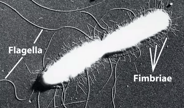 Figure: Electron micrograph of Salmonella typhi showing flagella and fimbriae (Image source: J.P.Duguid and J.F. Wilkinson)
Bacterial Flagella
Bacterial flagella are long, thin (about 20 nm), whip-like appendages that move the bacteria towards nutrients and other attractants. Like capsule and pili, flagella are external to the cell wall in some bacteria. Flagella are free at one end and attached to the cell at the other end. Flagellum can never be seen directly with the light microscope but only after staining with special flagella stains that increase their diameter. Flagella can be seen easily with the electron microscope.
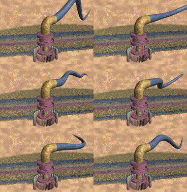 Figure: Flagellar motion in bacterial cells
Flagella are usually found in gram-negative bacilli. Gram-positive rods (e.g., Listeria species) and cocci (some Enterococcus species, Vagococcus species) also have flagella.
Most of the cocci (e.g. Staphylococci, Streptococci,etc.) don’t have flagella, so they are non-motile. Bacteria lacking flagella are called atrichous.
Why you need to understand bacterial flagella
Flagella are not just a morphological curiosity, they are directly relevant to clinical microbiology in four important ways:
1. Pathogenesis: flagella physically drive bacteria into host tissues. E. coli and Proteus mirabilis use peritrichous flagella to propel themselves up the urethra against urine flow, causing urinary tract infections. Helicobacter pylori uses its lophotrichous flagella to burrow through the viscous mucus layer of the stomach and reach the epithelial surface, a prerequisite for causing peptic ulcer disease.
2. Identification: the number and arrangement of flagella are taxonomically significant. Salmonella is serotyped partly based on its H (flagellar) antigen; this is what the "H" stands for in Salmonella O:H typing (e.g., Salmonella Typhimurium 4,5,12:i:1,2). The Widal test detects antibodies to Salmonella H antigens in typhoid fever diagnosis.
3. Motility patterns at the bench: when you examine a wet preparation under the microscope, the motility pattern tells you something about the organism:
- Darting "shooting star" motility → Vibrio cholerae (single polar flagellum)
- Swarming across the plate → Proteus mirabilis (peritrichous flagella)
- Corkscrew rotation → Spirochetes (endoflagella)
- Tumbling → Listeria monocytogenes at 25°C (peritrichous flagella)
4. Vaccine target — flagellin protein (the structural subunit of the filament) stimulates TLR5 on host immune cells. It is being investigated as a vaccine adjuvant and as a direct vaccine target for flagellated pathogens.
Structure
The long helical filament of bacterial flagella is composed of many subunits of a single protein, flagellin, arranged in several intertwined chains. A flagellum consists of several components and moves by rotation, much like a boat motor propeller. The base of the flagellum is structurally different from the filament.
The wider region at the base of the flagellum is called a hook. The hook connects the filament to the motor portion of the flagellum called a basal body.
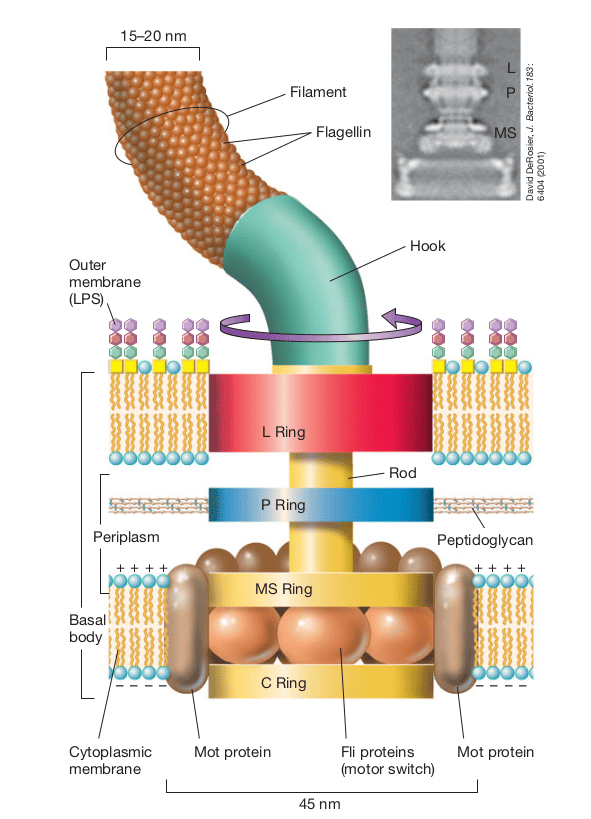 Figure: Structure of the bacterial flagella
The basal body is anchored in the cytoplasmic membrane and cell wall. It consists of a central rod that passes through a series of rings.
In gram-negative bacteria, three-set of rings are present in three different layers,
- L ring is anchored in the lipopolysaccharide layer.
- P ring is located in the periplasmic space and anchored in the cell wall’s peptidoglycan layer.
- MS ring is located within the cytoplasmic membrane. The smaller S-ring (stator ring) is attached to the M-ring or motor ring, forming the MS ring.
- C ring anchors the entire complex to the cell.
A series of proteins called Mot surrounds the inner pair of rings. These proteins drive the flagellar motor, causing rotation of the filament. Another set of proteins called Fli proteins functions as the motor switch, reversing the rotation of the flagella in response to intracellular signals.
The flagella of Gram-positive bacteria contain only two basal body rings; one ring is embedded in the peptidoglycan layer and another in the cell membrane.
How flagella actually move and why it matters
The analogy that makes this stick: Think of a bacterial flagellum as a boat propeller. The basal body is the motor, the hook is the drive shaft, and the filament is the propeller blade. When the motor spins counterclockwise (when viewed from behind), the helical flagellum acts like a corkscrew and propels the cell forward — this is called runs (smooth, directed movement toward an attractant). When the motor reverses to clockwise, the flagella bundle flies apart and the cell tumbles randomly — this is called tumbles (reorientation in a new direction).
The run-and-tumble mechanism: Bacteria alternate between runs and tumbles in a process called chemotaxis — biased random walk toward attractants (nutrients, oxygen) and away from repellents (toxins). When moving toward an attractant, runs become longer and tumbles become less frequent. This simple molecular mechanism allows bacteria to navigate their environment without a nervous system or eyes.
Why this matters clinically: E. coli chemotaxis toward uroepithelial cells is mediated by flagella. Chemicals produced by bladder cells attract the bacteria — flagella-mediated chemotaxis drives the organisms toward the very cells they need to colonise. Flagellaless mutants of uropathogenic E. coli are significantly less virulent in animal infection models.
Types of Flagellar Arrangement : What, Why, and How to Remember
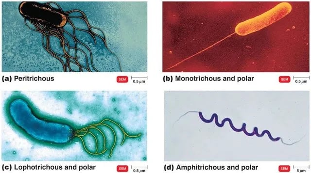 Figure: Flagellar arrangement in bacteria (Image source: Cedric Woudstra)
The mnemonic: **"A Monkey Lives Peacefully"**
| Letter | Flagellar type | Meaning | Classic example |
|---|---|---|---|
| A | Atrichous | No flagella — non-motile | Staphylococcus aureus, Klebsiella pneumoniae |
| M | Monotrichous | Single flagellum at one pole | Vibrio cholerae, Pseudomonas aeruginosa |
| L | Lophotrichous | Cluster of flagella at one pole | Pseudomonas fluorescens, Helicobacter pylori |
| P | Peritrichous | Flagella distributed all over the surface | E. coli, Salmonella typhi, Proteus mirabilis |
(Amphitrichous — flagella at both poles — can be added: "A Monkey Lives Peacefully, Always")
Atrichous (no flagella)
What: Bacteria with no flagella at all. Non-motile.
Why it matters: Non-motility is itself diagnostically significant. Klebsiella pneumoniae is non-motile — this helps differentiate it from Enterobacter aerogenes (motile), which produces very similar colony morphology. Shigella is non-motile — its lack of motility distinguishes it from Salmonella (motile) in the enteric pathogen identification workflow.
Exam tip: The three non-motile Enterobacteriaceae you will most often be tested on are Klebsiella, Shigella, and Yersinia pestis. Remember: KSY — "Keep Still, You!"
Monotrichous (single polar flagellum)
What: A single flagellum at one end of the cell.
The analogy: Monotrichous bacteria are like a canoe with one paddle at the stern, efficient forward propulsion in a single direction, with characteristic darting motility.
Why it matters: Vibrio cholerae — its single polar flagellum produces the characteristic "shooting star" or "darting" motility visible in a wet preparation of fresh stool. In cholera outbreaks before culture facilities were available, this motility pattern in direct stool microscopy was a rapid presumptive diagnostic clue. V. cholerae is also inhibited by specific antisera (serogroup O1 or O139), which stops motility — a rapid presumptive identification.
Pseudomonas aeruginosa — single polar flagellum; flagellin is an important virulence factor that triggers TLR5-mediated inflammation. In cystic fibrosis patients, mucoid strains of P. aeruginosa often lose flagella — reducing their immunogenicity and allowing chronic colonisation.
Clinical memory anchor: "One pole, one motor, one direction — like a torpedo" → Vibrio and Pseudomonas.
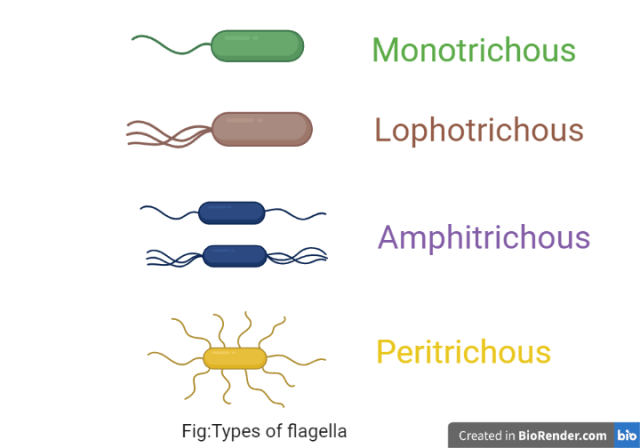 Figure: Types of flagella
Lophotrichous (tuft of flagella at one pole)
What: Multiple flagella arising from one end, forming a bundle.
The analogy: Lophotrichous bacteria are like a rowing team — many oars at one end pulling together. The flagellar bundle provides stronger propulsion than a single flagellum.
Why it matters: Helicobacter pylori — lophotrichous (4–7 sheathed flagella at one pole). These flagella are essential for penetrating the thick mucus layer protecting the stomach epithelium. Without functional flagella, H. pylori cannot colonise the stomach. The flagella are also sheathed (covered by a membrane extension of the outer membrane) — this sheath protects the flagellin from acid degradation in the stomach environment and reduces its TLR5 immunostimulatory activity, helping the bacteria evade innate immune recognition.
Why flagella sheathing matters: This is a unique feature of H. pylori that explains in part why the stomach immune response to H. pylori is inadequate to clear the infection — the sheathed flagella are less visible to the immune system.
Peritrichous (flagella all around the cell)
What: Multiple flagella distributed over the entire cell surface. During swimming, these bundle together at the rear to propel the cell forward.
The analogy: Peritrichous bacteria are like a rowing team surrounding a boat — multiple paddles pulling from all sides, then synchronising into a coordinated bundle for forward movement. When flagella run counterclockwise, they bundle and push; when one reverses, the bundle flies apart and the cell tumbles.
Why it matters: All motile members of the Enterobacteriaceae family have peritrichous flagella — E. coli, Salmonella, Proteus, Serratia, Enterobacter. This is a key characteristic of the family.
Proteus mirabilis demonstrates peritrichous flagella most dramatically: on blood agar, the cell elongates, massively increases its flagella number (from ~6 to ~500 per cell), and coordinates with neighbouring cells to swarm across the entire plate surface in concentric rings — a striking example of collective flagella-driven behaviour. Swarming is inhibited on MacConkey agar (bile salts) and CLED agar (electrolyte-deficiency suppresses motility) — this is why these media are preferred for urine cultures.
Exam tip: The Enterobacteriaceae motility rule: "Most Enterobacteriaceae are motile and peritrichous — except KSY (Klebsiella, Shigella, Yersinia pestis)."
Amphitrichous (flagella at both poles)
What: Single flagellum or a tuft at both poles.
Why it matters: Less common clinically. Alcaligenes faecalis is a classic example — an environmental gram-negative rod occasionally causing opportunistic infections in immunocompromised patients. Campylobacter was formerly classified as amphitrichous but its single polar flagella at each end are now considered bipolar monotrichous.
Clinical Applications of Flagella Knowledge
At the bench — motility patterns as identification clues
When you examine a wet preparation from a clinical specimen, different motility patterns give immediate presumptive clues:
| Motility pattern | Description | Organism to consider |
|---|---|---|
| Darting "shooting star" | Rapid, straight-line movement, sudden reversal | Vibrio cholerae |
| Swarming | Concentric rings spread across entire blood agar plate | Proteus mirabilis |
| Tumbling/rotating | End-over-end rotation, broth culture | Listeria monocytogenes (25°C) |
| Corkscrew (lashing) | Flexible, rapid cork-screw rotation | Spirochetes (Treponema, Leptospira) |
| Stately, slow | Slow, graceful movement | Clostridium spp. |
| Non-motile | No movement (true non-motility vs Brownian movement) | Klebsiella, Shigella |
The critical distinction — true motility vs Brownian movement: Students often confuse Brownian movement (random, jittery, non-directional vibration caused by water molecules hitting the bacteria) with true motility. True motility is directional and purposeful — the bacterium moves from point A to point B. Brownian movement is random, non-progressive oscillation with no net displacement. In a hanging drop or wet preparation, distinguish by watching whether the organism changes its position relative to surrounding debris — debris also shows Brownian movement but does not display directional travel.
In diagnosis — flagellar antigens
The H antigen of Salmonella is its flagellar antigen — composed of flagellin protein. The Kauffmann-White serotyping scheme for Salmonella uses both O (somatic/LPS) and H (flagellar) antigens. This is also why the Widal test detects both O and H agglutinins:
- Anti-O agglutinins appear earlier (days 6–8) and indicate active infection
- Anti-H agglutinins appear later (days 10–12), persist longer, and may reflect past infection or vaccination
Understanding what "H antigen" actually is — a flagellar protein — makes Widal test interpretation far more intuitive.
In treatment — flagella as a drug target
Flagellar assembly and the flagellar motor involve over 40 proteins. Several of these are being investigated as novel antibiotic targets, particularly for flagellated uropathogens and H. pylori. Compounds that inhibit flagellar assembly prevent colonisation without bactericidal activity — potentially reducing selection pressure for resistance.
Archaeal Flagella
Flagella are also present in major species of Archaea. Major genera of methanogens, extreme halophiles, thermoacidophiles, and hyperthermophiles are capable of swimming motility. Still, their speed is comparatively less than that of bacteria, probably because of the small diameter of flagella.
Differences between Bacterial and Archaeal flagella are tabulated here:
| Properties | Bacterial flagella | Archaeal flagella |
|---|---|---|
| Flagellar filament | Flagellar filament is made up of a single type of protein | Several different flagellin proteins are found. |
| Diameter of Flagella | The diameter of bacterial flagella is 15-20 nm, depending on the species. | Archaeal flagella is roughly half the diameter of bacterial flagella, measuring only 10–13 nm in width. |
| Source of energy for the rotation of flagella | Proton motive force | ATP |
Protozoa Having Flagella
Protozoa are a heterogeneous group with three different locomotion organs: flagella, cilia, and pseudopods. Certain protozoa, such as Leishmania and Trypanosoma, have flagellated forms called promastigotes and non-flagellated forms called amastigotes. Giardia lamblia and urogenital flagellate Trichomonas vaginalis also have flagella.
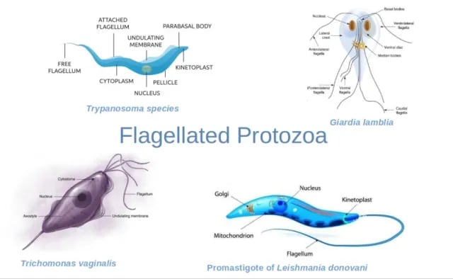 Figure: Some pathogenic flagellated protozoa
The trophozoite of Giardia lamblia contains four pairs of flagella. Trichomonas vaginalis is a pear-shaped flagellated protozoan possessing five flagella, four located at its anterior portion. The fifth flagellum is incorporated within the undulating membrane of the parasite.
Study Tips: How to Learn and Remember Flagella
The "AMLPA" framework for flagellar types
Write out this simple table from memory as your exam revision anchor:
A — Atrichous    → NONE        → Klebsiella, Shigella (non-motile)
M — Monotrichous → SINGLE POLE → Vibrio, Pseudomonas (darting motility)
L — Lophotrichous→ TUFT, POLE  → Helicobacter (burrows through mucus)
P — Peritrichous → ALL OVER    → E. coli, Salmonella, Proteus (all Enterobacteriaceae)
A — Amphitrichous→ BOTH POLES  → Alcaligenes
Three clinical stories that make flagella unforgettable
Story 1 — The UTI climber Imagine E. coli in the bladder. It has peritrichous flagella — motors spinning all around it. These flagella bundle together and it climbs from the bladder toward the kidney, against urine flow. This is pyelonephritis beginning. Non-motile Klebsiella cannot do this as efficiently — which is one reason E. coli causes more ascending UTIs than Klebsiella.
Story 2 — The stomach navigator H. pylori lives in a hostile acid environment that would kill most bacteria in seconds. Its lophotrichous sheathed flagella are its survival tool — they drive it through the thick mucus layer in seconds before the acid can denature its proteins. The sheath on the flagella hides flagellin from the immune system. Once it reaches the neutral pH under the mucus, it uses urease to neutralise local acid and establish a permanent niche. Without its flagella, it cannot make this journey — flagella-deficient H. pylori mutants cannot colonise the stomach.
Story 3 — The Proteus ghost Open an unselective blood agar plate that has been contaminated with Proteus mirabilis. The entire plate is covered with a thin, spreading, barely visible film — the swarming motility driven by massively upregulated peritrichous flagella. The putrid smell is unmistakeable. The organism you cannot see (because it has spread so thin) has colonised every millimetre of the plate. This swarming behaviour — invisible but pervasive — is precisely how Proteus behaves in catheterised patients, spreading from urinary catheters into the bladder without warning.
Key exam facts to memorise
- Flagellin is the structural protein of the filament — immunogenic, stimulates TLR5
- All motile Enterobacteriaceae are peritrichous (except none — all motile ones are peritrichous)
- Non-motile Enterobacteriaceae to memorise: Klebsiella, Shigella, Yersinia pestis
- H antigen in Salmonella serotyping = flagellar (Heat-labile) antigen
- Spirochetes have endoflagella (periplasmic) — not external flagella
- Mycoplasma has no flagella and uses gliding motility instead
- Swarming on blood agar = Proteus; on CLED or MacConkey it is inhibited
- H. pylori flagella are sheathed — unique feature that aids immune evasion
References and further readings
- Madigan, M. T., Bender, K. S., Buckley, D. H., Sattley, W. M., & Stahl, D. A. (2018). Brock Biology of Microorganisms (15th ed.). Pearson.
- Tille, P. M. (2017). Bailey & Scott's Diagnostic Microbiology (14th ed.). Mosby Elsevier.
- Haiko, J., & Westerlund-Wikström, B. (2013). The role of the bacterial flagellum in adhesion and virulence. Biology, 2(4), 1242–1267. https://doi.org/10.3390/biology2041242
- Josenhans, C., & Suerbaum, S. (2002). The role of motility as a virulence factor in bacteria. International Journal of Medical Microbiology, 291(8), 605–614. https://doi.org/10.1078/1438-4221-00173
Frequently Asked Questions
What is the difference between monotrichous, lophotrichous, amphitrichous, and peritrichous flagella?
What are the three structural components of a bacterial flagellum?
Why do all motile Enterobacteriaceae have peritrichous flagella?
How do H antigens differ from O antigens in Salmonella serotyping?
What makes Helicobacter pylori flagella different from other bacteria?
What is the difference between true bacterial motility and Brownian movement?
Why does Proteus mirabilis swarm on blood agar but not on MacConkey or CLED?
What is chemotaxis and how do bacterial flagella enable it?

Tankeshwar Acharya, MSc (Medical Microbiology)
Tankeshwar Acharya is an Assistant Professor in the Department of Microbiology at Patan Academy of Health Sciences (PAHS), Nepal, where he has been teaching and practicing clinical microbiology for over 14 years. He is the founder of Microbe Online, one of the leading free microbiology education resources on the web, covering bacteriology, mycology, parasitology, immunology, and clinical laboratory diagnostics written from direct experience in both the classroom and the diagnostic laboratory.