
Modified Kirby-Bauer Disc Diffusion Method: Procedure, Reading Rules & Common Errors
The full modified Kirby-Bauer procedure: inoculum prep, disc placement, and the three exceptions to standard zone reading that catch most students off guard, including the β-lactamase "heaped edge" rule.

On this page
Modified Kirby-Bauer disc diffusion test method is a reference method that could be used as a routine technique to test the susceptibility of a bacterial isolate in a clinical laboratory. The disc diffusion method was originally described in 1966, is well standardized, and has been widely evaluated.
Why This Matters
Disc diffusion is the method most clinical labs actually run, day to day, and it doesn't need the equipment automated MIC systems require, which is exactly why it remains the CLSI-recommended routine method in resource-limited settings as much as in well-funded ones. That also means small protocol deviations don't stay theoretical; they change real reports.
The Warm Incubator Problem: the procedure below notes that incubating above 35°C invalidates oxacillin/methicillin results. That's not a minor footnote. An incubator running even slightly warm can make a methicillin-resistant S. aureus (MRSA) isolate falsely appear oxacillin-susceptible and a falsely reassuring report can lead to a β-lactam being prescribed for an infection it won't touch. The protocol's precision exists because of consequences exactly like this one.
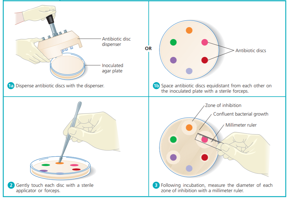 Figure: Kirby Bauer Disc Diffusion Method
Procedure for Modified Kirby Bauer method
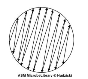 Figure: Swabbing pattern to ensure proper inoculation of the organism.
- Prepare the inoculum from the primary culture plate by touching with a loop the tops of each of 3 to 5 colonies of similar appearance, of the organism to be tested and transfer this growth to a tube of saline. If the inoculum has to be made from a pure culture, suspend a loopful of the confluent growth similarly.
Note: Multiple similar looking colonies should be picked to minimize the possibility of testing a non-representative colony such as picking a susceptible colony only and missing the resistant mutants dispersed in other colonies. - Compare the tube with the 0.5 McFarland turbidity standard (approximate cell density 1.5 x 10⁸ CFU/mL) and adjust the density of the test suspension to that of the standard by adding more bacteria or more sterile saline.
Remember: Proper adjustment of the turbidity of the inoculum is essential to ensure that the resulting lawn of growth is confluent or almost confluent. - Inoculate the plates by dipping a sterile swab into the inoculum. Remove excess inoculum by pressing and rotating the swab firmly against the side of the tube above the level of the liquid.
- Streak the swab all over the surface of the medium three times, rotating the plate through an angle of 60° after each application. Finally, pass the swab around the edge of the agar surface. The swab should follow as it is drawn across the plate (as shown in the figure). Discard the swab into an appropriate container.
- Leave the inoculum to dry for a few minutes (at least 3 to 5 minutes, but no more than 15 minutes) at room temperature with the lid closed.
- Place the appropriate antimicrobial-impregnated disks on the surface of the agar (antimicrobial disks can be purchased from any reputable suppliers)
- Antimicrobial discs can be placed on the inoculated plates using a pair of sterile forceps. It is convenient to use a template to place the discs uniformly or a sterile needle-tip may also be used to place the antibiotic discs on the plate. Alternatively, an antibiotic disc dispenser (as shown in image-2) can be used to apply the discs to the inoculated plate.
- Disks should not be placed closer than 24 mm (center to center) on the Mueller Hinton agar plate. Ordinarily, no more than 12 disks should be placed on a 150-mm plate or more than 5 disks on a 100-mm plate. Avoid placing disks close to the edge of the plate, as the zones will not be fully round and can be difficult to measure.
- Agar depth matters as much as disc spacing. Plates poured too thin or too thick will skew zone sizes regardless of how carefully everything else is done. See Mueller-Hinton Agar for the exact depth specification and plate volumes by plate size.
- Each disc should be gently pressed down to ensure complete contact with the agar surface so it does not fall when the plate is inverted during incubation. Do not push the disc into the agar.
- The plates should be placed in an incubator at 35 °C within 30 minutes of preparation. Temperatures above 35°C invalidate results for oxacillin/methicillin. Do not incubate in an atmosphere of carbon dioxide, as this will decrease the pH of the agar and cause errors from incorrect media pH.
- After overnight incubation, the diameter of each zone (including the diameter of the disc) should be measured and recorded in mm. The results should then be interpreted according to the antimicrobial susceptibility interpretation chart.
Measurement of the zone of inhibition
The measurements can be made
- with a ruler on the under-surface of the plate without opening the lid.
- If the medium is opaque, the zone can be measured by means of a pair of calipers.
- A template may be used to assess the final result of the susceptibility tests.
 Figure: Using antibiotic disc dispenser
The endpoint of inhibition is judged by the naked eye at the edge where the growth starts, but there are three exceptions:
- With sulfonamides and co-trimoxazole, slight growth occurs within the inhibition zone; such growth should be ignored.
- When β-lactamase-producing staphylococci are tested against penicillin, zones of inhibition are produced with a heaped-up, clearly defined edge; these are readily recognizable when compared with the sensitive control, and regardless of the size of the zone of inhibition, they should be reported as resistant.
- Certain Proteus species may swarm into the area of inhibition around some antibiotics, but the zone of inhibition is usually clearly outlined and the thin layer of swarming growth should be ignored.
Results
Results can be read after 16 to 18 hours of incubation (standard for most organisms). Following incubation, measure the zone sizes to the nearest millimeter (mm) using a ruler or caliper; include the diameter of the disk in the measurement.
Interpretation and Reporting of antimicrobial susceptibility results
- Using the published CLSI guidelines, determine the susceptibility or resistance of the organism to each drug tested. Note that there are different charts for different organisms.
- For each drug, indicate on the recording sheet whether the zone size is susceptible (S), intermediate (I), or resistant (R) based on the interpretation chart. Zone sizes are not reported to physicians.
Quality control of the disc diffusion test
A susceptibility result is only trustworthy if the test itself is working. The discs may have lost potency, the Mueller-Hinton agar may be the wrong depth or have the wrong cation content, the inoculum may be off, or the technique may have drifted. Quality control catches all of these by running reference strains whose expected zone sizes are already known.
The reference strains. Disc diffusion is controlled with well-characterized ATCC strains that have published, defined zone-diameter ranges for each antibiotic:
- Escherichia coli ATCC 25922, the main control for Enterobacterales.
- Staphylococcus aureus ATCC 25923, the control for gram-positive organisms.
- Pseudomonas aeruginosa ATCC 27853, the control for non-Enterobacterales gram-negative rods.
- Escherichia coli ATCC 35218, a beta-lactamase producer, used to control beta-lactam and beta-lactamase-inhibitor combinations (for example, amoxicillin-clavulanate).
- Enterococcus faecalis ATCC 29212, used with certain agents and for media checks.
Fastidious organisms have their own control strains, for example Haemophilus influenzae ATCC 49247 and Streptococcus pneumoniae ATCC 49619.
How it works. The control strain is set up exactly like a patient isolate, same medium, same inoculum, same discs, same incubation, and its zone diameters are measured and compared with the published CLSI ranges. If every zone falls within range, the discs, medium, and technique are all validated, and patient results from that run can be reported. If a zone falls outside its range, the run has failed: patient results are held until the cause is found and corrected.
What an out-of-range result points to. A failed control zone is a clue to where the test went wrong:
- Zones too large: agar too thin, inoculum too light, or discs too concentrated.
- Zones too small: agar too thick, inoculum too heavy, discs that have lost potency, or delayed disc application.
- A specific drug consistently out of range: usually a problem with that disc lot.
- Systematic drift across many drugs: often a medium problem, wrong agar depth (the medium should be about 4 mm deep), incorrect calcium or magnesium content (which especially affects Pseudomonas aeruginosa with aminoglycosides and colistin), or an incorrect pH.
This is why the control strain is so useful: its expected answer is known, so the direction and pattern of its failure point straight to the fault.
How often QC is run. Traditionally, the control strains are tested daily. Once a laboratory can show good, consistent performance, typically 20 or 30 consecutive days of in-range results for each antibiotic-organism combination, it may reduce testing to weekly, reverting to daily if any failure occurs or when a new lot or shipment of discs or media is introduced. Current CLSI guidance frames this as daily testing, or a reduced frequency governed by the laboratory's own individualized quality control plan. Either way, the principle is the same: test often enough to catch a problem before it reaches a patient result, and always test with each new lot of materials.
Quality control of susceptibility testing is one part of the laboratory's overall quality control. For what these reference strains are and which to use, see Quality Control Strains and ATCC in Microbiology. For how susceptibility QC fits within the whole quality system, see Quality Control in the Microbiology Laboratory.
Limitations
If performed precisely according to standard protocol, disk diffusion method yields data that can reliably predict the in vivo effectiveness of the drug in question but it has a few limitations too:
- Does not provide accurate information about the minimum inhibitory concentration (MIC)
- Does not provide reliable results with some antibiotic/organism combinations, such as for penicillin G in Neisseria meningitidis and S. pneumoniae.
Standard Kirby-Bauer vs. the Stokes Method
Both are disc diffusion methods reading the same kind of zone; the difference is how each controls for variability. Standard Kirby-Bauer relies on tightly standardizing every variable independently: inoculum density, agar depth, disc potency, incubation temperature.
The Stokes method takes a different approach: it runs a known control strain on the same plate as the test organism, so the comparison is made directly against a reference rather than against a fixed mm chart. That makes Stokes more forgiving of small batch-to-batch variation in media or disc potency, which is part of why it remains popular in labs without tightly controlled reagent supply chains.
Learning & Remembering
The Warm Incubator Problem: see "Why This Matters" above, how a few degrees of incubator drift can turn a resistant MRSA isolate into a falsely reassuring oxacillin-susceptible report.
The Heaped Edge: Most resistant zones are simply smaller. But when β-lactamase-producing staphylococci are tested against penicillin, the zone can look deceptively close to normal size, except for a telltale heaped-up, sharply defined edge where the enzyme has degraded the drug right at the boundary. CLSI guidance is explicit: report this as resistant regardless of zone size, because the edge, not the diameter, is the tell here.
One sentence that captures it: A zone of inhibition only means what the protocol says it means. Change the temperature, the agar depth, or the inoculum, and the same isolate can report as a different organism entirely.
Exam facts
| Question | Answer |
|---|---|
| Minimum disc spacing (center to center)? | 24 mm |
| Max discs on a 150 mm plate / 100 mm plate? | 12 / 5 |
| Standardized agar depth? | ~4 mm |
| Incubation temperature? | 35°C; above this invalidates oxacillin/methicillin results |
| Drying time before applying discs? | 3–5 minutes minimum, no more than 15 |
| Name the three exceptions to standard zone-edge reading | Sulfonamide/co-trimoxazole haze (ignore), β-lactamase-producing staph vs. penicillin (heaped edge = resistant regardless of size), Proteus swarming (ignore the thin swarm layer) |
| What does disc diffusion NOT provide? | An exact MIC value |
References and further reading
- Cappuccino, J. G., & Welsh, C. T. Microbiology: A Laboratory Manual, 11th ed. Pearson; 2016.
- CLSI. M100—Performance Standards for Antimicrobial Susceptibility Testing. Clinical and Laboratory Standards Institute; current annual edition.
- CLSI. M02—Performance Standards for Antimicrobial Disk Susceptibility Tests. Clinical and Laboratory Standards Institute; current edition.
Frequently Asked Questions
Why does incubating above 35°C invalidate oxacillin/methicillin results?
Why is a heaped-up zone edge reported as resistant even if the zone looks otherwise normal-sized?
Why does agar depth matter for disc diffusion accuracy?
What's the difference between standard Kirby-Bauer and the Stokes method?
Standard Kirby-Bauer relies on tightly standardizing inoculum density, agar depth, disc potency, and incubation temperature independently. The Stokes method instead runs a known control strain on the same plate as the test organism, comparing results directly rather than against a fixed chart, making it more forgiving of batch-to-batch variation.
Why are sulfonamide and co-trimoxazole zones read differently from other antibiotics?
Can disc diffusion provide an exact MIC value?
No. Disc diffusion only categorizes isolates as Susceptible, Intermediate, or Resistant. For an exact MIC value, methods like E-test or broth/agar dilution are needed.

Tankeshwar Acharya, MSc (Medical Microbiology)
Tankeshwar Acharya is an Assistant Professor in the Department of Microbiology at Patan Academy of Health Sciences (PAHS), Nepal, where he has been teaching and practicing clinical microbiology for over 14 years. He is the founder of Microbe Online, one of the leading free microbiology education resources on the web, covering bacteriology, mycology, parasitology, immunology, and clinical laboratory diagnostics written from direct experience in both the classroom and the diagnostic laboratory.
Comments
No comments yet. Be the first to share your thoughts.
Leave a comment
All comments are reviewed before they appear.